. 2009 Aug;5(8):e1000461.
doi: 10.1371/journal.pcbi.1000461. Epub 2009 Aug 13.

How thioredoxin dissociates its mixed disulfide

Affiliations

How thioredoxin dissociates its mixed disulfide

Goedele Roos et al. PLoS Comput Biol. 2009 Aug.

Abstract

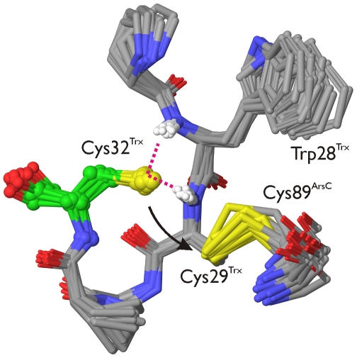
The dissociation mechanism of the thioredoxin (Trx) mixed disulfide complexes is unknown and has been debated for more than twenty years. Specifically, opposing arguments for the activation of the nucleophilic cysteine as a thiolate during the dissociation of the complex have been put forward. As a key model, the complex between Trx and its endogenous substrate, arsenate reductase (ArsC), was used. In this structure, a Cys29(Trx)-Cys89(ArsC) intermediate disulfide is formed by the nucleophilic attack of Cys29(Trx) on the exposed Cys82(ArsC)-Cys89(ArsC) in oxidized ArsC. With theoretical reactivity analysis, molecular dynamics simulations, and biochemical complex formation experiments with Cys-mutants, Trx mixed disulfide dissociation was studied. We observed that the conformational changes around the intermediate disulfide bring Cys32(Trx) in contact with Cys29(Trx). Cys32(Trx) is activated for its nucleophilic attack by hydrogen bonds, and Cys32(Trx) is found to be more reactive than Cys82(ArsC). Additionally, Cys32(Trx) directs its nucleophilic attack on the more susceptible Cys29(Trx) and not on Cys89(ArsC). This multidisciplinary approach provides fresh insights into a universal thiol/disulfide exchange reaction mechanism that results in reduced substrate and oxidized Trx.


Conflict of interest statement

The authors have declared that no competing interests exist.

Figures

Figure 1
Figure 1. In the structure of the Trx-ArsC mixed disulfide complex the functionally key disulfide between Cys29Trx and Cys89ArsC is formed.
The Bs_Trx-ArsC complex (2IPA) with the side chains of residues Cys29Trx, Cys32Trx, Trp28Trx, Arg16ArsC, Cys82ArsC and Cys89ArsC in stick representation is shown. The Trx α1-helix is shown in blue; the ArsC looped-out redox helix between Cys82ArsC and Cys89ArsC in pink.
Figure 2
Figure 2. Bs_Trx reduces Bs_ArsC via an intermediate Trx-ArsC complex.
A Cys29Trx of reduced Bs_Trx (2GZY) nucleophilically attacks Cys89ArsC of the Cys82ArsC-Cys89ArsC disulfide of oxidized Bs_ArsC (1Z2E), leading to the formation of the mixed Cys29Trx-Cys89ArsC disulfide (2IPA). (B and C) Cys32Trx performs a nucleophilic attack on Cys29Trx, leading to the release of reduced Bs_ArsC (1Z2D) and oxidized Bs_Trx (2GZZ).
Figure 3
Figure 3. Experimental pKa’s are correlated with the calculated NPA charge.
NPA-charge-pKa calibration curve (black circles) obtained A for a series of small thiolate molecules (the experimental pKa's are obtained from ref. [74]) and B for a series of cysteine residues of Trx and ArsC systems. The data points of the calibration curve B (a–g) are tabulated in Table S1 in Supplemental Data and are also indicated on curve A. trifluoromethanethiol (1), methanethiol (2), mercaptoethanol (3), cysteine (4), trifluoroethanethiol (5), benzenemethanethiol (6) and thioacetic acid (7). Cys89 S. aureus ArsC (a), Cys77 B. subtilis resA , (b), Cys10 S. aureus ArsC (c), Cys32 E. coli Trx1 (d), Cys29 S. aureus P31T C32S Trx1 (e), Cys73 R. capsulatus Trx2 (f) and Cys35 E. coli Trx1 (g).
Figure 4
Figure 4. Model systems of Bs_Trx, Bs_ArsC and Bs_Trx-ArsC complex for pKa and reactivity analysis.
A Reduced Bs_Trx (2GZY) modelled by the Trp28Trx-Cys32Trx active site and the Lys33-Glu45 α1-helix (blue). B Oxidized Bs_ArsC (1Z2E) represented by the Cys82ArsC-Cys89ArsC looped-out redox helix (ArsC_ox). C Model of Bs_Trx-ArsC (Trx_ArsC_1), in which no hydrogen bond interactions with Cys32Trx nor with Cys82ArsC are present, represented by the Trp28Trx-Cys32Trx Trx active site and the Cys82ArsC-Cys89ArsC ArsC redox helix (pink) and Thr11ArsC. D Model of Bs_Trx_ArsC (Trx-ArsC_2), in which the Cys82ArsCSγ—Arg16ArsCNη1 and the Cys32TrxSγ—Cys29TrxN hydrogen bonds are present. The Trp28Trx-Cys32Trx Trx active site and the Cys82ArsC-Cys89ArsC ArsC redox helix (pink) and Thr11ArsC and Arg16ArsC are included. E Trx_ArsC_1_trunc and Trx_ArsC_2_trunc model systems including the Trp28Trx-Cys32Trx active site of Trx and Cys89ArsC of the ArsC. F Trx_ArsC_2_trunc+helix and Trx_ArsC_1_trunc+helix model systems including everything described under (E.) and the Trx Lys33Trx-Glu45Trx α1-helix (blue).
Figure 5
Figure 5. Hydrogen bonding of Cys32Trx in the mixed disulfide of Bs_Trx-ArsC.
Time course in the MD simulation of the distance between A Cys32TrxSγ and Trp28TrxN and between B Cys32TrxSγ and Cys29TrxN, with ionized (black) and neutral (red) Cys82ArsC. C Example of one MD snapshot with the Cys32TrxSγ—Cys29TrxN and Cys32TrxSγ—Trp28TrxN hydrogen bonds introduced, when Cys82ArsC is deprotonated.
Figure 6
Figure 6. A conformational change brings Cys32Trx in contact with Cys29Trx.
Time course in the MD simulation (ionized Cys82ArsC) of the distance between Cys29TrxSγ and Cys32TrxSγ (red) and Cys32TrxSγ and Trp28TrxN (A, black) and Cys32TrxSγ and Cys29TrxN (B, black). C Superposition of the Trx active site (Trx28Trx-Cys32Trx) and the ArsC redox helix (Cys82ArsC-Cys89ArsC) of the Bs_Trx-ArsC complex at 0 ns (purple) and 14.5 ns (green) simulation time showing the conformational change associated with the approach of Cys32Trx to Cys29Trx during the MD simulation (ionized Cys82ArsC).
Figure 7
Figure 7. Cys32Trx is primed for nucleophilic attack in the Trx-ArsC activated complex.
Selected snapshots from an MD simulation of the Trx-ArsC complex (ionized Cys82ArsC), presenting the structural basis of the activation of Cys32Trx (green carbons, ball and sticks) for its nucleophilic attack onto Cys29Trx. In these snapshots, Cys32TrxSγ is hydrogen-bonded (magenta dotted lines) to the amide NH groups of both Trp28Trx and Cys29Trx. Also, the sulphur atoms of Cys32Trx and Cys29Trx are within 4.5 Å of each other. The hydrogen bonds satisfy the geometric criteria: Cys32TrxSγ—Trp28TrxN≤4 Å and Cys32TrxSγ—Cys29NTrx≤4 Å, with the corresponding angles between Sγ and the N−H vectors being ≥150 degrees. The hydrogen-bonds donated to the sulphur of Cys32Trx lower its pKa to 7.4 (Table 4), corresponding to a significant population of the thiolate form of Cys32Trx. This thiolate being close to Cys29Trx, it is in effect primed for nucleophilic attack onto Cys29Trx (black arrow). In combination with the supporting reaction analysis, pKa calculations and complex formation experiments (main text), the conformations and interactions shown here are proposed to underpin the dissociation mechanism of the Tx-ArsC complex.
Figure 8
Figure 8. The putative role of Asp23Trx in the deprotonation of Cys32Trx revisited.
A Water is sometimes proposed to assist the deprotonation of Cys32Trx by Asp23Trx. The position of the water molecule in the Bs_Trx-ArsC complex was obtained by modeling. B Evaluation of the wild type Sa_Trx-Sa_ArsCTrip and the Sa_Trx D23A-Sa_ArsCTrip complex formation on a non-reducing SDS-PAGE (pH 7.5). No stable complex is detected. (1. Sa_Trx+Sa_ArsCTrip; 2. Sa_Trx D23A+Sa_ArsCTrip 3. Positive control band of the Sa_Trx-ArsC complex [1]). (C.) Reversed phase-analysis of the reaction products after complex formation between Sa_Trx D23A and Sa_ArsCTrip. E D Time course in the MD simulation (ionized Cys82ArsC) of the distance between Asp23TrxOδ1 and Cys32TrxSγ (red) and Cys32TrxSγ and Trp28TrxN (D, black) and Cys32TrxSγ and Cys29TrxN (E, black).

Similar articles

Cited by

References

    1. Messens J, Van Molle I, Vanhaesebrouck P, Limbourg M, Van Belle K, et al. How thioredoxin can reduce a buried disulphide bond. J Mol Biol. 2004;339:527–537. - PubMed
    1. Aslund F, Berndt KD, Holmgren A. Redox potentials of glutaredoxins and other thiol-disulfide oxidoreductases of the thioredoxin superfamily determined by direct protein-protein redox equilibria. J Biol Chem. 1997;272:30780–30786. - PubMed
    1. Cheng Z, Arscott LD, Ballou DP, Williams CH., Jr. The relationship of the redox potentials of thioredoxin and thioredoxin reductase from Drosophila melanogaster to the enzymatic mechanism: reduced thioredoxin is the reductant of glutathione in Drosophila. Biochemistry. 2007;46:7875–7885. - PubMed
    1. Martin JL. Thioredoxin–a fold for all reasons. Structure. 1995;3:245–250. - PubMed
    1. Eklund H, Gleason FK, Holmgren A. Structural and functional relations among thioredoxins of different species. Proteins. 1991;11:13–28. - PubMed

Publication types