Molecular plasticity of E-cadherin and sialyl lewis x expression, in two comparative models of mammary tumorigenesis
- PMID: 19675678
- PMCID: PMC2722091
- DOI: 10.1371/journal.pone.0006636
Molecular plasticity of E-cadherin and sialyl lewis x expression, in two comparative models of mammary tumorigenesis
Abstract
Background: The process of metastasis involves a series of steps and interactions between the tumor embolus and the microenvironment. Key alterations in adhesion molecules are known to dictate progression from the invasive to malignant phenotype followed by colonization at a distant site. The invasive phenotype results from the loss of expression of the E-cadherin adhesion molecule, whereas the malignant phenotype is associated with an increased expression of the carbohydrate ligand-binding epitopes, (e.g. Sialyl Lewis (x/a)) that bind endothelial E-selectin of the lymphatics and vasculature.
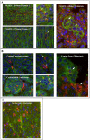
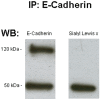
Methodology: Our study analyzed the expression of two adhesion molecules, E-cadherin and Sialyl Lewis x (sLe(x)), in both a canine mammary carcinoma and human inflammatory breast cancer (IBC) model, using double labelled immunofluorescence staining.
Results: Our results demonstrate that canine mammary carcinoma and human IBC exhibit an inversely correlated cellular expression of E-cadherin and sLe(x) within the same tumor embolus.
Conclusions: Our results in these two comparative models (canine and human) suggest the existence of a biologically coordinated mechanism of E-cadherin and sLe(x) expression (i.e. molecular plasticity) essential for tumor establishment and metastatic progression.
Conflict of interest statement
Figures


Similar articles
-
Sialyl Lewis x expression in canine malignant mammary tumours: correlation with clinicopathological features and E-Cadherin expression.BMC Cancer. 2007 Jul 6;7:124. doi: 10.1186/1471-2407-7-124. BMC Cancer. 2007. PMID: 17617904 Free PMC article.
-
Cooperative role of E-cadherin and sialyl-Lewis X/A-deficient MUC1 in the passive dissemination of tumor emboli in inflammatory breast carcinoma.Oncogene. 2002 May 16;21(22):3631-43. doi: 10.1038/sj.onc.1205389. Oncogene. 2002. PMID: 12032865
-
Expression of sialyl lewis X, sialyl Lewis A, E-cadherin and cathepsin-D in human breast cancer: immunohistochemical analysis in mammary carcinoma in situ, invasive carcinomas and their lymph node metastasis.Anticancer Res. 2005 May-Jun;25(3A):1615-22. Anticancer Res. 2005. PMID: 16033070
-
Molecular mechanism for cancer-associated induction of sialyl Lewis X and sialyl Lewis A expression-The Warburg effect revisited.Glycoconj J. 2004;20(5):353-64. doi: 10.1023/B:GLYC.0000033631.35357.41. Glycoconj J. 2004. PMID: 15229399 Review.
-
In vitro experimental studies of sialyl Lewis x and sialyl Lewis a on endothelial and carcinoma cells: crucial glycans on selectin ligands.Glycoconj J. 1997 Aug;14(5):593-600. doi: 10.1023/a:1018536509950. Glycoconj J. 1997. PMID: 9298692 Review.
Cited by
-
Mucin-Type O-GalNAc Glycosylation in Health and Disease.Adv Exp Med Biol. 2021;1325:25-60. doi: 10.1007/978-3-030-70115-4_2. Adv Exp Med Biol. 2021. PMID: 34495529
-
Tumour-associated carbohydrate antigens in breast cancer.Breast Cancer Res. 2010;12(3):204. doi: 10.1186/bcr2577. Epub 2010 Jun 8. Breast Cancer Res. 2010. PMID: 20550729 Free PMC article. Review.
-
Glycosylation in cancer: mechanisms and clinical implications.Nat Rev Cancer. 2015 Sep;15(9):540-55. doi: 10.1038/nrc3982. Epub 2015 Aug 20. Nat Rev Cancer. 2015. PMID: 26289314 Review.
References
-
- Woodhouse EC, Chuaqui RF, Liotta LA. General Mechanisms of Metastasis. Cancer. 1997;80:1529–1537. - PubMed
-
- Liotta LA, Kohn EC. The Microenvironment of the Tumour-host Interface. Nature. 2001;411:375–379. - PubMed
-
- Joyce CT, Kalluri R. Mechanisms of Metastasis: Epithelial-to-Mesenchymal Transition and Contribution of Tumor Microenvironment. J Cell Biochem. 2007;101:816–829. - PubMed
-
- Liotta LA, Steeg PS, Stetler-Stevenson WG. Cancer metastasis and angiogenesis: an imbalance of positive and negative regulation. Cell. 1991;64:327–336. - PubMed
Publication types
MeSH terms
Substances
LinkOut - more resources
Full Text Sources

