. 2010 Jan;16(1):179-89.
doi: 10.1089/ten.TEA.2009.0164.

Bone grafts engineered from human adipose-derived stem cells in perfusion bioreactor culture

Affiliations

Bone grafts engineered from human adipose-derived stem cells in perfusion bioreactor culture

Mirjam Fröhlich et al. Tissue Eng Part A. 2010 Jan.

Abstract

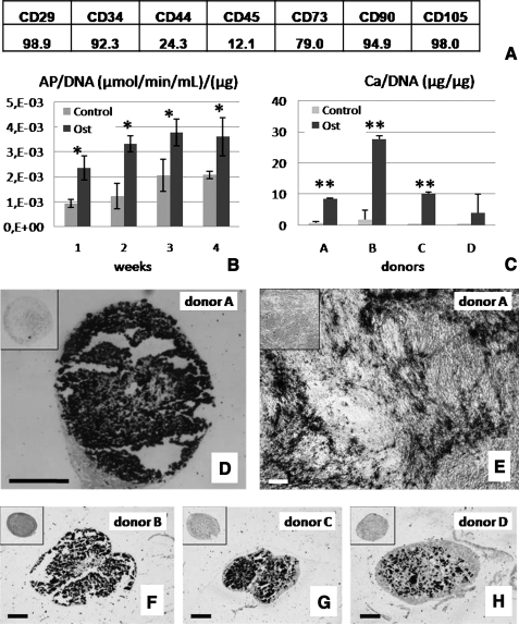
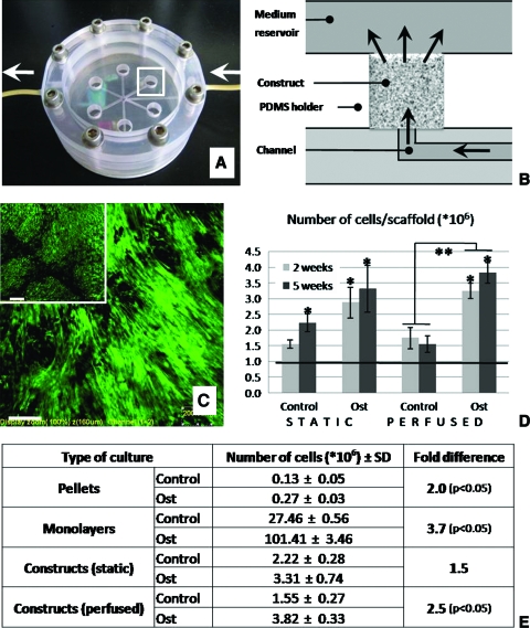
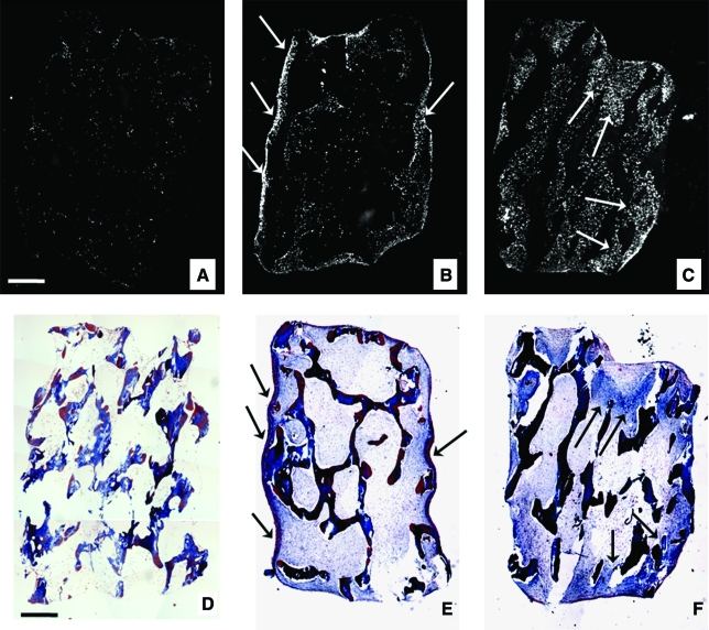
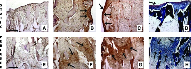
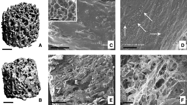
We report engineering of half-centimeter-sized bone constructs created in vitro using human adipose-derived stem cells (hASCs), decellularized bone scaffolds, and perfusion bioreactors. The hASCs are easily accessible, can be used in an autologous fashion, are rapidly expanded in culture, and are capable of osteogenic differentiation. hASCs from four donors were characterized for their osteogenic capacity, and one representative cell population was used for tissue engineering experiments. Culture-expanded hASCs were seeded on fully decellularized native bone scaffolds (4 mm diameter x 4 mm thick), providing the necessary structural and mechanical environment for osteogenic differentiation, and cultured in bioreactors with medium perfusion. The interstitial flow velocity was set to a level necessary to maintain cell viability and function throughout the construct volume (400 microm/s), via enhanced mass transport. After 5 weeks of cultivation, the addition of osteogenic supplements (dexamethasone, sodium-beta-glycerophosphate, and ascorbic acid-2-phosphate) to culture medium significantly increased the construct cellularity and the amounts of bone matrix components (collagen, bone sialoprotein, and bone osteopontin). Medium perfusion markedly improved the distribution of cells and bone matrix in engineered constructs. In summary, a combination of hASCs, decellularized bone scaffold, perfusion culture, and osteogenic supplements resulted in the formation of compact and viable bone tissue constructs.


Figures

FIG. 1.
FIG. 1.
Osteogenic capacity of human adipose-derived stem cells (hASCs). (A) The presence of antigens in the starting population of hASCs. (B) Alkaline phosphatase activity of hASCs was higher in osteogenic (Ost) than in unsupplemented control medium (Control) (data are shown for donor A, n = 3, *p < 0.05). (C) Calcium contents of hASC pellets from four different donors (A, B, C, and D) cultured in osteogenic medium (Ost) and control medium (Control) (n = 3–4, **p < 0.001). (D, F–H) von Kossa staining of the central regions of osteoinduced pellets of donors A, B, C, and D (black: calcium deposits). Insets show negative stains for pellets cultured in control medium. (E) von Kossa staining of osteoinduced cell monolayers for donor A is shown for comparison. Scale bar is 250 μm for all images.
FIG. 2.
FIG. 2.
Cell content and viability during 5 weeks of culture: four culture settings and two culture media. (A) Perfusion bioreactor used in the study enables culturing of six constructs simultaneously. The region indicated by a white rectangle is shown in panel B in full detail. Arrows indicate directions of medium flow. (B) Medium flows throughout the scaffold, as indicated by arrows. The schematic corresponds to the region indicated by rectangle in the figure A. (C) After 5 weeks of culture, tissue constructs from both the perfused and static groups (inset) contained viable cells. Live/dead assay of the central regions of the constructs is shown (green indicates live cells and red would indicate dead cells). Scale bar is 200 μm. (D) The number of cells in constructs increased up to three times during the culture period in comparison to the initial cell numbers (denoted by a line) (n = 3, *p < 0.05). Osteogenic medium resulted in higher amounts of cells than control medium (**p < 0.05). (E) Number of cells was higher under osteogenic conditions when compared with control conditions in all types of cultures performed. Color images available online at www.liebertonline.com/ten.
FIG. 3.
FIG. 3.
Cell and matrix distribution within constructs: effects of perfusion. Constructs immediately after seeding (A, D), and after 5 weeks of culture in osteogenic medium under either static conditions (B, E) or with medium perfusion (C, F). Constructs were stained with (A–C) 4′-6-diamidino-2-phenylindole to visualize cell distribution (cells shown in white) and (D–F) trichrome to observe the presence of collagen-rich matrix (blue). (A) Seeding resulted in even initial distribution of cells throughout the scaffold. After 5 weeks of static culture, cells were found mostly in the outer regions of the constructs (B). After 5 weeks of culture with medium perfusion, cells were evenly distributed throughout the construct volume (C). After 5 weeks of culture, the regions of newly formed collagen matrix (indicated by arrows in E and F) colocalized with the regions of higher cell densities (indicated by arrows in B and C). Scale bar: 0.5 mm (A, D). Color images available online at www.liebertonline.com/ten.
FIG. 4.
FIG. 4.
Presence of collagen, bone sialoprotein (BSP), and osteopontin within the hASCs constructs. Absence of osteogenic supplements resulted in lack of bone matrix markers in static as well as in perfused cultures: result for BSP is shown (A, E). Deposition of BSP (B, F), osteopontin (C, G), and collagen (D, H) in constructs cultured for 5 weeks under osteogenic conditions, either statically (B–D) or with medium perfusion (F–H). Strong presence of all three bone markers (indicated by arrows) was observed throughout the perfused constructs (central regions are shown in F, G, H) and only at the periphery of statically cultured constructs (construct edges are shown in B, C, D). Scale bar: 250 μm (A, E). Color images available online at www.liebertonline.com/ten.
FIG. 5.
FIG. 5.
Morphology and mineral content of tissue constructs cultured with perfusion of osteogenic medium. Microcomputed tomography images of the initial (unseeded) scaffold (A) and a tissue construct cultured for 5 weeks (B) show the new mineral deposition during cultivation. Scanning electron microscopy images (C–F) show that cells and deposited matrix filled the pore spaces of initial scaffold (inset in C), both in the outer (C, D) and the inner construct regions (E, F). Small round formations (arrows in D) are consistent with the formation of hydroxyapatite crystals (D). Scale bar: 1 mm (A, B, C, E) and 25 μm (D, F).

References

    1. Salgado A.J. Coutinho O.P. Reis R.L. Bone tissue engineering: state of the art and future trends. Macromol Biosci. 2004;4:743. - PubMed
    1. Giannoudis P.V. Dinopoulos H. Tsiridis E. Bone substitutes: an update. Injury. 2005;36:20. - PubMed
    1. Lecanda F. Avioli L.V. Cheng S.L. Regulation of bone matrix protein expression and induction of differentiation of human osteoblasts and human bone marrow stromal cells by bone morphogenetic protein-2. J Cell Biochem. 1997;67:386. - PubMed
    1. Bruder S.P. Jaiswal N. Haynesworth S.E. Growth kinetics, self-renewal, and the osteogenic potential of purified human mesenchymal stem cells during extensive subcultivation and following cryopreservation. J Cell Biochem. 1997;64:278. - PubMed
    1. Meinel L. Karageorgiou V. Fajardo R. Snyder B. Shinde-Patil V. Zichner L. Kaplan D. Langer R. Vunjak-Novakovic G. Bone tissue engineering using human mesenchymal stem cells: effects of scaffold material and medium flow. Ann Biomed Eng. 2004;32:112. - PubMed

Publication types

Substances