. 2009 Aug 13;63(3):316-28.
doi: 10.1016/j.neuron.2009.07.019.

Overexpression of IGF-1 in muscle attenuates disease in a mouse model of spinal and bulbar muscular atrophy

Affiliations

Overexpression of IGF-1 in muscle attenuates disease in a mouse model of spinal and bulbar muscular atrophy

Isabella Palazzolo et al. Neuron. .

Abstract

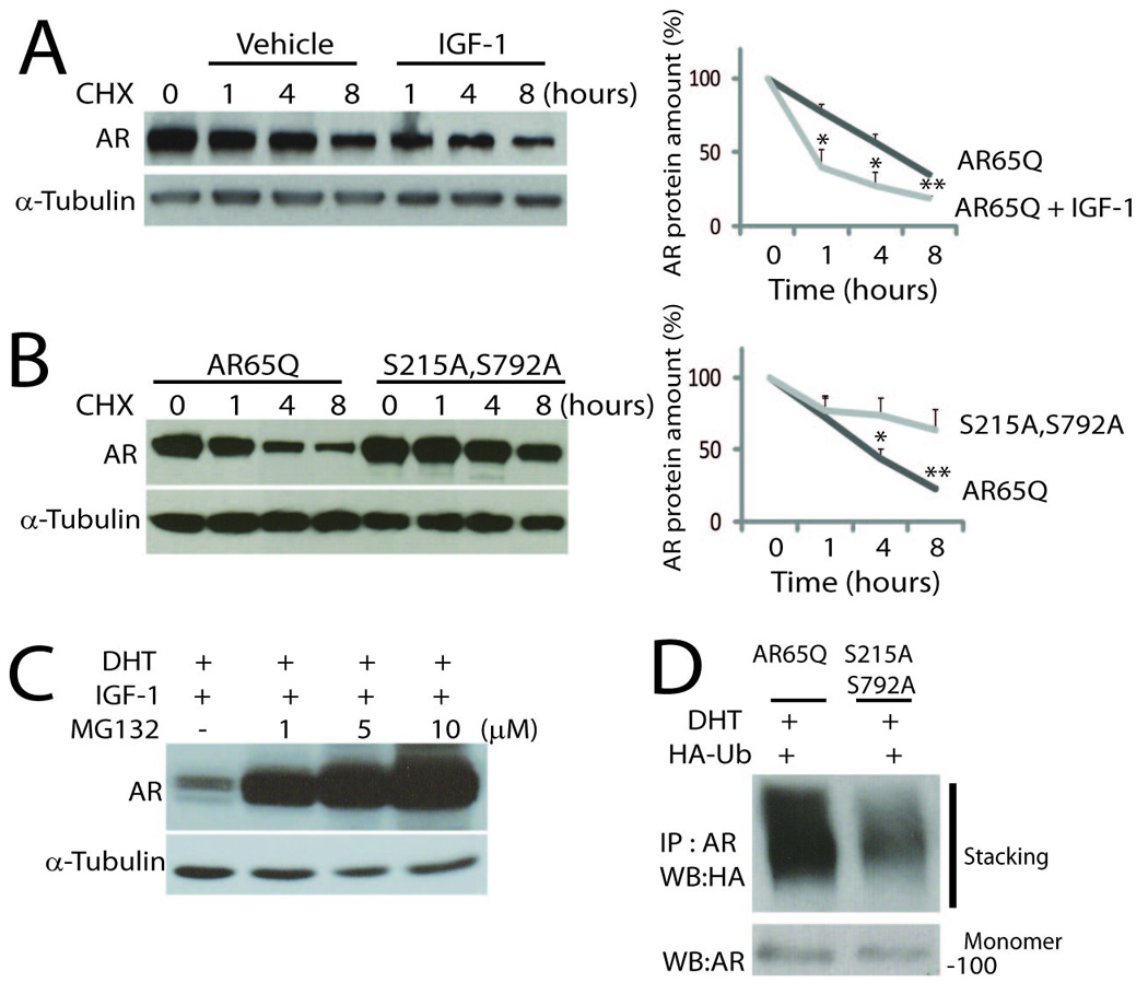
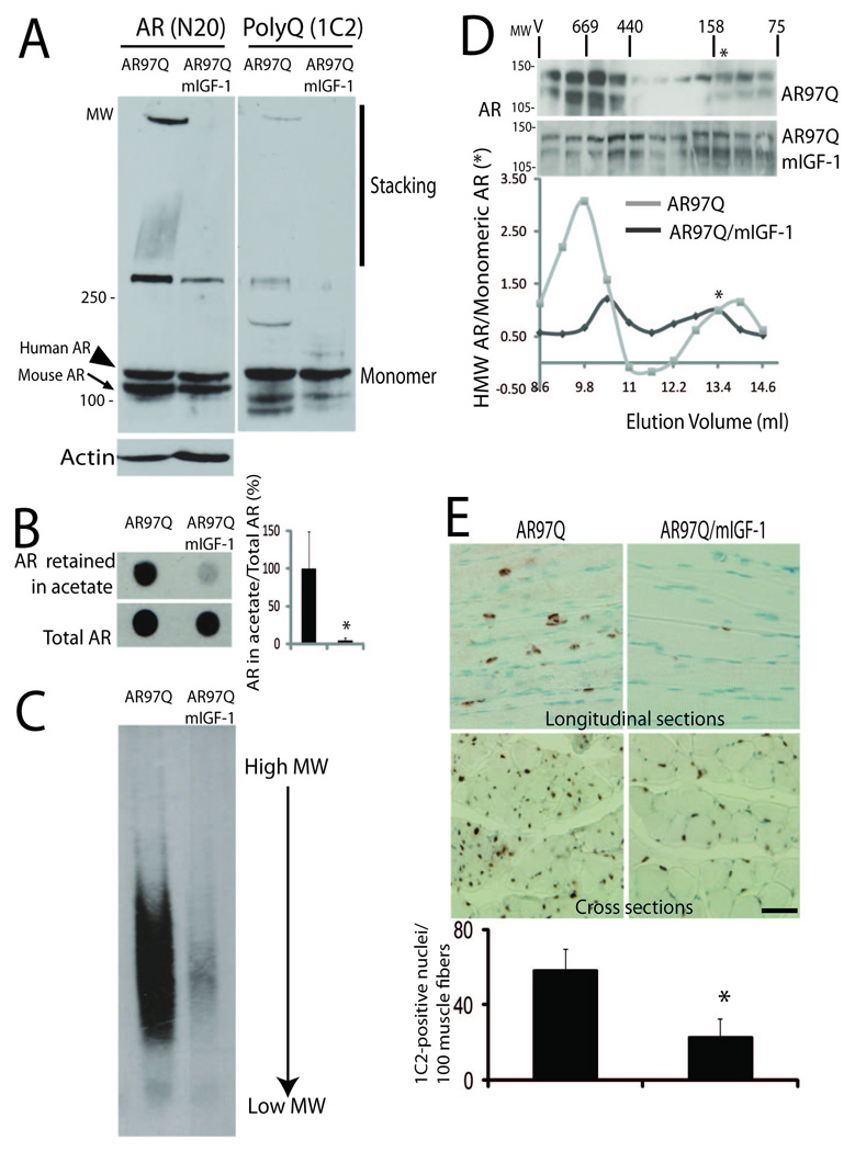
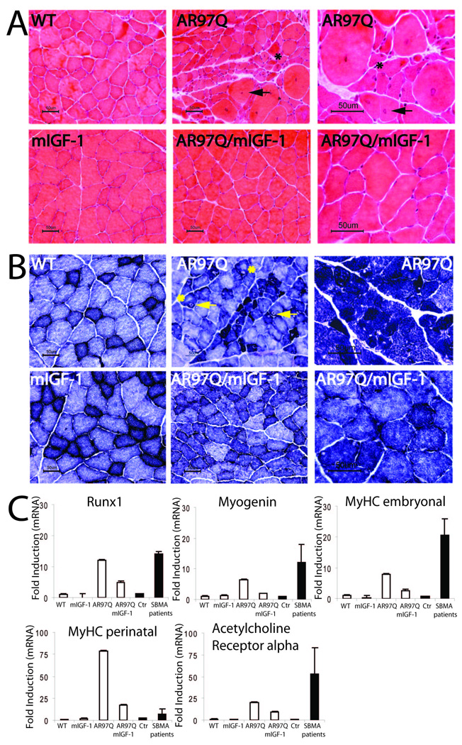
Expansion of a polyglutamine tract in the androgen receptor (AR) causes spinal and bulbar muscular atrophy (SBMA). We previously showed that Akt-mediated phosphorylation of AR reduces ligand binding and attenuates the mutant AR toxicity. Here, we show that in culture insulin-like growth factor 1 (IGF-1) reduces AR aggregation and increases AR clearance via the ubiquitin-proteasome system through phosphorylation of AR by Akt. In vivo, SBMA transgenic mice overexpressing a muscle-specific isoform of IGF-1 selectively in skeletal muscle show evidence of increased Akt activation and AR phosphorylation and decreased AR aggregation. Augmentation of IGF-1/Akt signaling rescues behavioral and histopathological abnormalities, extends the life span, and reduces both muscle and spinal cord pathology of SBMA mice. This study establishes IGF-1/Akt-mediated inactivation of mutant AR as a strategy to counteract disease in vivo and demonstrates that skeletal muscle is a viable target tissue for therapeutic intervention in SBMA.


Figures

Figure 1
Figure 1. IGF-1 Decreases AR Aggregation in Cultured Cells through Activation of the PI3K/Akt Pathway and Phosphorylation of AR by Akt
(A) Analysis of AR aggregation in COS1 cells transfected with vector expressing mutant AR with 65 glutamine residues (AR65Q) and treated with DHT, LY294002 (LY), and IGF-1 by western blotting (top panel) and filter retardation assay (bottom panel). DHT increased the accumulation of both high molecular weight (HMW) AR species (black bars) and monomeric AR (white bars). IGF-1 decreased both the basal and ligand-induced accumulation of HMW and monomeric AR, and this effect was reduced by the PI3K inhibitor LY294002. Graphs, mean ± s.e.m., n = 4, (top panel) * p = 0.01 relative to non-stimulated AR65Q–expressing cells (black bars), and # p = 0.004 relative to DHT-treated cells (white bars), (bottom panel) * p = 0.003, (post-hoc t test). AR was detected with antibody to AR (N20). α-Tubulin is shown as loading control. MW, molecular weight. (Right panel) Phosphorylation of Akt was analyzed by western blotting under the same experimental conditions with anti-phospho-serine 473 and anti-total Akt antibodies. Phosphorylation of Akt is represented as the ratio of the signals detected with anti-phospho-serine and total Akt antibodies. Graph, mean ± s.e.m., n = 4, * p = 0.05, # p = 0.01. (B) Western analysis (upper panel) and filter retardation assay (bottom panel) showed that DHT increased the accumulation of both HMW AR species (black bars) and monomeric AR (white bars) in COS1 cells expressing AR65Q and AR65Q with alanine substitutions for serine 215 and serine 792 (S215A,S792A), but not in cells expressing AR with aspartate substitutions (S215D,S792D). Graphs, mean ± s.e.m., n = 3, (top panel) * p = 0.01, (bottom panel) * p = 0.05, NS non-significant relative to the non-stimulated sample expressing AR65Q (post-hoc t test). (C) Western analysis of HEK293T cells treated with DHT and IGF-1 revealed that IGF-1 decreases the accumulation of HMW (black bars) and monomeric (white bars) AR in cells expressing AR65Q, but not the alanine-substituted receptor. Graph, mean ± s.e.m., n = 3, * p = 0.01 and NS non-significant (post-hoc t test) relative to the corresponding DHT-treated sample. In (A, B, and C) the first sample is represented as 100%.
Figure 2
Figure 2. IGF-1 Promotes Mutant AR Clearance Through the Ubiquitin-Proteasome System
(A) Western analysis of MN-1 cells transfected with AR65Q and incubated with cycloheximide (CHX) for 1, 4, and 8 hours in the presence and absence of IGF-1 showed that IGF-1 increases the rate of AR clearance. Graph, mean ± s.e.m., n = 3, * p = 0.03, ** p = 0.001 (student’s t test). (B) Western analysis of HEK293T cells transfected as indicated and treated with CHX showed that the clearance of the alanine-substituted receptor is slower than AR65Q. Graph, mean ± s.e.m., n = 5, * p = 0.04, **p = 0.01. (C) Western analysis of MN-1 cells expressing AR65Q and treated with DHT, IGF-1, and the proteasome inhibitor MG132 showed that the IGF-1-induced clearance of AR65Q is blocked by MG132 in a dose-dependent fashion. Shown is one experiment representative of 3. (D) Immunoprecipitation with anti-AR antibody of HEK293T cells expressing the indicated AR65Q versions together with HA-tagged ubiquitin (HA-Ub), followed by western analysis with anti-HA or AR antibodies, showed reduced ubiquitylation of the alanine-substituted receptor. Shown is one experiment representative of 4.
Figure 3
Figure 3. mIGF-1 Stimulates Phosphorylation of Akt and AR in SBMA Muscle
(A) Rat mIGF-1 (left panel) and human AR (right panel) mRNA levels were measured by quantitative real-time PCR in skeletal muscle of 12 weeks old mice. mIGF-1 is expressed in both AR97Q/mIGF-1 and mIGF-1 mice. AR97Q is expressed in both AR97Q and AR97Q/mIGF-1 mice. Data were normalized to phosphoglycerate kinase (PGK1) mRNA. Data are represented relative to mIGF-1 mice = 100% (left) and to AR97Q mice = 100% (right). Graphs, mean ± s.e.m.; n = 3. (B) Western analysis to detect phosphorylation at serine 473 of Akt (left panel) and serine 215 of AR (right panel) was performed using specific phospho-serine antibodies in the skeletal muscle of 6 (left) and 12 (right) weeks old mice. Total Akt and AR were measured as loading control. Graphs, mean ± s.e.m., n = 5 right panel, n = 3 left panel, * p = 0.04. (C) Western analysis of skeletal muscle from AR97Q and AR97Q/mIGF-1 mice injected with the proteasome inhibitor velcade showed increased phosphorylation of human expanded polyglutamine AR in mice overexpressing mIGF-1. Graphs, mean ± s.e.m., n = 3, * p = 0.01. In (B and C) the first sample is represented as 100%.
Figure 4
Figure 4. mIGF-1 Reduces AR Aggregation in SBMA Muscle
(A) Western blotting and (B) filter retardation assay analyses showed a reduction in the accumulation of high molecular weight AR species in the skeletal muscle of AR97Q/mIGF-1 mice at 12 weeks of age as compared to AR97Q mice. Endogenous and expanded polyglutamine AR were detected with N20 antibody, and expanded polyglutamine AR with 1C2 antibody. Actin was used as a loading control. Shown is one experiment representative of 4 in (A) and of 3 in (B). Graphs, mean ± s.e.m., n = 3, * p = 0.001. (C) 1.5% SDS-agarose gel analysis revealed that the amount of AR aggregates in the muscle of AR97Q mice was reduced in AR97Q/mIGF-1 mice. (D) Size-exclusion chromatography (SEC) and western blotting analysis of muscle lysates showed that IGF-1 reduces the amount of AR eluting off as high molecular weight species (fractions 1–4) in AR97Q/mIGF-1 mice compared to AR97Q mice. The horizontal MW markers (kDa) were obtained by SEC analysis of protein standards. V, void volume. The graph shows the ratio between the high molecular weight AR species and monomeric AR, which eluted off as fraction 9 (asterisk). Shown is one experiment, which was representative of 3. (E) Immunohistochemical analysis using 1C2 antibody on longitudinal and cross sections of AR97Q and AR97Q/mIGF-1 muscle showed a reduction of diffuse nuclear staining and nuclear inclusions in AR97Q/mIGF-1 compared to AR97Q mice. Graphs, mean ± s.e.m.; n = 5, * p = 0.04.Bar, 2 µm.
Figure 5
Figure 5. mIGF-1 Delays Disease Onset and Extends Disease Duration and Survival of SBMA Mice
Kaplan-Meier analysis of disease onset (A), disease duration (B) and survival (C) in AR97Q and AR97Q/mIGF-1 mice. (A) The median disease onset - the week in which the mouse starts to lose 5% body weight - was delayed by 10.5 weeks (arrow) in AR97Q/mIGF-1 mice relative to AR97Q mice. (B) The median disease duration - the time interval from disease onset to death - was extended by 20 weeks (arrow) in AR97Q/mIGF-1 mice compared to AR97Q mice. (C) Survival analysis showed that the median lifespan of AR97Q mice was extended by 30 weeks (arrow) in AR97Q/mIGF-1 mice. AR97Q n = 18, AR97Q/mIGF-1 n = 19. Censored data are indicated by the symbol “I” (See Experimental Procedures).
Figure 6
Figure 6. Overexpression of mIGF-1 Attenuates Behavioral Abnormalities in SBMA Mice
(A and B) Body weight analysis showed that AR97Q mice progressively lose body weight starting from 8 weeks of age. AR97Q/mIGF-1 mice have normal body weight up to 12 weeks of age, and do not show body weight loss until 16 weeks of age. Bars, s.e.m., n = 15. (C) Hanging wire (n = 15) and (D) rotarod (n = 7) analysis showed progressive deterioration in performance of AR97Q mice, which was delayed and improved in AR97Q/mIGF-1 mice. Bars, s.e.m. (E) Footprints of 12 weeks old mice showed that AR97Q mice dragged hind legs, whereas AR97Q/mIGF-1 mice did not show defects in coordination of steps. Red, front paws; blue, hind paws. Shown is one experiment representative of three.
Figure 7
Figure 7. mIGF-1 Ameliorates SBMA Muscle Pathology
(A) Hematoxylin and eosin and (B) nicotinamide adenine dinucleotide staining of transverse sections of quadriceps of 12 weeks old mice. (A) Arrows = fibers with central nuclei; asterisks = angulated and grouped fibers. (B) Arrows = target fibers; asterisks = moth eaten fibers. Shown is one experiment representative of 3. (C) The mRNA levels of runx1, myogenin, myosin heavy chain (MyHC) embryonal and perinatal, and acetylcholine receptor alpha were measured by real-time PCR in the skeletal muscle of 12 weeks old mice and SBMA patients. Data from mice and patients were normalized to PGK1 and beta-glucoronidase mRNA, respectively. Data are represented relative to wild type mice or control human samples = 1. Graphs, mean ± s.e.m., n = 3 mice and n = 4 patients).
Figure 8
Figure 8. mIGF-1 Reduces Spinal Cord Pathology in SBMA Mice
(A) Nissl-stained transverse sections of ventral spinal cords of 12 weeks old mice. Quantitative analysis showed that the number of motor neurons/section is increased in AR97Q/mIGF-1 mice as compared to AR97Q mice. Graphs, mean ± s.e.m., n = 3. (B) Western analysis showed that expression of choline acetyltransferase (ChAT) is increased in the spinal cord of 12 weeks old AR97Q/mIGF-1 mice as compared to AR97Q mice. Graphs, mean ± s.e.m., n = 3. The first sample is represented as 100%. (C) Histograms of the cross-sectional area of motor neurons (See Experimental Procedures). The distribution of motor neuron area is altered in AR97Q mice, but not in AR97Q/mIGF-1 mice relative to wild type mice. Graphs, mean ± s.e.m., n = 3 animals. (D) Western analysis revealed that AR aggregation is decreased in the spinal cord of AR97Q/mIGF-1 mice as compared to AR97Q mice at 12 weeks of age. AR was detected with N20 antibody. Actin is shown as loading control. Shown is one experiment representative of 3. (E) Immunohistochemical analysis using 1C2 antibody on cross sections of AR97Q and AR97Q/mIGF-1 ventral spinal cord showed a reduction of nuclear inclusions in AR97Q/mIGF-1 relative to AR97Q mice. Graphs, mean ± s.e.m., n = 4. Bar, 2 µm.

Comment in

Similar articles

Cited by

References

    1. Alessi DR, Andjelkovic M, Caudwell B, Cron P, Morrice N, Cohen P, Hemmings BA. Mechanism of activation of protein kinase B by insulin and IGF-1. EMBO J. 1996;15:6541–6551. - PMC - PubMed
    1. Bagriantsev SN, Kushnirov VV, Liebman SW. Analysis of amyloid aggregates using agarose gel electrophoresis. Methods Enzymol. 2006;412:33–48. - PubMed
    1. Banno H, Katsuno M, Suzuki K, Takeuchi Y, Kawashima M, Suga N, Takamori M, Ito M, Nakamura T, Matsuo K, et al. Phase 2 trial of leuprorelin in patients with spinal and bulbar muscular atrophy. Ann Neurol. 2009;65:140–150. - PubMed
    1. Bodine SC, Stitt TN, Gonzalez M, Kline WO, Stover GL, Bauerlein R, Zlotchenko E, Scrimgeour A, Lawrence JC, Glass DJ, Yancopoulos GD. Akt/mTOR pathway is a crucial regulator of skeletal muscle hypertrophy and can prevent muscle atrophy in vivo. Nat Cell Biol. 2001;3:1014–1019. - PubMed
    1. Bonuccelli G, Sotgia F, Capozza F, Gazzerro E, Minetti C, Lisanti MP. Localized treatment with a novel FDA-approved proteasome inhibitor blocks the degradation of dystrophin and dystrophin-associated proteins in mdx mice. Cell Cycle. 2007;6:1242–1248. - PubMed

Publication types

MeSH terms