

Mechanism of cytotoxicity mediated by the C31 fragment of the amyloid precursor protein
- PMID: 19679105
- PMCID: PMC2838490
- DOI: 10.1016/j.bbrc.2009.08.042
Mechanism of cytotoxicity mediated by the C31 fragment of the amyloid precursor protein
Abstract
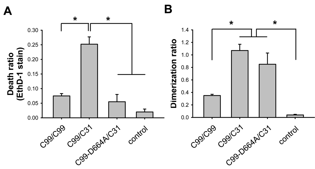
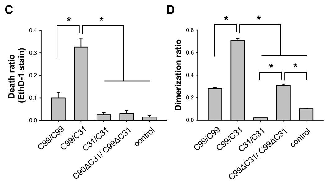
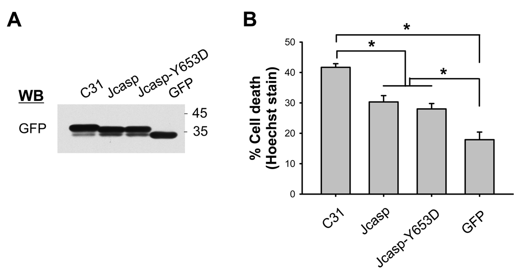
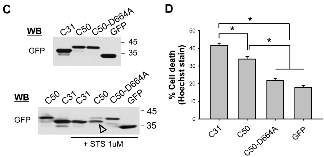
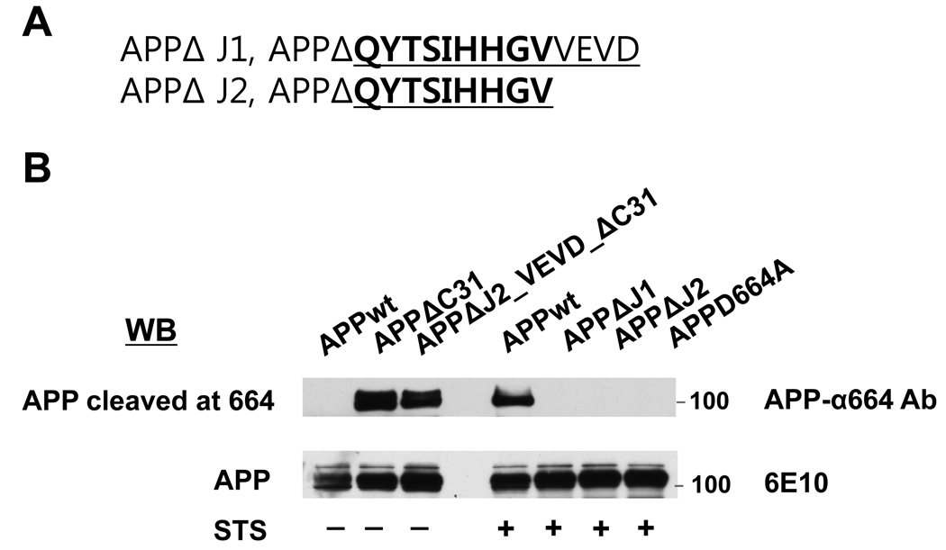
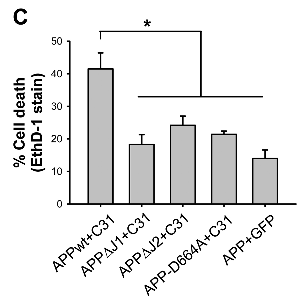
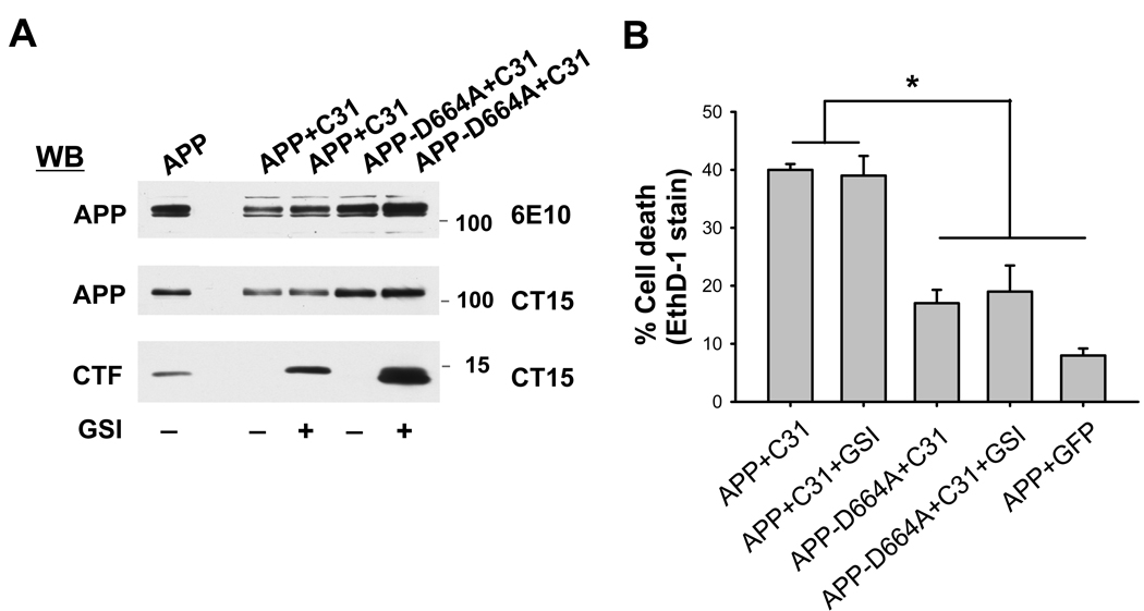
The cytoplasmic tail of the amyloid precursor protein (APP) contains two putatively cytotoxic peptides, Jcasp and C31, derived by caspase cleavage of APP. Jcasp is a fragment starting from the epsilon-secretase site to position 664, while C31 is a fragment from position 665 to the C-terminus. Our studies now showed that compared to C31, Jcasp appeared to play a minor role in cytotoxicity. In particular, inhibition of Jcasp generation by treatment of gamma-secretase inhibitor did not lead to any attenuation of C31-induced toxicity. Secondly, because C31 toxicity is largely absent in cells lacking endogenous APP, we determined, using a split beta-galactosidase complementary assay to monitor protein-protein interactions, the presence of APP associated complexes. Our results demonstrated that both APP homomeric and C31/APP heteromeric complexes were correlated with cell death, indicating that C31 complexes with APP to recruit the interacting partners that initiate the signals related to cellular toxicity.
Figures
Similar articles
-
Caspase cleavage of the amyloid precursor protein modulates amyloid beta-protein toxicity.J Neurochem. 2003 Nov;87(3):733-41. doi: 10.1046/j.1471-4159.2003.02059.x. J Neurochem. 2003. PMID: 14535955
-
beta-Secretase cleavage of the amyloid precursor protein mediates neuronal apoptosis caused by familial Alzheimer's disease mutations.Brain Res Mol Brain Res. 2001 Dec 16;97(1):103-13. doi: 10.1016/s0169-328x(01)00294-7. Brain Res Mol Brain Res. 2001. PMID: 11744168
-
Alternative Selection of β-Site APP-Cleaving Enzyme 1 (BACE1) Cleavage Sites in Amyloid β-Protein Precursor (APP) Harboring Protective and Pathogenic Mutations within the Aβ Sequence.J Biol Chem. 2016 Nov 11;291(46):24041-24053. doi: 10.1074/jbc.M116.744722. Epub 2016 Sep 29. J Biol Chem. 2016. PMID: 27687728 Free PMC article.
-
Inhibition of amyloid precursor protein processing enhances gemcitabine-mediated cytotoxicity in pancreatic cancer cells.J Biol Chem. 2013 Oct 18;288(42):30114-30124. doi: 10.1074/jbc.M113.459255. Epub 2013 Sep 10. J Biol Chem. 2013. PMID: 24022491 Free PMC article.
-
A mechanism for beta-amyloid overproduction in Alzheimer's disease: precursor-independent generation of beta-amyloid via antisense RNA-primed mRNA synthesis.FEBS Lett. 1996 Jul 22;390(2):124-8. doi: 10.1016/0014-5793(96)00663-1. FEBS Lett. 1996. PMID: 8706841 Review.
Cited by
-
Physiological functions of APP family proteins.Cold Spring Harb Perspect Med. 2012 Feb;2(2):a006288. doi: 10.1101/cshperspect.a006288. Cold Spring Harb Perspect Med. 2012. PMID: 22355794 Free PMC article. Review.
-
Cross-linking of cell surface amyloid precursor protein leads to increased β-amyloid peptide production in hippocampal neurons: implications for Alzheimer's disease.J Neurosci. 2012 Aug 1;32(31):10674-85. doi: 10.1523/JNEUROSCI.6473-11.2012. J Neurosci. 2012. PMID: 22855816 Free PMC article.
-
APP processing induced by herpes simplex virus type 1 (HSV-1) yields several APP fragments in human and rat neuronal cells.PLoS One. 2010 Nov 15;5(11):e13989. doi: 10.1371/journal.pone.0013989. PLoS One. 2010. PMID: 21085580 Free PMC article.
-
Do anti-amyloid beta protein antibody cross reactivities confound Alzheimer disease research?J Negat Results Biomed. 2017 Jan 26;16(1):1. doi: 10.1186/s12952-017-0066-3. J Negat Results Biomed. 2017. PMID: 28126004 Free PMC article. Review.
-
The multifaceted nature of amyloid precursor protein and its proteolytic fragments: friends and foes.Acta Neuropathol. 2015 Jan;129(1):1-19. doi: 10.1007/s00401-014-1347-2. Epub 2014 Oct 7. Acta Neuropathol. 2015. PMID: 25287911 Free PMC article. Review.
References
-
- Selkoe DJ. Alzheimer's disease: genes, proteins, and therapy. Physiol. Rev. 2001;81:741–766. - PubMed
-
- Terry RD, Masliah E, Salmon DP, Butters N, DeTeresa R, Hill R, Hansen LA, Katzman R. Physical basis of cognitive alterations in Alzheimer’s disease: synapse loss is the major correlate of cognitive impairment. Ann. Neurol. 1991;30:572–580. - PubMed
-
- Lu DC, Rabizadeh S, Chandra S, Shayya RF, Ellerby LM, Ye X, Salvesen GS, Koo EH, Bredesen DE. A second cytotoxic proteolytic peptide derived from amyloid beta-protein precursor. Nat. Med. 2000;6:397–404. - PubMed
-
- Galvan V, Gorostiza OF, Banwait S, Ataie M, Logvinova AV, Sitaraman S, Carlson E, Sagi SA, Chevallier N, Jin K, Greenberg DA, Bredesen DE. Reversal of Alzhemier’s-like pathology and behavior in human APP transgenic mice by mutation of Asp664. Proc. Natl. Acad. Sci. USA. 2006;103:7130–7135. - PMC - PubMed
Publication types
MeSH terms
Substances
Grants and funding
LinkOut - more resources
Full Text Sources
