. 2010 Apr;20(4):884-97.
doi: 10.1093/cercor/bhp154. Epub 2009 Aug 13.

The effect of variation in expression of the candidate dyslexia susceptibility gene homolog Kiaa0319 on neuronal migration and dendritic morphology in the rat

Affiliations

The effect of variation in expression of the candidate dyslexia susceptibility gene homolog Kiaa0319 on neuronal migration and dendritic morphology in the rat

Veronica J Peschansky et al. Cereb Cortex. 2010 Apr.

Abstract

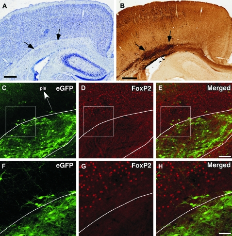
We investigated the postnatal effects of embryonic knockdown and overexpression of the candidate dyslexia gene homolog Kiaa0319. We used in utero electroporation to transfect cells in E15/16 rat neocortical ventricular zone with either 1) small hairpin RNA (shRNA) vectors targeting Kiaa0319, 2) a KIAA0319 expression construct, 3) Kiaa0319 shRNA along with KIAA0319 expression construct ("rescue"), or 4) a scrambled version of Kiaa0319 shRNA. Knockdown, but not overexpression, of Kiaa0319 resulted in periventricular heterotopias that contained large numbers of both transfected and non-transfected neurons. This suggested that Kiaa0319 shRNA disrupts neuronal migration by cell autonomous as well as non-cell autonomous mechanisms. Of the Kiaa0319 shRNA-transfected neurons that migrated into the cortical plate, most migrated to their appropriate lamina. In contrast, neurons transfected with the KIAA0319 expression vector attained laminar positions subjacent to their expected positions. Neurons transfected with Kiaa0319 shRNA exhibited apical, but not basal, dendrite hypertrophy, which was rescued by overexpression of KIAA0319. The results provide additional supportive evidence linking candidate dyslexia susceptibility genes to migrational disturbances during brain development, and extends the role of Kiaa0319 to include growth and differentiation of dendrites.


Figures

Figure 1.
Figure 1.
In situ hybridization of Kiaa0319 in embryonic rat brains. Photomontages of in situ hybridization of Kiaa0319 antisense probes in E14/15 (A,D), E16/17 (B,E), and E19/20 (C,F) rat embryos. (G) High power photomicrographs of developing cerebral cortex indicating CP, subventricular zone (SVZ) and VZ/IZ. Kiaa0319 is expressed highly in the CP, striatum, and hippocampus at all ages. There is increased expression in the mitral cell layer of the olfactory bulb at E19/20, and generalized moderate expression in the brain stem at all ages. Bar in panels A–F = 1 mm. Bar in panel G = 100 μm.
Figure 2.
Figure 2.
Neuronal migration following embryonic knockdown or overexpression of Kiaa0319. (AD) Position of transfected neurons in 2 representative sections from brains embryonically transfected with plasmids containing Kiaa0319 shRNA (A), scrambled Kiaa0319 shRNA (B), KIAA0319 protein (C), or Kiaa0319 shRNA along with KIAA0319 protein (D). (E) Photomicrograph of cerebral cortex of Nissl-stained section illustrating region of PVH (arrows). This animal was embryonically transfected with Kiaa0319 shRNA + eGFP. Bar = 250 μm. (F) Photomicrograph of section adjacent to Panel E immunohistochemically stained for eGFP. Transfected neurons are located within the PVH. Bar = 250 μm. (G and H) High-power photomicrograph of PVH (arrows) illustrated in panels (E) and (F). Bar = 125 μm.
Figure 3.
Figure 3.
Nestin-positive fibers located in PVH in the brain of a rat embryonically transfected with Kiaa0319 shRNA. (A) Brightfield photomontage of Nissl-stained section showing PVH (small arrows). This section also has an ectopic collection of neurons in the molecular layer, which is an artifact of the injection (large arrow). Bar = 500 μm. (B) Brightfield photomontage of section adjacent to (A) immunohistochemically stained for GFP illustrating PVH (arrows). Bar = 250 μm. (C) Brightfield photomontage of section adjacent to (B) (arrows for orientation with B) immunohistochemically stained for Nestin. Nestin stains fibers in the PVH as well as blood vessels throughout the brain. Box indicates region illustrated in (D). Bar = 250 μm. (D) High-power brightfield photomicrograph of region denoted in (C) illustrating dense plexus of Nestin-positive fibers (arrows). Bar = 25 μm. (E) High-power brightfield photomicrograph of region homologous to that in (D). Compared with (D), there are far fewer Nestin-positive fibers (arrows). (F) (Mall arrows), blood vessels (large arrows), and eGFP-positive neurons (arrowheads) present. There are no cells that are colabeled. Bar = 100 μm.
Figure 4.
Figure 4.
Confocal microscopy of lamina specific markers Cux1 and Ctgf in the brain of rats embryonically transfected with shRNA targeted against Kiaa0319. The white line delineates the border between a PVH and the white matter. The 4 panels illustrate cells transfected with eGFP (A), cells immunopositive for the upper lamina specific marker Cux1 (B), cells immunopositive for the subplate specific marker Ctgf (C) and a merged panel (D). There are eGFP (shRNA–transfected) cells that are Cux1 positive (straight sided arrowheads), and some that are not (concave arrowheads). There are, in addition, a large number of Cux1-positive cells that are not colabeled with eGFP (arrows). This suggests that these nontransfected cells arrive in the PVH by non–cell autonomous mechanisms. There are no cells that colabel with Ctgf+ cells. Bar = 100 μm.
Figure 5.
Figure 5.
Distribution of the lower lamina specific marker Foxp2 in the brain of a rat embryonically transfected with Kiaa0319 shRNA. (A) Photomicrograph of cerebral cortex of Nissl-stained section illustrating region of PVH (arrows). This animal was embryonically transfected with Kiaa0319 shRNA + eGFP. Bar = 500 μm. (B) Photomicrograph of section adjacent to panel (A) immunohistochemically stained for eGFP. Transfected neurons are located within the PVH. Bar = 500 μm. (C, D, and E) Confocal microscopic images of PVH (white lines) seen in (A) and (B). The 3 panels illustrate cells transfected with eGFP and Kiaa0319 shRNA (C), cells immunopositive for the lower lamina specific marker Foxp2 (D), and a merged panel (E). There are no cells double labeled for eGFP and Foxp2, nor are there any Foxp2 labeled cells in the PVH. This suggests that lower lamina cells are not disrupted by embryonic transfection with Kiaa0319 shRNA. Arrow in (C) is for orientation. Box in each panel indicates region magnified in panels (F, G, and H). Bar = 200 μm. (F, G, and H) Magnified regions of panels (C, D, and E), respectively. Bar = 100 μm.
Figure 6.
Figure 6.
Non–cell autonomous effects of Kiaa0319 shRNA transfection. Distribution of cells in the border between the subventricular zone (SVZ) and the IZ (AC) and the VZ/SVZ border (DF) 4 h after an injection of BrdU at E18/19 in rats transfected at E15/16 with Kiaa0319 shRNA + eGFP. There are few cells double labeled for eGFP and BrdU (arrow), indicating that these are 2 separate populations. Z-axis movie of panels C and F are available as supplements. Bar = 20 μm. (GN) Distribution of Cux1- and E18/19 BrdU+ cells in the layer 2/3 (GJ) and the PVH (delineated by dashed white lines; KN) of a rat embryonically transfected with Kiaa0319 shRNA + eGFP at E15/16. There are large numbers of eGFP+ (G and K), Cux1+ (H and L), and BrdU+ (I and M) cells in both layer 2/3 and the PVH. A significant subset of cells in both in layer 2 and the PVH are both Cux1+ and BrdU+. These are seen as purple cells in the merged panels (J and N; arrows). As in Figure 3, there is another subset of cells that colabel for both eGFP and Cux1 (yellow cells in the merged panel, arrowheads). There are, however, no cells colabeled for eGFP and BrdU in either layer 2/3 or the PVH. The lack of double-labeled cells in the PVH support the notion that there are non–cell autonomous effects of embryonic transfections with Kiaa0319 shRNA. Bar = 200 μm.
Figure 7.
Figure 7.
GABAergic interneurons are present in PVHs. Distribution of Calretinin-positive (AC) and Parvalbumin-positive (EF) interneurons in a PVH (dashed white lines) from a brain embryonically transfected with Kiaa0319 shRNA. There are no neurons that colabel for eGFP (arrowheads, neurons that were transfected with Kiaa0319 shRNA) and either of the 2 GABAergic antibodies (arrows). Bar = 100 μm.
Figure 8.
Figure 8.
Migration distance analysis following embryonic knockdown and/or overexpression of Kiaa0319. Quantitative migration analysis of the percent of neurons migrating (X-axis) against the normalized depth of the cerebral cortex. Photomicrograph of cerebral cortex is included as an aid for laminar delineation. There are significantly more unmigrated neurons in the shRNA group (blue) when compared with the other 3 groups. There is no difference in the upper layer migration between the Kiaa0319 shRNA and Kiaa0319 scrambled (green) groups, nor is there a difference between the Overexpression (red) and Rescue (light blue) groups. Neurons in brains embryonically transfected with KIAA0319 protein migrate below their expected laminar position based on comparison with the scrambled group.
Figure 9.
Figure 9.
Neuronal morphology and quantitative analysis following embryonic knockdown and/or overexpression of Kiaa0319. (AD) Representative tracing of neurons embryonically transfected with plasmids expressing Kiaa0319 shRNA (A), KIAA0319 protein (B), scrambled Kiaa0319 shRNA (C), and both Kiaa0319 shRNA and KIAA0319 protein (D). (E) Sholl analysis of apical dendrites for each condition. The dendrites of Kiaa0319 shRNA–transfected neurons are longer within 200 μm of the cell body when compared with the other 3 groups. (F) Sholl analysis of basal dendrites for each condition reveals no significant differences among the 4 groups. (G) Quantitative analysis of specific dendritic features confirms the Sholl analysis. Apical dendrites of neurons embryonically transfected with Kiaa0319 shRNA have more nodes, ends, and overall length when compared with the scrambled and rescue groups. In contrast, there are no significant differences in any of these measures in the basal dendrites. There is no difference in the number of basal dendrites. *Differs from Kiaa0319 shRNA, P < 0.05.

References

    1. Allen KM, Gleeson JG, Shoup SM, Walsh CA. A YAC contig in Xq22.3-q23, from DXS287 to DXS8088, spanning the brain- specific genes doublecortin (DCX) and PAK3. Genomics. 1998;52:214–218. - PubMed
    1. Anderson S, Marin O, Horn C, Jennings K, Rubenstein J. Distinct cortical migrations from the medial and lateral ganglionic eminences. Development. 2001;128:353–363. - PubMed
    1. Andrews W, Barber M, Hernadez-Miranda LR, Xian J, Rakic S, Sundaresan V, Rabbitts TH, Pannell R, Rabbitts P, Thompson H, et al. The role of Slit-Robo signaling in the generation, migration and morphological differentiation of cortical interneurons. Dev Biol. 2008;313:648–658. - PubMed
    1. Bai J, Ramos RL, Ackman JB, Thomas AM, Lee RV, LoTurco JJ. RNAi reveals doublecortin is required for radial migration in rat neocortex. Nat Neurosci. 2003;6:1277–1283. - PubMed
    1. Banerjee M, Worth D, Prowse DM, Nikolic M. Pak1 phosphorylation on t212 affects microtubules in cells undergoing mitosis. Curr Biol. 2002;12:1233–1239. - PubMed

Publication types

MeSH terms