

IFN-producing killer dendritic cells are antigen-presenting cells endowed with T-cell cross-priming capacity
- PMID: 19679552
- PMCID: PMC2761009
- DOI: 10.1158/0008-5472.CAN-09-0508
IFN-producing killer dendritic cells are antigen-presenting cells endowed with T-cell cross-priming capacity
Abstract
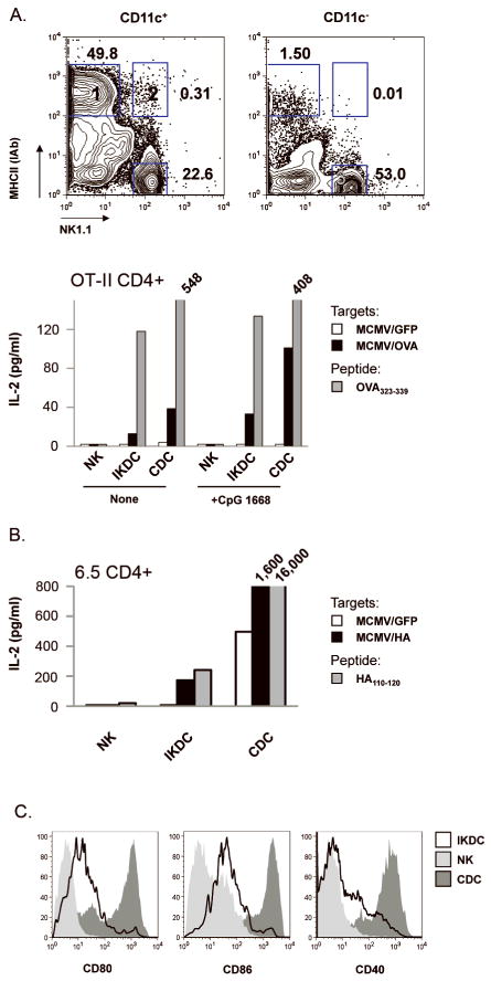
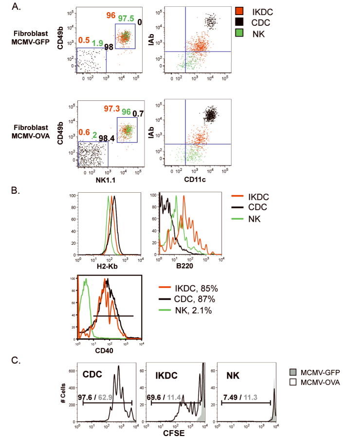
IFN-producing killer dendritic cells (IKDC) represent a recently discovered cell type in the immune system that possesses a number of functions contributing to innate and adaptive immunity, including production of type 1 and 2 IFNs, interleukin (IL)-12, natural killing, and ultimately antigen presentation to naïve T cells. Here, we compared in vitro and in vivo responses of mouse IKDC, conventional dendritic cells (DC), and natural killer (NK) cells to murine cytomegalovirus infection and found distinct functions among these cell subsets. Upon recognition of infected fibroblasts, IKDC, as well as NK, produced high level of IFN-gamma, but unlike NK, IKDC simultaneously produced IL-12p40 and up-regulated MHC class II (MHC-II) and costimulatory molecules. Using MHC-II molecule expression as a phenotypic marker to distinguish activated IKDC from activated NK, we further showed that highly purified MHC-II(+) IKDC but not NK cross-present MHC class I-restricted antigens derived from MCMV-infected targets to CD8(+) T cells in vitro and in vivo. Our findings emphasize the unique nature of IKDC as a killer antigen-presenting cell directly linking innate and adaptive immunity.
Conflict of interest statement
The authors declare that they have no competing financial interests.
Figures


Similar articles
-
The dendritic cell-like functions of IFN-producing killer dendritic cells reside in the CD11b+ subset and are licensed by tumor cells.Cancer Res. 2009 Aug 15;69(16):6590-7. doi: 10.1158/0008-5472.CAN-08-4473. Cancer Res. 2009. PMID: 19679551
-
Trans-presentation of IL-15 dictates IFN-producing killer dendritic cells effector functions.J Immunol. 2008 Jun 15;180(12):7887-97. doi: 10.4049/jimmunol.180.12.7887. J Immunol. 2008. PMID: 18523252
-
Migratory and antigen presentation functions of IFN-producing killer dendritic cells.Cancer Res. 2009 Aug 15;69(16):6598-606. doi: 10.1158/0008-5472.CAN-09-0501. Epub 2009 Aug 4. Cancer Res. 2009. PMID: 19654308
-
Licensing of killer dendritic cells in mouse and humans: functional similarities between IKDC and human blood γδ T-lymphocytes.J Immunotoxicol. 2012 Jul-Sep;9(3):259-66. doi: 10.3109/1547691X.2012.685528. Epub 2012 May 27. J Immunotoxicol. 2012. PMID: 22632132 Review.
-
Interferon-gamma: biologic functions and HCV therapy (type I/II) (1 of 2 parts).Clin Ter. 2006 Jul-Aug;157(4):377-86. Clin Ter. 2006. Retraction in: Clin Ter. 2008 May-Jun;159(3):207. PMID: 17051976 Retracted. Review.
Cited by
-
Tolerogen-induced interferon-producing killer dendritic cells (IKDCs) protect against EAE.J Autoimmun. 2011 Dec;37(4):328-41. doi: 10.1016/j.jaut.2011.09.005. Epub 2011 Oct 22. J Autoimmun. 2011. PMID: 22018711 Free PMC article.
-
Characterization of tumoricidal activities mediated by a novel immune cell regimen composing interferon-producing killer dendritic cells and tumor-specific cytotoxic T lymphocytes.BMC Cancer. 2024 Mar 28;24(1):395. doi: 10.1186/s12885-024-12101-3. BMC Cancer. 2024. PMID: 38549061 Free PMC article.
-
Effective antitumor immunity against murine gliomas using dendritic cells transduced with hTERTC27 recombinant adenovirus.Oncol Rep. 2012 Apr;27(4):1163-9. doi: 10.3892/or.2011.1619. Epub 2011 Dec 30. Oncol Rep. 2012. PMID: 22210010 Free PMC article.
-
A role for pre-mNK cells in tumor progression.J Immunother Cancer. 2016 Mar 15;4:16. doi: 10.1186/s40425-016-0120-6. eCollection 2016. J Immunother Cancer. 2016. PMID: 26981246 Free PMC article.
-
Immunomodulatory effects of cyclophosphamide and implementations for vaccine design.Semin Immunopathol. 2011 Jul;33(4):369-83. doi: 10.1007/s00281-011-0245-0. Epub 2011 May 25. Semin Immunopathol. 2011. PMID: 21611872 Review.
References
-
- Chan CW, Crafton E, Fan HN, et al. Interferon-producing killer dendritic cells provide a link between innate and adaptive immunity. Nature medicine. 2006;12(2):207–13. - PubMed
-
- Taieb J, Chaput N, Menard C, et al. A novel dendritic cell subset involved in tumor immunosurveillance. Nature medicine. 2006;12(2):214–9. - PubMed
-
- Pillarisetty VG, Katz SC, Bleier JI, Shah AB, Dematteo RP. Natural killer dendritic cells have both antigen presenting and lytic function and in response to CpG produce IFN-gamma via autocrine IL-12. J Immunol. 2005;174(5):2612–8. - PubMed
-
- Spits H, Lanier LL. Natural killer or dendritic: what's in a name? Immunity. 2007;26(1):11–6. - PubMed
Publication types
MeSH terms
Substances
Grants and funding
LinkOut - more resources
Full Text Sources
Medical
Molecular Biology Databases
Research Materials
