. 2009 Nov 1;18(21):4130-40.
doi: 10.1093/hmg/ddp362. Epub 2009 Aug 12.

Depletion of zebrafish Tcap leads to muscular dystrophy via disrupting sarcomere-membrane interaction, not sarcomere assembly

Affiliations

Depletion of zebrafish Tcap leads to muscular dystrophy via disrupting sarcomere-membrane interaction, not sarcomere assembly

Ruilin Zhang et al. Hum Mol Genet. .

Abstract

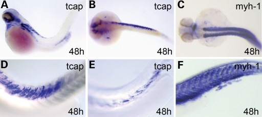
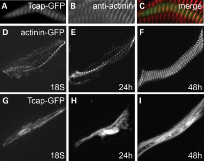
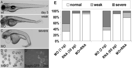
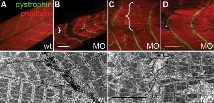
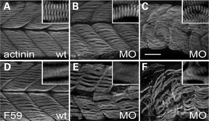
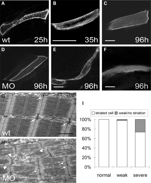
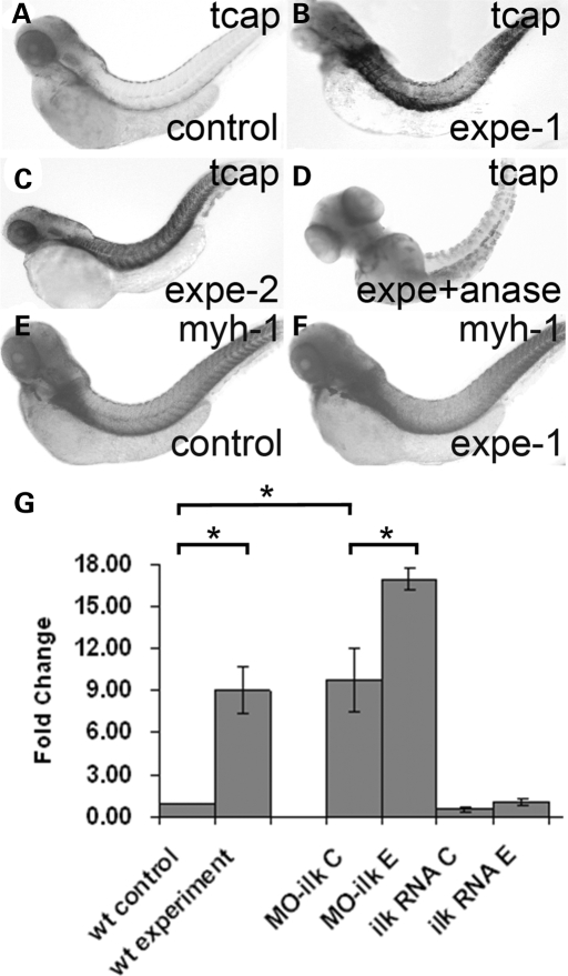
Tcap/telethonin encodes a Z-disc protein that plays important roles in sarcomere assembly, sarcomere-membrane interaction and stretch sensing. It remains unclear why mutations in Tcap lead to limb-girdle muscular dystrophy 2G (LGMD2G) in human patients. Here, we cloned tcap in zebrafish and conducted genetic studies. We show that tcap is functionally conserved, as the Tcap protein appears in the sarcomeric Z-disc and reduction of Tcap resulted in muscular dystrophy-like phenotypes including deformed muscle structure and impaired swimming ability. However, the observations that Tcap integrates into the sarcomere at a stage after the Z-disc becomes periodic, and that the sarcomere remains intact in tcap morphants, suggest that defective sarcomere assembly does not contribute to this particular type of muscular dystrophy. Instead, a defective interaction between the sarcomere and plasma membrane was detected, which was further underscored by the disrupted development of the T-tubule system. Pertinent to a potential function in stretch sensor signaling, zebrafish tcap exhibits a variable expression pattern during somitogenesis. The variable expression is inducible by stretch force, and the expression level of Tcap is negatively regulated by integrin-link kinase (ILK), a protein kinase that is involved in stretch sensing signaling. Together, our genetic studies of tcap in zebrafish suggested that pathogenesis in LGMD2G is due to a disruption of sarcomere-T-tubular interaction, but not of sarcomere assembly per se. In addition, our data prompted a novel hypothesis that predicts that the transcription level of Tcap can be regulated by the stretch force to ensure proper sarcomere-membrane interaction in striated muscles.


Figures

Figure 1.
Figure 1.
tcap expression pattern revealed by whole mount in situ hybridization. (AF) Tcap had a variable expression pattern in the somite at 48 hpf, different from the bilateral and ubiquitous expression of sarcomeric gene like myh1 (C and F). Tcap transcripts can be enriched in the anterior (A), posterior (not shown), dorsal (D) or ventral (E) part of the body, and even the unilateral (B). (A and D–F) Lateral view, anterior to the left. (B and C) Dorsal view, anterior to the left.
Figure 2.
Figure 2.
Tcap is a Z-disc protein but assembles after α-actinin becomes periodic. (AC) Tcap-GFP fusion protein showed a striated pattern (A, green) and co-localized with the Z-disc protein α-actinin (B, anti-actinin, red; C, overlap). (DI) α-actinin2-GFP revealed the processes of sarcomere assembly (D, 18 somite; E, 24 hpf) including lateral alignment (F, 48 hpf). Tcap-GFP still disperses in the cytoplasm when α-actinin2 already restricts to periodic Z-lines (G). Shown in (D–F) are different cells from (G–I). Tcap-GFP was restricted to mature Z-disc after 24 hpf (H and I).
Figure 3.
Figure 3.
Tcap MO result in specific phenotypes. (AC) Representative pictures of day 5 wild-type fish (A), weak and severe phenotypes of tcap morphants. A weak phenotype included shorter body length and a slight body curvature (B), less sensitive to touch and abnormal swimming pattern. A severe phenotype included severe body curvature (C), muscular disarray, much slower response to touch, and even lost of swimming ability. Both groups showed eye and jaw hypoplasia, and pericardiac edema can be occasionally detected in severe group. (D) Co-injection of tcap ATG morpholino can abolish the expression of N-terminal-Tcap-GFP chimeric mRNA, indicating a high knockdown efficacy. (D), bright field. (D′) Green fluorescence. The GFP fluorescence level in the fish group injected with both MO and RNA (left in D and D′) is much weaker than the fish group injected with RNA only (right in D and D′). (E) Phenotypes in tcap morphants can be rescued by co-injection of tcap RNA. The graph shows a dosage-dependent effect of MO-Tcap, and co-injection of tcap RNA can reduce the morphant percentage at both concentrations of MO-Tcap.
Figure 4.
Figure 4.
Tcap knockdown leads to muscular dystrophy-like phenotypes. (AD) Representative pictures of anti-dystrophin antibody stains myoseptum in green and phalloidin stains F-actin in red. Unlike wild-type fish which have a V-shaped myoseptum and well-organized myofibers (A), tcap morphants usually exhibit a U-shaped myoseptum, which is discontinuous or missed in some regions (brace in B and C) and the myofibers grow through adjacent somites. Disrupted myofiber organization with uneven lengths as well as detachment from the myoseptum (asterisk in B and D) are often observed. Scale bar, 50 µm. (E and F) Representative electron microscopy pictures showing a disrupted extracellular matrix structure in a tcap morphant (F) in comparison to wild-type fish (E). Scale bar, 5 µm.
Figure 5.
Figure 5.
Tcap knockdown did not affect sarcomere assembly. (AC) Anti-α-actinin staining of wild-type fish (A) and tcap morphants with either a weak (B) or severe (C) phenotype. Although the myofibrils in tcap morphants appeared wavy, disoriented and uneven in length, they still had a well-organized striated pattern (inset), indicating undisrupted Z-disc assembly. (DF) F59 staining of wild-type fish (D) and tcap morphants with either a weak (E) or severe (F) phenotype, which suggested that thick filament assembly was not disrupted either. Scale bar, 50 µm.
Figure 6.
Figure 6.
Tcap knockdown affects T-tubule development. (AC) Time course of T-tubule development in wild-type fish. Membrane-targeted GFP under the control of β-actin enhancer was injected to fluorescently label the sarcolemma that invaginated and formed the striated T-tubule system. The invagination initiated at around 25 hpf sporadically (A), continued through all direction (B) and finally formed an organized striated pattern at 96 hpf (C). Scale bar, 20 µm. (DF) Representative pictures of a cell with normal striations (D), and cells with either weak striation (E) or no striation (F) in tcap morphants that have been co-injected with a membrane-targeted GFP construct. Scale bar, 20 µm. (GH) Representative pictures of electron microscopy showing T-tubule and Z-disc alignment in wild-type fish (G) or tcap morphants (H). Unlike wild-type fish with well-aligned T-tubules and Z-discs (arrow), the T-tubules in some regions of morphants were misaligned or missing (arrowhead). Scale bar, 1 µm. (I) Graphic summary of the percentage of striated cell, cell with weak striation or no striation in different classes of tcap morphants.
Figure 7.
Figure 7.
tcap expression level can be altered by stretch movement and modulated by ILK. (AD) tcap expression levels can be altered by two treatments mimicking increased stretch force. Shown are whole mount in situ hybridization at 4 dpf. Under two different experimental treatments that restrict the surrounding space of fish embryos in order to increase stretch force (for detail refer to methods), the tcap expression level was increased on day 4 (B and C) compared with that in control fish (A). Co-treatment with anesthesia ablated the upregulation of Tcap expression (D). This upregulation was not observed in other sarcomeric genes such as myh-1 (E and F). (G) ILK negatively regulated Tcap level. Shown is a graphic summary of the result of quantitative RT–PCR. The expression of Tcap in treated embryos is significantly increased compared with that in wild-type fish (P = 0.02). Reduction of ilk by injection of morpholino significantly increased Tcap expression (P = 0.03) while overexpression of ilk by injection of mRNA decreased Tcap expression (P = 0.09). The inducibility of Tcap expression is still maintained in either manipulation of ILK. *P < 0.05.

References

    1. Dalkilic I., Kunkel L.M. Muscular dystrophies: genes to pathogenesis. Curr. Opin. Genet. Dev. 2003;13:231–238. - PubMed
    1. Davies K.E., Nowak K.J. Molecular mechanisms of muscular dystrophies: old and new players. Nat. Rev. Mol. Cell. Biol. 2006;7:762–773. - PubMed
    1. Koenig M., Monaco A.P., Kunkel L.M. The complete sequence of dystrophin predicts a rod-shaped cytoskeletal protein. Cell. 1988;53:219–228. - PubMed
    1. Ervasti J.M., Ohlendieck K., Kahl S.D., Gaver M.G., Campbell K.P. Deficiency of a glycoprotein component of the dystrophin complex in dystrophic muscle. Nature. 1990;345:315–319. - PubMed
    1. Petrof B.J., Shrager J.B., Stedman H.H., Kelly A.M., Sweeney H.L. Dystrophin protects the sarcolemma from stresses developed during muscle contraction. Proc. Natl Acad. Sci. USA. 1993;90:3710–3714. - PMC - PubMed

Publication types

MeSH terms