. 2009 Sep 16;28(18):2812-24.
doi: 10.1038/emboj.2009.212. Epub 2009 Aug 13.

Thymus-specific deletion of insulin induces autoimmune diabetes

Affiliations

Thymus-specific deletion of insulin induces autoimmune diabetes

Yong Fan et al. EMBO J. .

Abstract

Insulin expression in the thymus has been implicated in regulating the negative selection of autoreactive T cells and in mediating the central immune tolerance towards pancreatic beta-cells. To further explore the function of this ectopic insulin expression, we knocked out the mouse Ins2 gene specifically in the Aire-expressing medullary thymic epithelial cells (mTECs), without affecting its expression in the beta-cells. When further crossed to the Ins1 knockout background, both male and female pups (designated as ID-TEC mice for insulin-deleted mTEC) developed diabetes spontaneously around 3 weeks after birth. beta-cell-specific autoimmune destruction was observed, as well as islet-specific T cell infiltration. The presence of insulin-specific effector T cells was shown using ELISPOT assays and adoptive T cell transfer experiments. Results from thymus transplantation experiments proved further that depletion of Ins2 expression in mTECs was sufficient to break central tolerance and induce anti-insulin autoimmunity. Our observations may explain the rare cases of type 1 diabetes onset in very young children carrying diabetes-resistant HLA class II alleles. ID-TEC mice could serve as a new model for studying this pathology.


Conflict of interest statement

The authors declare that they have no conflict of interest.

Figures

Figure 1
Figure 1
Analysis of Ins2 expression in specific thymic stromal cells of Ins1 knockout mice. (A) Isolation of thymic stromal cells. Left panel, fluorescence-activated cell sorting of bone marrow-derived antigen presenting cells. CD45+, MHC II+ cells were further gated for CD4, CD11c+ thymic dendritic cells (R1), and CD4,CD11c macrophages and B cells (R2). Right panel, CD45, EpCAM+ stromal cells were sorted for medullary epithelial cells (R3: Ly51low, UEAhigh) and cortical epithelial cells (R4: Ly51high, UEAlow). The same R1–R4 designation has been used throughout the paper unless otherwise specified. (B) RT–PCR analysis of the mouse Ins2 gene expression in specific thymic antigen presenting cells (R1–R4). The house keeping Hprt gene expression was used as control. K-2/8, cytokeratin 2/8.
Figure 2
Figure 2
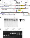
Genetic modification of the mouse Ins2 gene for thymus-specific deletion. (A) A targeting construct with two loxp sites flanking the mouse Ins2 gene and an frt-tagged Neo cassette for selection was used to modify the Ins2 gene through homologous recombination (black dash line). Neo cassette within the loxp-tagged Ins2 allele was deleted from the genome by FLP recombinase. A AseI, B: BamHI, E: EcoRI, red bar: 5′ external probe. (B) Southern blot analysis of Ins2-targeted embryonic stem cell clones. Genomic DNA was isolated, digested using AseI and probed with the 5′ external probe. * indicates a loxp-tagged Ins2 embryonic stem cell clone. As the result of the genetic modifications, an AseI site was introduced to the loxp-tagged Ins2 allele (Ins2l), which appears as the extra 10-kb band on the blot, in addition to the 20-kb band from the wild-type allele (Ins2+). (C) Agarose gel showing PCR-based genotyping of mice. +, wild type; l, loxp-tagged Ins2 (Ins2l).
Figure 3
Figure 3
Characterization of Aire–Cre transgene expression. (A) Upper panel, schematic drawing of the Aire–Cre construct. A 23-kb and an 11-kb genomic fragment were subcloned from the 5′ and 3′ of mouse Aire gene, respectively, and engineered to regulate the Cre gene expression. Lower panel, RT–PCR analysis of Aire–Cre transgene expression. (B) RT–PCR analysis of Cre expression in specific thymic stromal cells (R1–R4) isolated from the Aire–Cre transgenic mice. (C) Aire–Cre:Rosa26R-lacZ thymus stained with anti-LacZ antibody (green) and CD45 antibody (red). LacZ+ and CD45+ cells (yellow). Arrows indicate LacZ+, CD45 thymic epithelial cells. (D) Absence of Cre recombinase expression in pancreatic β-cells of Aire–Cre mice. Pancreas samples from six-week-old Aire–Cre:Rosa26R-lacZ, Rosa26R-lacZ (negative control), and Rip–Cre:Rosa26R-lacZ (rat insulin promoter, positive control) mice stained with anti-insulin (red) and anti-LacZ (green) antibodies. (E) Efficient deletion of the floxed Ins2 allele in mTEC cells collected from Aire–Cre:Ins1−/+:Ins2l/l mice. Genomic DNA was isolated from different thymic stromal cells (R1–R4) and pancreatic islets and the presence of the floxed Ins2 allele was analysed using PCR, with the same primer pairs depicted in Figure 2B. PCR amplification of the mouse Ica1 gene was used as control of the genomic DNA input. BM, bone marrow; H, heart; IS, islets; Kd, kidney; Lu, lung; Lv, liver; Pan, pancreas; Sp, spleen; Th, thymus.
Figure 4
Figure 4
Thymic insulin expression in ID-TEC mice. (A) RT–PCR analysis of thymic insulin expression in ID-TEC mice. (B) Real-time PCR quantification of thymic insulin expression in ID-TEC and control mice. (Error bar: s.e.m.) Mk, molecular size marker.
Figure 5
Figure 5
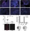
ID-TEC mice develop spontaneous diabetes within 3 weeks after birth. (A) Normal islet development of ID-TEC mice at birth. Pancreata from control and ID-TEC mice were collected at postnatal day 1, and stained using anti-insulin (green) and glucagon (red) antibodies. (B) Plasma insulin levels of 10-day-old ID-TEC pups. (C) Pancreatic insulin contents of 10-day-old ID-TEC pups. Pancreata were collected from three litters and insulin content in each pancreas was measured and plotted.—mean insulin levels; Δ, control littermates; filled red circle, ID-TEC pups. (D) Blood glucose levels of ID-TEC (red line, n=7) and control mice (blue line, n=11). (E) Plasma insulin levels of diabetic, 4-week-old ID-TEC mice. (F) Pancreatic sections stained with anti-insulin (green) and anti-glucagon (red) antibodies.
Figure 6
Figure 6
Diabetes developed in ID-TEC mice is autoimmune in nature. (AF) Pancreata from ID-TEC mice were collected at postnatal day 21. Pancreatic sections were stained with anti-CD4 (red) (A–C), anti-CD8 (D), anti-B220 (E) and anti-F4/80 (F) antibodies (red). Higher magnification ( × 400) of a damaged islet with infiltrating T cells is shown in (B). Sections (A), (B), (D) and (F) were counter stained with anti-insulin antibody (green), whereas section (C) was counter stained with anti-glucagon antibody (green). Nuclei were stained with Hoechst 33342 in all the sections (blue). Damaged islets are outlined by dashes. (G) Sera collected from 3–8-week-old diabetic ID-TEC mice and age-matched controls (1:50 dilution) were subjected to insulin autoantibody (IAA) assay. Data are presented as absorbance at 450 nm (OD450). —, mean value. **P<0.01. (H) Sera collected from 6–8-week-old diabetic ID-TEC mice or control mice were used to stain islet sections from C57BL/6 Rag1−/− mice (1:5 dilution). Bound mouse antibody was revealed with goat anti-mouse IgG. (I) Presence of autoreactive T cells responding specifically to insulin in ID-TEC mice. Suspensions of splenocytes were pooled from three animals, and cultured overnight with whole insulin protein, whole GAD65 protein, insulin B-chain peptide 9–23 (Ins B9–23), or GAD65 peptide 206–220 (GAD 206–220). The frequency of IFN-γ-producing cells was measured by ELISPOT assays in triplicate. The upper panel shows one representative result from three independent assays. Filled bar, ID-TEC mice; open bar, controls. Representative microscopic pictures of ELISPOT assay using insulin as antigen are shown in the lower panel: ID-TEC splenocytes (left), controls (right).
Figure 7
Figure 7
ID-TEC thymus is sufficient to transfer islet autoimmunity. (A) Representative FACS results showing the successful T cell reconstitution in nude mice with thymus transplantation underneath the kidney capsule. (BE)At 16 weeks after thymus transplantation, pancreata were collected and stained with anti-CD4 (red) and anti-insulin (green) antibodies. (B) A representative pancreas section from nude mice transplanted with thymi from littermate controls, in which Ins2 was intact, showing the absence of T cell infiltration. (C–E) Representative pancreata collected from nude mice transplanted with ID-TEC thymi.
Figure 8
Figure 8
Both CD4+ and CD8+ T cells can transfer islet autoimmunity in immune-compromised Rag1−/− mice. (A) Anti-insulin autoimmunity can be transferred to C57BL/6 Rag1−/− hosts, which carry the same MHC haplotype (H2b) as ID-TEC mice, by adoptive transfer of ID-TEC splenocytes. At 3 weeks after reconstitution, splenocytes were pooled from two animals, cultured overnight with either whole insulin or GAD 206–220 peptide as negative control. One representative result from three independent ELISPOT assays is shown. C, controls; I, ID-TEC (B) Detection of T cell infiltration in Rag1−/− pancreata after adoptive transfer of ID-TEC splenocytes. At 4 weeks after the transfer, pancreata were collected and stained with anti-CD4 (red) and anti-insulin antibody (green). (C, D) Pancreata were collected from Rag1−/− mice adoptively transferred with either CD4+CD25 T cells (C) or CD8+ T cells (D), and stained with anti-CD4 (C) or anti-CD8 (D) antibody (red). Insulin staining is shown in green.
Figure 9
Figure 9
Unimpaired negative selection of HY autoantigens and normal Treg cell development in male ID-TEC mice. (A) Thymi collected from male ID-TEC mice were separated as in Figure 1 (R1, thymic dendritic cells; R2, macrophage per B-cell; R3, mTEC). Isolated cells were subjected to RT–PCR analysis of expression of the genes encoding the immunodominant male-specific autoantigens (Uty, Smcy and Dby). (B) FACS analysis of Uty-specific CD8+ T cells in the spleens of male and female ID-TEC mice. CD8+ T cells were isolated from the spleens using magnetic beads and stained with anti-CD3 and anti-CD8 antibodies, together with Db/Uty pentamers (ProImmune). Representative FACS data are shown, which were gated on the CD3+CD8+ population. (C) ELISPOT analysis of IFN-γ-secreting CD4+ T cell clones stimulated with the immunodominant male-specific HY Dby peptide. Data are presented as mean±s.e.m. Lower panel, representative microscopic pictures of the ELISPOT assay with male (left) or female (right) ID-TEC splenocytes are shown. (D, E). Pancreata collected from Rag1−/− mice adoptively transferred with CD4+CD25 effector T cells and CD8+ T cells, with (D, E) or without (F, G) an equal number of CD4+CD25+ Treg cells, were stained with either anti-CD4 (D, F) or anti-CD8 (E, G) antibodies (red), counter-stained with anti-insulin antibody (green) and with Hoechst 33342 for nuclei (blue).

Comment in

  • Insulin teaches a new lesson in tolerance.
    Kay TW, Krishnamurthy B, Brodnicki TC, Mannering SI. Kay TW, et al. EMBO J. 2009 Sep 16;28(18):2687-8. doi: 10.1038/emboj.2009.251. EMBO J. 2009. PMID: 19759522 Free PMC article. No abstract available.

Similar articles

Cited by

References

    1. Abiru N, Maniatis AK, Yu L, Miao D, Moriyama H, Wegmann D, Eisenbarth GS (2001) Peptide and major histocompatibility complex-specific breaking of humoral tolerance to native insulin with the B9-23 peptide in diabetes-prone and normal mice. Diabetes 50: 1274–1281 - PubMed
    1. Achenbach P, Koczwara1 K, Knopff1 A, Naserke H, Ziegler AG, Bonifacio E (2004) Mature high-affinity immune responses to (pro)insulin anticipate the autoimmune cascade that leads to type 1 diabetes. J Clin Invest 114: 589–597 - PMC - PubMed
    1. Alleva DG, Crowe PD, Jin L, Kwok WW, Ling N, Gottschalk M, Conlon PJ, Gottlieb PA, Putnam AL, Gaur A (2001) A disease-associated cellular immune response in type 1 diabetes to an immunodominant epitope of insulin. J Clin Invest 107: 173–180 - PMC - PubMed
    1. Anderson MS, Venanzi ES, Chen Z, Berzins SP, Benoist C, Mathis D (2005) The cellular mechanism of Aire control of T cell tolerance. Immunity 23: 227–239 - PubMed
    1. Anderson MS, Venanzi ES, Klein L, Chen Z, Berzins SP, Turley SJ, von Boehmer H, Bronson R, Dierich A, Benoist C, Mathis D (2002) Projection of an immunological self shadow within the thymus by the Aire protein. Science 298: 1395–1401 - PubMed

Publication types