

Review
doi: 10.1038/nrmicro2199.
The type III secretion system of Pseudomonas aeruginosa: infection by injection
Affiliations
- PMID: 19680249
- PMCID: PMC2766515
- DOI: 10.1038/nrmicro2199
Item in Clipboard
Review
The type III secretion system of Pseudomonas aeruginosa: infection by injection
Nat Rev Microbiol.
2009 Sep.
Abstract
The Gram-negative bacterium Pseudomonas aeruginosa uses a complex type III secretion apparatus to inject effector proteins into host cells. The configuration of this secretion machinery, the activities of the proteins that are injected by it and the consequences of this process for infection are now being elucidated. This Review summarizes our current knowledge of P. aeruginosa type III secretion, including the secretion and translocation machinery, the regulation of this machinery, and the associated chaperones and effector proteins. The features of this interesting secretion system have important implications for the pathogenesis of P. aeruginosa infections and for other type III secretion systems.
Figures
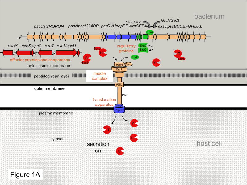
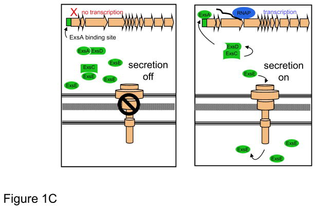
(A) The T3SS may be functionally divided into five components: the needle complex; the translocation apparatus, regulatory proteins, effector proteins, and chaperones. These five parts work together to inject effector proteins into host cells in a highly regulated manner. (C) Linkage of T3SS transcription to protein secretion is achieved through the interactions of four proteins: ExsA, ExsC, ExsD, and ExsE. When secretion is turned off, ExsE accumulates within the bacterium and binds ExsC, allowing ExsD to bind ExsA, and thus preventing transcription of type III secretion genes. When secretion is activated, the regulatory protein ExsE is secreted from the cell, allowing ExsC to bind ExsD. Sequestration of ExsD frees the transcriptional activator ExsA, resulting in unimpeded transcription of type III secretion genes. RNAP, RNA polymerase
(A) The T3SS may be functionally divided into five components: the needle complex; the translocation apparatus, regulatory proteins, effector proteins, and chaperones. These five parts work together to inject effector proteins into host cells in a highly regulated manner. (C) Linkage of T3SS transcription to protein secretion is achieved through the interactions of four proteins: ExsA, ExsC, ExsD, and ExsE. When secretion is turned off, ExsE accumulates within the bacterium and binds ExsC, allowing ExsD to bind ExsA, and thus preventing transcription of type III secretion genes. When secretion is activated, the regulatory protein ExsE is secreted from the cell, allowing ExsC to bind ExsD. Sequestration of ExsD frees the transcriptional activator ExsA, resulting in unimpeded transcription of type III secretion genes. RNAP, RNA polymerase
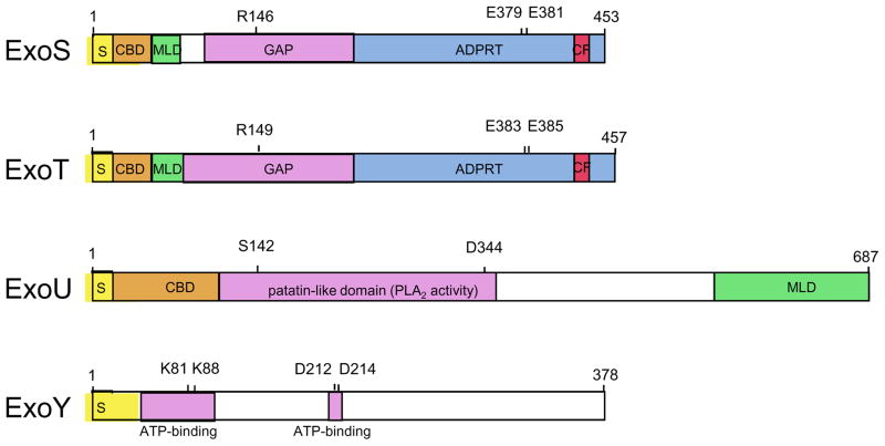
ExoS is a 453 amino acid bi-functional toxin that has both GAP and ADPRT activity. Arg146 is required for GAP activity and both Glu379 and Glu381 are required for efficient catalytic addition of the ADP-ribose moiety of NAD+ to substrate . Within the ADPRT domain, residues 418 to 429 comprise a binding site for the eukaryotic cofactor 14-3-3 that is necessary for activation of the ADPRT activity of ExoS. ExoT is a 457 amino acid protein that is closely related to ExoS. Arg149 is required for the GAP activity of ExoT and residues Glu383 and Glu385 are crucial for its ADPRT activity . Residues 422–433 are thought to comprise the site of cofactor 14-3-3 binding. ExoU is a 687 amino acid protein that contains a patatin-like domain necessary for PLA2 activity. Residues Ser142 and Asp344 are required for this activity , , . ExoY is a 378 amino acid adenylate cyclase. Residues Lys81, Lys88, Asp212, and Asp214 are required for its activity and are thought to be necessary for interactions between ExoY and ATP. See text for additional details. S, secretion signal; CBD, chaperone binding site; MLD, membrane localization domain; GAP, GTPase activating protein activity; ADPRT, ADP-ribosyl transferase activity; PLA2, phospholipase A2; CF, cofactor binding site. (Adapted from 176)
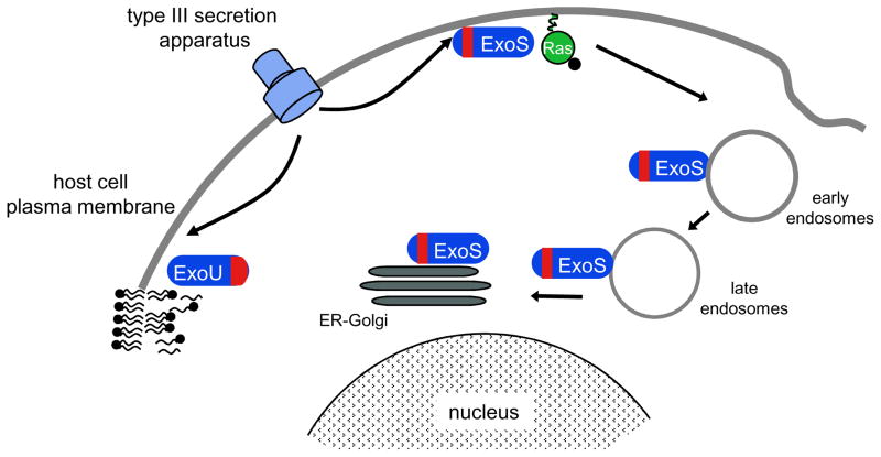
The N-terminal MLD of ExoS (shown in red) initially targets this effector protein to the plasma membrane and subsequently to early and late endosomes and finally to the endoplasmic reticulum (ER) and Golgi. Membrane localization is necessary for efficient ADP ribosylation of membrane-associated Ras. Following translocation into mammalian cells, ExoU is targeted to the plasma membrane by a C-terminal MLD. At this location, the enzymatic activity of ExoU is thought to cleave phospholipids within the plasma membrane. (Adapted from 65)
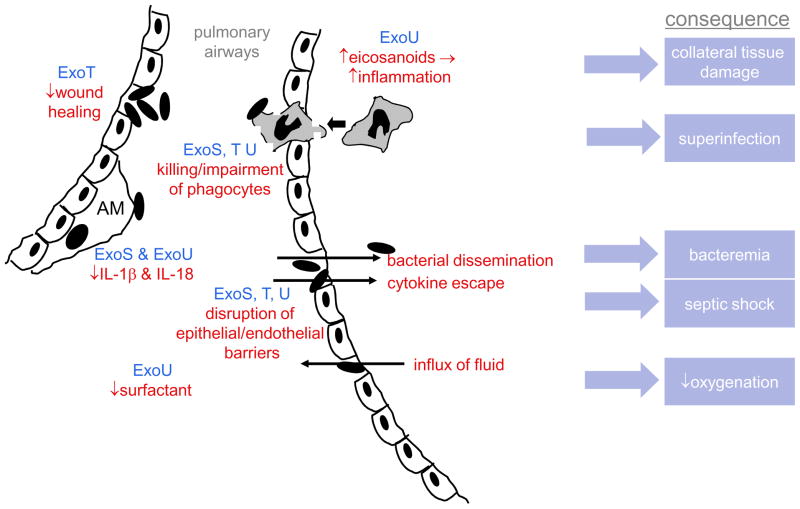
In early infection, P. aeruginosa exploits breaches in the epithelial mucosa, which is facilitated by ExoT-mediated inhibition of wound healing. ExoU and ExoS block IL-1β and IL-18 production by alveolar macrophages, blunting or biasing the early inflammatory response. When subsequent development of an inflammatory response does occur, it is excessively amplified by ExoU-induced eicosanoid release and causes collateral damage to host tissues. However the recruited phagocytes are unable to eradicate P. aeruginosa because they are killed or impaired by ExoS, ExoT, and ExoU; the resulting paucity of functional phagocytes makes the lungs prone to superinfection by other bacteria. ExoS, ExoT, and ExoU also disrupt epithelial/endothelial barriers, allowing bacteria and proinflammatory cytokines to escape to the bloodstream, leading to bacteremia and septic shock, respectively. These same breaches allow protein-rich fluid to flow into the air spaces of the lung, which together with ExoU-mediated decreases in pulmonary surfactant, causes decreased lung compliance and oxygenation. AM=alveolar macrophage
References
-
- Stryjewski ME, Sexton DJ. In: Severe infections caused by Pseudomonas aeruginosa. Hauser AR, Rello J, editors. Kluwer Academic Publishers; Boston: 2003. pp. 1–15.
-
- Engel JN. In: Severe infections caused by Pseudomonas aeruginosa. Hauser AR, Rello J, editors. Kluwer Academic Publishers; Boston: 2003. pp. 201–229.
-
- Yahr TL, Goranson J, Frank DW. Exoenzyme S of Pseudomonas aeruginosa is secreted by a type III secretion pathway. Mol Microbiol. 1996;22:991–1003. This is the first study to show that P. aeruginosa had a type III secretion system. - PubMed
-
- Pastor A, Chabert J, Louwagie M, Garin J, Attree I. PscF is a major component of the Pseudomonas aeruginosa type III secretion needle. FEMS Microbiol Lett. 2005;253:95–101. This is the first report in which the P. aeruginosa type III secretion needle complexes were visualized. - PubMed
Publication types
MeSH terms
Substances
Grants and funding
LinkOut - more resources
Full Text Sources
Other Literature Sources
