. 2009 Dec;16(12):1681-93.
doi: 10.1038/cdd.2009.108. Epub 2009 Aug 14.

TRPC channel-mediated neuroprotection by PDGF involves Pyk2/ERK/CREB pathway

Affiliations

TRPC channel-mediated neuroprotection by PDGF involves Pyk2/ERK/CREB pathway

H Yao et al. Cell Death Differ. 2009 Dec.

Abstract

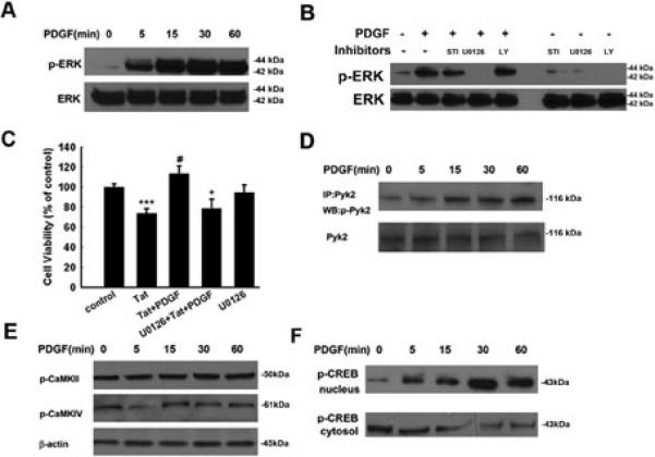
Platelet-derived growth factor-BB (PDGF) has been reported to provide tropic support for neurons in the central nervous system. The protective role of PDGF on dopaminergic neurons, especially in the context of HIV-associated dementia (HAD), however, remains largely unknown. Here, we show that exogenous PDGF was neuroprotective against toxicity induced by HIV-1 Tat in primary midbrain neurons. Furthermore, we report the involvement of transient receptor potential canonical (TRPC) channels in PDGF-mediated neuroprotection. TRPC channels are Ca(2+)-permeable, nonselective cation channels with a variety of physiological functions. Blocking TRPC channels with either a blocker or short-interfering RNAs (specific for TRPC 5 and 6) in primary neurons resulted in suppression of both PDGF-mediated neuroprotection as well as elevations in intracellular Ca(2+). PDGF-mediated neuroprotection involved parallel but distinct ERK/CREB and PI3K/Akt pathways. TRPC channel blocking also resulted in suppression of PDGF-induced Pyk2/ERK/CREB activation, but not Akt activation. Relevance of these findings in vivo was further corroborated by intrastriatal injections of PDGF and HIV-1 Tat in mice. Administration of PDGF was able to rescue the dopaminergic neurons in the substantia nigra from Tat-induced neurotoxicity. This effect was attenuated by pre-treatment of mice with the TRP blocker, thus underscoring the novel role of TRPC channels in the neuroprotection mediated by PDGF.


Figures

Figure 1
Figure 1. PDGF exerts neuroprotection against Tat toxicity
(A) Effect of HIV Tat on the cell survival of rat primary midbrain neurons using the MTT assay. (B) PDGF protects rat primary midbrain neurons against Tat neurotoxicity using the MTT assay. (C) Immunostaining of rat primary midbrain neurons treated with Tat &/or PDGF for 24 h with anti-MAP-2 antibody. Scale bar: 50 μm. (D) Densitometric scan of neuritis (from panel C) expressed as a ratio of neurite length/neuron. (E) Immunostaining of rat primary midbrain neurons treated with Tat &/or PDGF using anti-TH-Ab. Scale bar: 50 μm. (F) Densitometric scan of neuritis (from panel E) using the same method as in (D). Rat primary midbrain neurons treated with Tat &/or PDGF were stained for apoptosis marker Annexin V-FITC using flow cytometric analysis. (G) Hoechst 33342 staining monitored by fluorescence microscopy. (H) Quantification of Annexin V-positive (G) and Hoechst-positive (H) cells as the percentage of total cells. All the data in these figures are presented as mean±SEM of four individual experiments. **p<0.01; ***p<0.001 vs control group; #p<0.05; ##p<0.01 vs Tat (14 nM) group.
Figure 2
Figure 2. PDGF-βR is critical for PDGF-mediated neuroprotection
PDGF receptor antagonist blocked PDGF-βR phosphorylation induced by PDGF as determined by co-immunoprecipitation (A) and also significantly inhibited PDGF-mediated neuroprotection (B). The data are presented as mean±SEM of four individual experiments. **p<0.01 vs control group; ##p<0.01 vs Tat-treated group; +p<0.05 vs both PDGF & Tat treated group; ^p<0.05 vs both PDGF alone treated group. (C) Western blot analysis of whole cell lysates from rat primary midbrain neurons transfected with siRNAs against PDGF-βR or nonsense (Non) siRNA, using antibodies specific for PDGF-βR. Data are representative of three independent experiments. (D) Transfection of rat primary midbrain neurons with siRNAs specific for PDGF-βR or nonsense (Non) siRNA abolished PDGF-mediated neuroprotection. The data are presented as mean±SEM of four individual experiments. **p<0.01 vs control group; ##p< 0.01 vs Tat-treated group; +p<0.05 vs both PDGF & Tat treated group.
Figure 3
Figure 3. TRPC channels are required for PDGF-mediated neuroprotection
(A) Cell viability of rat primary neurons exposed to Tat &/or PDGF in the absence or presence of indicated drugs. (SKF96365: 20 μM; EGTA: 2 mM; 2ApB: 100 μM; Xest-C: 1 μM; U73122: 1 μM; U73343: 1 μM; OAG: 50 μM). All the data in these figures are presented as mean ± SEM of four individual experiments. **p<0.01 vs control group; ##p<0.01 vs Tat-treated group; +p<0.05; ++p<0.01 vs both PDGF &Tat treated group. (B) Western blot analysis of whole cell lysates from rat primary midbrain neurons transfected with siRNAs against TRPC1, 5, 6 or nonsense (Non) siRNA using antibodies specific for either TRPC1, 5 or 6. Data are representative of three independent experiments. (C) Rat primary neurons transfected with TRPC1, 5 or 6 siRNAs were monitored for PDGF-mediated cell viability. The data are presented as mean±SEM of four individual experiments. ***p<0.001 vs control group; ##p< 0.01 vs Tat-treated group; ++p<0.01, +++p<0.001 vs both PDGF & Tat treated group. (D) Co-localization of TRPC5/6 and PDGF-βR in rat primary midbrain neuronal cultures. Midbrain neurons grown for 10 days were double-stained for TRPC5/6 (green) and PDGF-βR (red). DAPI was the nuclei counter stain (blue). Confocal images were taken at a magnification of 60 × (objective lens) with an Eclipse C1 Plus confocal microscope (Nikon). Scale bar: 50 μm.
Figure 4
Figure 4. TRPC channels contribute to PDGF-induced intracellular Ca2+ elevations
(A) Rat primary neurons loaded with Fluo-4 [Ca2+]i sensitive fluorophores before and after PDGF treatment were recorded within a single field using a Fluoview 300 confocal microscope (left panel, numbers in the panels indicate time in seconds) and differential interference contrast (right panel). Scale bar: 100μm. (B) Changes in intracellular [Ca2+]i levels in neurons following PDGF treatment were measured using the Fluo-4/Fura Red ratio, and the change in ratio is illustrated from a typical neuron. (C) Changes in fluorescence amplitude (Fluo-4/Fura Red) in rat primary neurons exposed to PDGF in the absence or presence of the indicated drugs (STI-571:1 μM; SKF96365: 20 μM; EGTA: 2 mM; 2ApB: 100 μM; Xest-C: 1 μM; U73122: 1 μM; U73343: 1 μM; Nifedipine: 10 μM). All the data in these figures are presented as mean ± SEM of four individual experiments. ***p<0.001 vs control group; ###p<0.001 vs Tat-treated group. (D) PDGF-mediated calcium influx in rat primary neurons transfected with either TRPC1, 5 or 6 siRNA. The data are presented as mean±SEM of four individual experiments. ***p<0.001 vs control group; ###p< 0.001 vs PDGF-treated group.
Figure 5
Figure 5. PDGF-induced neuroprotection involves activation of ERK
(A) PDGF induced time-dependent and sustained phosphorylation of ERK. (B) PDGF maintained increased phosphorylation of ERK for at least 1 h, and this effect was abolished by PDGF receptor antagonist STI-571, MEK inhibitor U0126 but not by PI3-K inhibitor LY294002; STI: STI-571: LY: LY294002. (C) Pretreatment of neurons with MEK inhibitor U0126 (20 μM) for 1 h significantly attenuated the protective effect of PDGF. Data are presented as mean ± SEM of four individual experiments. ***p<0.001 vs control group; #p<0.05 vs Tat-treated group; +p<0.05 vs both PDGF & Tat treated group. PDGF exposure resulted in increased time-dependent phosphorylation of Pyk2 (D), but not of CaMKII and CaMKIV (E) in rat primary neurons. PDGF exposure resulted in increased time-dependent phosphorylation of CREB in the nuclear fraction with a concomitant decrease in the cytosolic fraction in rat primary neurons (F).
Figure 6
Figure 6. TRPC channels are critical for PDGF-induced ERK/CREB activation
Rat primary neurons exposed to PDGF in the presence of TRPC blocker and EGTA were monitored for PDGF-mediated ERK, Pyk2 and CREB activation (A-C; upper panels). Densitometric analysis of pERK/ERK, pPyk2/Pyk2 and pCREB/CREB from a representative immunoblot is presented (A-C; lower panels). Data are presented as mean ± SEM of four individual experiments. *p<0.05;**p<0.01 vs control group; #p<0.05,##p<0.01 vs PDGF group.
Figure 7
Figure 7. TRPC5 & 6 are essential for PDGF-mediated phosphorylation of Pyk2, ERK and CREB
Effect of TRPC1, 5 or 6 suppression on PDGF-mediated phosphorylation of ERK, Pyk2 & CREB in rat primary neurons (A-C; upper panels). Densitometric analyses of PDGF-mediated phosphorylation of ERK, Pyk2 and CREB in the presence or absence of siRNAs (A-C; lower panels). Data are presented as mean ± SEM of three individual experiments. *p<0.05; **p<0.01; ***p<0.001 vs control group; #p<0.05, ##p<0.01 vs PDGF with nonsense siRNA group.
Figure 8
Figure 8. TRPC channels are not critical for PDGF-induced Akt activation
(A) PDGF induced time-dependent activation of Akt. (B) PDGF maintained sustained phosphorylation of Akt for at least 1 h and this effect was attenuated by both the PDGF receptor antagonist STI-571 and PI3-K inhibitor LY294002. (C) Pretreatment of neurons with LY294002 (20 μM) for 1 h resulted in inhibition of PDGF-mediated protection using MTT analysis. Data are presented as mean ± SEM of four individual experiments. ***p<0.001 vs control group; #p<0.05 vs Tat-treated group; +p<0.05 vs both PDGF & Tat treated group. (D) Infection of neurons with the dominant-interfering Akt (DN-Akt) resulted in abrogation of PDGF-mediated neuroprotection. Infection with WT Akt (WT-Akt) construct had no effect on cell survival. Data are presented as mean ± SEM of four individual experiments. *p< 0.05 vs adenovirus alone group; ##p< 0.01 vs Tat-treated group. (E) Rat primary neurons exposed to PDGF in the presence of absence of TRP blocker and EGTA was monitored for PDGF-mediated Akt activation (upper panel). Densitometric analyses of pAkt/Akt from the representative immunoblots is presented (lower panel). Data are presented as mean ± SEM of four individual experiments. ***p<0.001 vs control group. (F) Rat primary neurons transfected with TRPC1, 5 or 6 siRNAs were monitored for PDGF-mediated Akt phosphorylation (upper panel). Densitometric analyses of PDGF-mediated phosphorylation of Akt in rat neurons depleted of TRPC1, 5 or 6 from a representative immunoblot (lower panel). Data are presented as mean ± SEM of three individual experiments. *p<0.05; **p<0.01 vs control group.
Figure 9
Figure 9. PDGF protects dopaminergic neurons against Tat-induced in the substantia nigra
Representative mesencephalic sections from different groups of mice treated with PDGF and/or HIV Tat in the presence or absence of TRPC blocker SKF96365 were examined for TH positive neurons counted by stereology. There was increased loss of TH-positive neurons in the substantia nigra of Tat alone treated group of mice compared with the saline controls. Pre-treatment with PDGF resulted in amelioration of Tat toxicity in the substantia nigra neurons, and this effect was significantly attenuated in mice pre-treated with SKF 96365 (0.2 μmol). *p<0.05 vs saline group; #p<0.05 vs Tat; +p<0.05 vs PDGF+Tat. SN: Substantia Nigra. Scale bar: 300μm. SKF:SKF 96365 (0.2 μmol).
Figure 10
Figure 10. Schematic illustration demonstrating putative signaling pathways involved in PDGF-mediated neuroprotection in rat primary neurons
PDGF-mediated engagement of the PDGF-β receptor stimulates the PLC/IP3 R pathway, which in turn, activates TRPC channels resulting in elevation of [Ca2+]i transients. [Ca2+]i elevation in turn results in activation of Pyk2/ERK pathways leading to CREB activation and consequently neuronal survival. In addition to this, PDGF/PDGF-βR axis can also activate another distinct pathway - PI3K/Akt, which can also lead to potentiation of neuronal survival.

References

    1. Nath A, Anderson C, Jones M, Maragos W, Booze R, Mactutus C, et al. Neurotoxicity and dysfunction of dopaminergic systems associated with AIDS dementia. J Psychopharmacol. 2000;14(3):222–227. - PubMed
    1. Mocchetti I, Nosheny RL, Tanda G, Ren K, Meyer EM. Brain-derived neurotrophic factor prevents human immunodeficiency virus type 1 protein gp120 neurotoxicity in the rat nigrostriatal system. Ann N Y Acad Sci. 2007 Dec;1122:144–154. - PubMed
    1. Zauli G, Secchiero P, Rodella L, Gibellini D, Mirandola P, Mazzoni M, et al. HIV-1 Tat-mediated inhibition of the tyrosine hydroxylase gene expression in dopaminergic neuronal cells. J Biol Chem. 2000 Feb 11;275(6):4159–4165. - PubMed
    1. Eugenin EA, D'Aversa TG, Lopez L, Calderon TM, Berman JW. MCP-1 (CCL2) protects human neurons and astrocytes from NMDA or HIV-tat-induced apoptosis. J Neurochem. 2003 Jun;85(5):1299–1311. - PubMed
    1. Liu Y, Jones M, Hingtgen CM, Bu G, Laribee N, Tanzi RE, et al. Uptake of HIV-1 tat protein mediated by low-density lipoprotein receptor-related protein disrupts the neuronal metabolic balance of the receptor ligands. Nat Med. 2000 Dec;6(12):1380–1387. - PubMed

Publication types

MeSH terms

Substances