. 2009 Aug;5(8):e1000545.
doi: 10.1371/journal.ppat.1000545. Epub 2009 Aug 14.

Mycobacterium tuberculosis WhiB3 maintains redox homeostasis by regulating virulence lipid anabolism to modulate macrophage response

Affiliations

Mycobacterium tuberculosis WhiB3 maintains redox homeostasis by regulating virulence lipid anabolism to modulate macrophage response

Amit Singh et al. PLoS Pathog. 2009 Aug.

Abstract

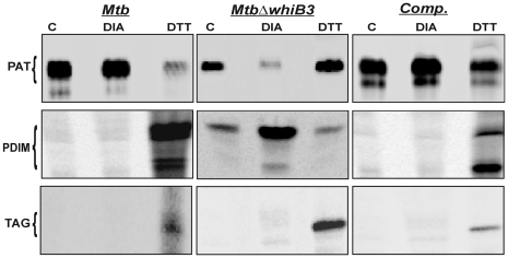
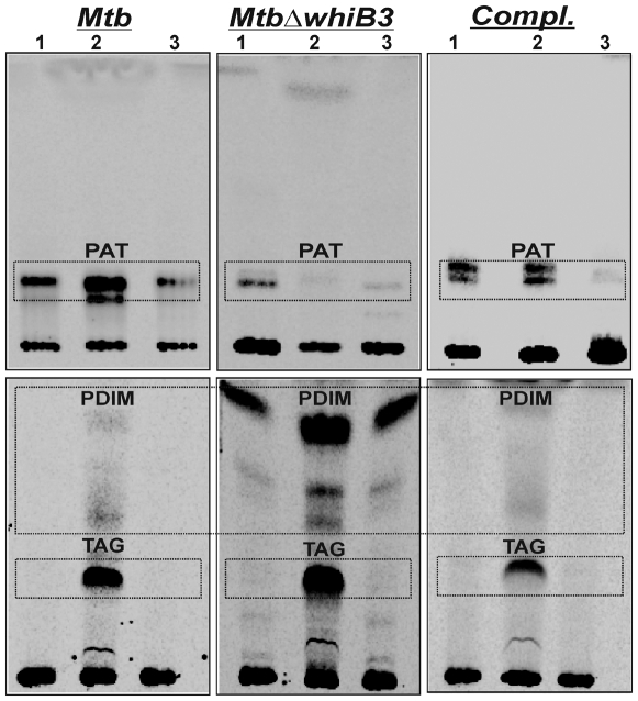
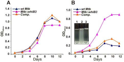
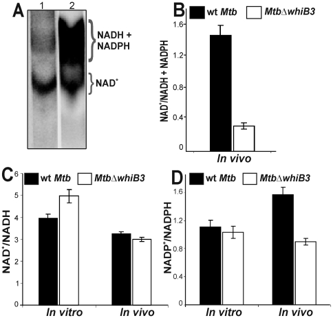
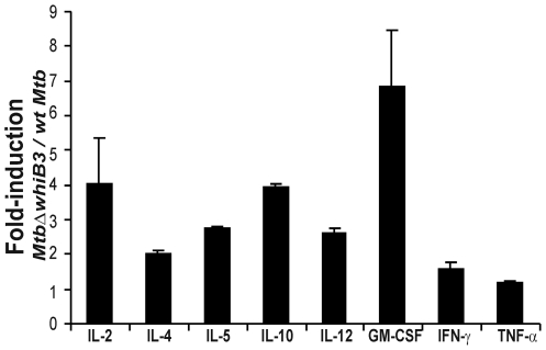
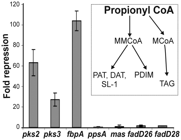
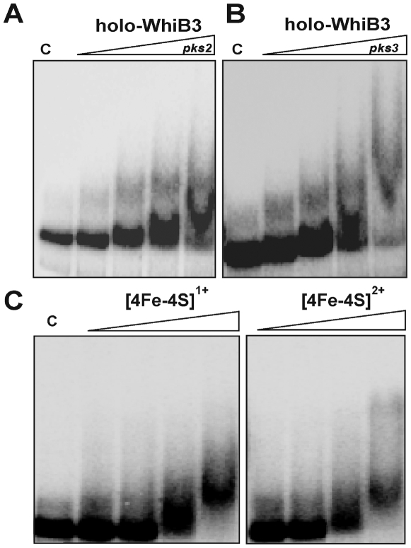
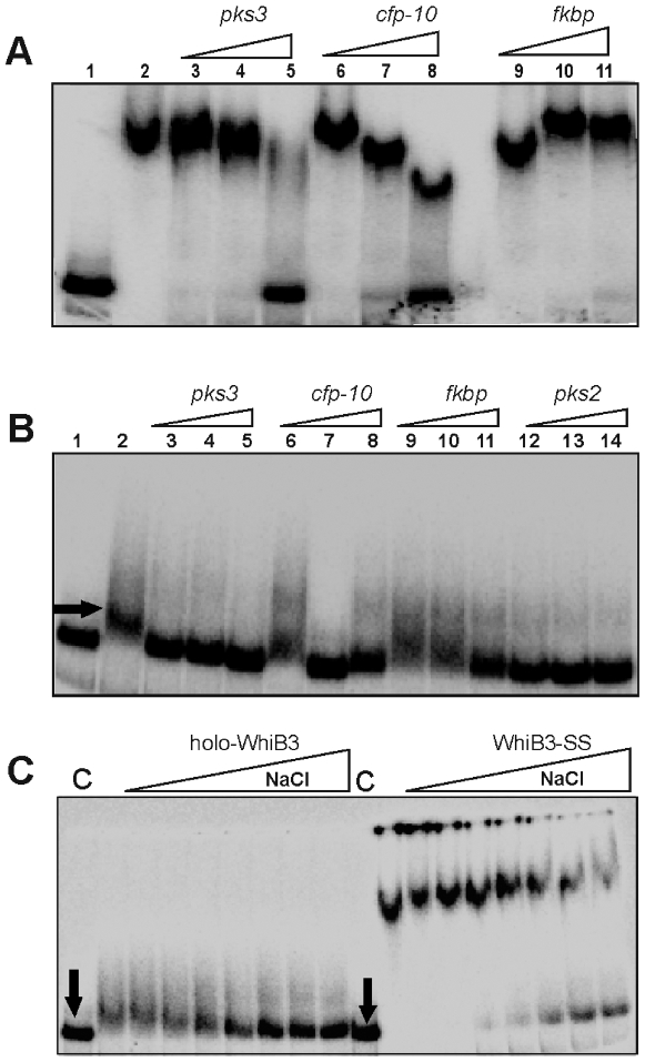
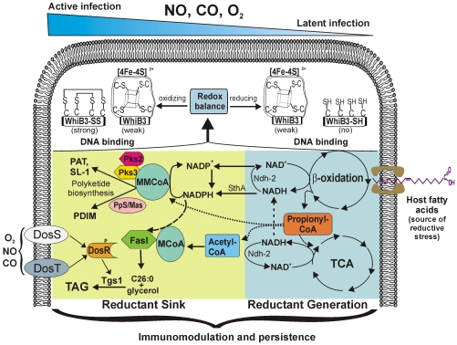
The metabolic events associated with maintaining redox homeostasis in Mycobacterium tuberculosis (Mtb) during infection are poorly understood. Here, we discovered a novel redox switching mechanism by which Mtb WhiB3 under defined oxidizing and reducing conditions differentially modulates the assimilation of propionate into the complex virulence polyketides polyacyltrehaloses (PAT), sulfolipids (SL-1), phthiocerol dimycocerosates (PDIM), and the storage lipid triacylglycerol (TAG) that is under control of the DosR/S/T dormancy system. We developed an in vivo radio-labeling technique and demonstrated for the first time the lipid profile changes of Mtb residing in macrophages, and identified WhiB3 as a physiological regulator of virulence lipid anabolism. Importantly, MtbDeltawhiB3 shows enhanced growth on medium containing toxic levels of propionate, thereby implicating WhiB3 in detoxifying excess propionate. Strikingly, the accumulation of reducing equivalents in MtbDeltawhiB3 isolated from macrophages suggests that WhiB3 maintains intracellular redox homeostasis upon infection, and that intrabacterial lipid anabolism functions as a reductant sink. MtbDeltawhiB3 infected macrophages produce higher levels of pro- and anti-inflammatory cytokines, indicating that WhiB3-mediated regulation of lipids is required for controlling the innate immune response. Lastly, WhiB3 binds to pks2 and pks3 promoter DNA independent of the presence or redox state of its [4Fe-4S] cluster. Interestingly, reduction of the apo-WhiB3 Cys thiols abolished DNA binding, whereas oxidation stimulated DNA binding. These results confirmed that WhiB3 DNA binding is reversibly regulated by a thiol-disulfide redox switch. These results introduce a new paradigmatic mechanism that describes how WhiB3 facilitates metabolic switching to fatty acids by regulating Mtb lipid anabolism in response to oxido-reductive stress associated with infection, for maintaining redox balance. The link between the WhiB3 virulence pathway and DosR/S/T signaling pathway conceptually advances our understanding of the metabolic adaptation and redox-based signaling events exploited by Mtb to maintain long-term persistence.


Conflict of interest statement

The authors have declared that no competing interests exist.

Figures

Figure 1
Figure 1. Mtb WhiB3 regulates cell shape and size.
(A) wt Mtb and MtbΔwhiB3 were cultured in 7H9 liquid media containing 0.1% Tween 80 till stationary phase. At an OD600 nm = 4.5 the aggregation phenotype was examined by allowing the cells to settle for 10 min. MtbΔwhiB3 cells settled rapidly whereas wt Mtb remained dispersed (B) Spot colony rugosity of wt Mtb and MtbΔwhiB3 was analyzed by spotting identical volumes (30 µl) and equal number of cells (3×10⁶) on Dubos complete medium. Cells were allowed to grow for 4 weeks and photographs were taken at 7× magnification using a Zeiss stereo microscope. (C and D) SEM analysis demonstrating that WhiB3 affects cell length. Low magnification SEM (C, inset) illustrates the severe clumping of MtbΔwhiB3 cells. Approximately 5 SEM fields (10 cells/field) were analyzed to determine the cell sizes of wt Mtb and MtbΔwhiB3. (E) TEM analysis showing hyperstaining of MtbΔwhiB3 cells as compared to wt Mtb cells. Inset; low magnification image.
Figure 2
Figure 2. Mtb WhiB3 regulates biosynthesis of complex virulence lipids.
Wt Mtb, MtbΔwhiB3, and MtbΔwhiB3tetRO:whiB3 (Comp.) were cultured in 7H9 liquid media containing 0.1% Tween 80, and total lipids labeled using [14C] propionate. In each lane, silica TLC plates were loaded with 100,000 cpm of [14C] propionate-derived total lipids and various lipid fractions were analysed. (A) Polar lipid fraction was resolved using 10% methanol in chloroform as the solvent. (B) PAT lipids were analyzed by developing TLC plates in petroleum ether∶acetone (92∶8). (C) PDIM and TAG were analyzed by developing TLC plates in petroleum ether∶acetone (98∶2). (D) SL-1 and DAT were analyzed by developing TLC plates in chloroform∶ethanol∶water (90∶10∶1). (E) TMM and TDM were analyzed by loading 100,000 cpm of [1, 2-14C] acetate-derived total lipids on a silica TLC plate and developed using chloroform∶methanol∶acetone∶acetic acid (80∶20∶6∶1). Lanes (1) wt Mtb, (2) MtbΔwhiB3 and (3) complemented MtbΔwhiB3tetRO:whiB3 strain (Comp). Phosphorimaging (imageQuant software; GE Healthcare) were used to examine the relative distribution of 14C among the lipid fractions.
Figure 3
Figure 3. Mtb WhiB3 regulates biosynthesis of pathogenic lipids in response to oxido-reductive stress.
Complex virulence lipids were analyzed by metabolic labeling using [14C] propionate under oxidizing (5 mM diamide) or reducing (5 mM DTT) conditions for 24 h. PAT, PDIM and TAG production was analyzed by spotting equal count in each case (50,000 cpm) on silica TLC and resolved using solvent systems as described in Figure 2. C; untreated cells.
Figure 4
Figure 4. Mtb WhiB3 regulates biosynthesis of pathogenic lipids within resting macrophages.
Raw 264.7 cells (5×10⁸) were independently infected with lightly sonicated wt Mtb, MtbΔwhiB3 and MtbΔwhiB3 tetRO:whiB3 at a MOI of 10∶1 for 4 h. Total lipids from Mtb growing within macrophages were metabolically labeled by the addition of [14C] propionate for 2 days, and analyzed by TLC (see Materials and Methods). PAT, PDIM and TAG were analyzed by loading equal counts (20,000 cpm) on TLC plates, and developed using solvents as described in Fig. 2 legend. Lipids isolated from Mtb growing in Lane (1) 7H9 medium, (2) Raw264.7 and (3) DMEM medium.
Figure 5
Figure 5. Mtb WhiB3 modulates propionate toxicity.
Cultures of Mtb were synchronized to OD600 nm = 0.04 and grown in 7H9 broth containing 10 mM (A) or 20 mM (B) sodium propionate. Culture turbidity (OD600 nm) was measured at the indicated time points. Results are representative of two independent experiments showing similar results. Inset; liquid cultures of wt Mtb (1), MtbΔwhiB3 (2) and Comp (3) grown for 10 days in 20 mM propionate.
Figure 6
Figure 6. Mtb WhiB3 maintains the intrabacterial NAD+/NADH and NADP+/NADPH poise.
(A) TLC analysis of oxidized and reduced pyridine nucleotides labeled with [14C] nicotinamide. Wt Mtb (lane 1) and MtbΔwhiB3 (lane 2) cells isolated from infected macrophages (∼108 cells) were labeled with [14C] nicotinamide for 24 h in 7H9 basal medium containing acetate as a carbon source and analyzed by TLC by loading equal cpm in each lane. Note the strong labeling of reduced pyridine nucleotides from MtbΔwhiB3 isolated from macrophages. (B) Densitometric analysis of the relative abundance of nucleotides in (A). Experiments were performed four times and similar observations were recorded. Cells grown in vitro or isolated from the infected macrophages were analyzed by enzymatic assays using (C) alcohol dehydrogenase for NAD+/NADH estimation and (D) glucose-6-phosphate dehydrogenase for NADP+/NADPH analysis. Data shown is the average of two independent experiments.
Figure 7
Figure 7. MtbΔwhiB3 alters macrophage inflammatory cytokine production.
Raw 264.7 macrophages were infected with well dispersed cells of wt Mtb and MtbΔwhiB3 at a MOI of 10∶1 for 24 h and the concentration of cytokine in the supernatant representing the Th1 and Th2 immune responses were quantified using the Bio-Plex multiplex Human Cytokine Th1/Th2 Assay kit (BioRad Laboratories).
Figure 8
Figure 8. Mtb WhiB3 regulates the expression of polyketide biosynthetic genes.
Total RNA was isolated from logarithmically grown cells of wt Mtb and MtbΔwhiB3 and the expression of PAT, DAT, SL-1 and PDIM biosynthetic genes was analyzed by real time PCR. Inset; the pathway describing the incorporation of a common precursor propionyl-CoA into PAT, DAT, SL-1, PDIM and TAG. RT-PCR data suggests that the repression of PAT, DAT and SL-1 biosynthetic genes in MtbΔwhiB3 results in the accumulation of propionyl CoA, which is then diverted to PDIM and TAG production.
Figure 9
Figure 9. Mtb WhiB3 is a Fe-S cluster containing DNA binding protein.
The [4Fe-4S]2+ form of WhiB3 was utilized for EMSA. EMSA reactions were performed under anaerobic conditions using 0.2 nM 32P labeled (A) pks2, or (B) pks3 promoter DNA. The WhiB3 [4Fe-4S]2+ concentrations used for EMSA were 0, 100, 200, 400 and 800 nM. (C) The redox status of the 4Fe-4S cluster does not influence WhiB3 DNA binding. Identical concentrations (800 nM) of WhiB3 [4Fe-4S]2+ and dithionite-reduced WhiB3 [4Fe-4S]1+ were analyzed for their ability to bind pks3 promoter DNA.
Figure 10
Figure 10. A thiol-disulfide redox switch reversibly regulates WhiB3 DNA binding.
(A) The inhibitory effect of DTT on apo-WhiB3 binding to pks3 promoter DNA is reversed by diamide. (B) Dose-dependent effect of diamide on DNA binding. EMSA was performed using diamide (50, 100, 200, 400, 800 and 1000 µM), pks3 promoter DNA, and apo-WhiB3 (800 nM).
Figure 11
Figure 11. WhiB3 DNA binding and sequence specificity.
(A) Sequence specificity of WhiB3-SS DNA binding. 0.2 nM 32P labeled pks3 promoter DNA with 800 nM of WhiB3-SS were used in the EMSA analysis. Lane 1; free probe, lane 2; WhiB3-SS:DNA complex. WhiB3 DNA binding was competed using 200 (lane; 3, 6 and 9), 400 (lane; 4, 7 and 10) and 800 (lane; 5, 8 and 11) fold molar excess of unlabeled competitor DNA [pks3 (specific), cfp-10 (non-specific), mammalian FKBP ORF (non-specific)]. (B) Lane 1; free probe, lane 2; holo-WhiB3:DNA complex. Competition assay for sequence specificity of holo-WhiB3 (800 nM) using pks3 promoter (specific, lane; 3, 4 and 5), cfp-10 ORF (non- specific, lane; 6, 7 and 8), mammalian fkbp ORF (non- specific, lane; 9, 10 and 11), and pks2 promoter (specific, lane, 12, 13 and 14) as competitor DNA. These results demonstrate that WhiB3 has low sequence discrimination. Our unpublished data have shown that WhiB3 binds strongly to linear and supercoiled pUC19 DNA, as well as HindIII digested λ-DNA. This, together with the cfp-10, ftsQ, and FKBP control DNA binding experiments provides strong evidence for the non-specific DNA binding activity of WhiB3. Arrow indicates the minor retardation of pks3 promoter due to weak binding of holo-WhiB3. (C) Oxidized apo-WhiB3 (WhiB3-SS) binds DNA stronger than holo-WhiB3. NaCl was used to examine and compare the contribution of the [4Fe-4S]2+ cluster or disulphide bonds on the strength of DNA (pks3 promoter) binding. EMSA was performed under anaerobic conditions using 800 nM holo-WhiB3 or WhiB3-SS in the presence of 0, 100, 200, 300, 400, 600, 800, 1000 mM of NaCl. C; DNA binding without NaCl. Arrows; free probe. In each experiment, a promoter fragment containing 330 bp sequences upstream of pks3 ATG was used as radiolabeled probe.
Figure 12
Figure 12. The reductive stress dissipation model for Mtb persistence.
Since gradients of NO, CO, O2 and host fatty acids generated within microenvironments of the lungs are most likely encountered by Mtb during infection , we propose that an intracellular Mtb redox imbalance is caused by the catabolism of highly reducing host lipids, which influences endogenous Mtb polyketide anabolism. In this model the DNA binding activity of WhiB3 is activated by post-translational modifications of the WhiB3 Cys thiols in response to redox stress to generate a distinct cellular response, which modulates the production of inflammatory polyketides and storage lipids (TAG) to maintain intracellular redox homeostasis, and to modulate virulence. A crucial component of the model is the strong reducing power of host fatty acids, and inhibition of respiration by NO, CO and hypoxia, which results in the accumulation of reducing equivalents in Mtb to generate reductive stress. In order to dispose of excess reductants, the bacilli anabolize PAT, SL-1, PDIM and TAG, which function as an electron sink. Notably, reductive stress is further enhanced by NO, CO, or hypoxia. Since TAG is also induced upon exposure to NO, CO and hypoxia via the DosR/S/T dormancy system, our results suggest cross-talk between the WhiB3 and Dos dormancy pathways resulting in TAG anabolism to dissipate reducing equivalents. The lipid defects exhibited by MtbΔwhiB3 during infection suggest that WhiB3 functions as a nano-switch to systematically regulate the production of virulence lipids to maintain redox homeostasis and persistence.

Similar articles

Cited by

References

    1. Dye C, Scheele S, Dolin P, Pathania V, Raviglione MC, et al. Global Burden of Tuberculosis: Estimated Incidence, Prevalence, and Mortality by Country. JAMA. 1999;282:677–686. - PubMed
    1. Chan J, Fujiwara T, Brennan P, McNeil M, Turco SJ, et al. Microbial glycolipids: possible virulence factors that scavenge oxygen radicals. Proc Natl Acad Sci U S A. 1989;86:2453–2457. - PMC - PubMed
    1. Reed MB, Domenech P, Manca C, Su H, Barczak AK, et al. A glycolipid of hypervirulent tuberculosis strains that inhibits the innate immune response. Nature. 2004;431:84–87. - PubMed
    1. Sherman DR, Sabo PJ, Hickey MJ, Arain TM, Mahairas GG, et al. Disparate responses to oxidative stress in saprophytic and pathogenic mycobacteria. Proc Natl Acad Sci U S A. 1995;92:6625–6629. - PMC - PubMed
    1. Camacho LR, Constant P, Raynaud C, Laneelle MA, Triccas JA, et al. Analysis of the phthiocerol dimycocerosate locus of Mycobacterium tuberculosis. Evidence that this lipid is involved in the cell wall permeability barrier. J Biol Chem. 2001;276:19845–19854. - PubMed

Publication types

MeSH terms