

Perfusion imaging of the right perisylvian neural network in acute spatial neglect
- PMID: 19680470
- PMCID: PMC2726039
- DOI: 10.3389/neuro.09.015.2009
Perfusion imaging of the right perisylvian neural network in acute spatial neglect
Abstract
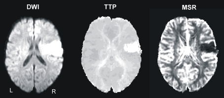
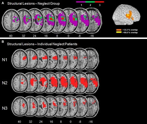
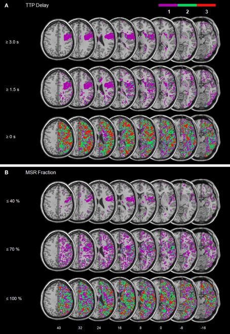
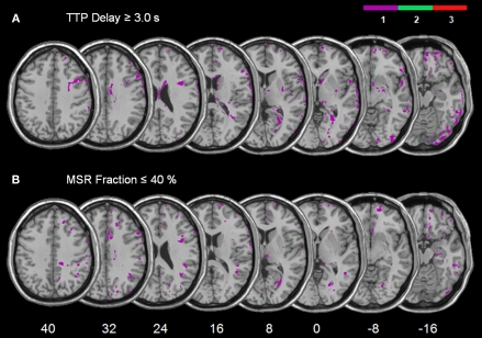
Recent studies have suggested a tightly connected perisylvian neural network associated with spatial neglect. Here we investigated whether structural damage in one part of the network typically is accompanied with functional damage in other, structurally intact areas of this network. By combining normalized fluid-attenuated inversion-recovery (FLAIR) imaging, diffusion-weighted imaging (DWI), and perfusion-weighted imaging (PWI) we asked whether or not lesions centering on fronto-temporal regions co-occur with abnormal perfusion in structurally intact parietal cortex. With thresholds applied to delineate behaviourally relevant malperfusion of brain tissue, the analysis of normalized time-to-peak (TTP) and maximal signal reduction (MSR) perfusion maps did not reveal significant changes outside the area of structural damage. In particular, we found no abnormal perfusion in the structurally intact inferior parietal lobule (IPL) and/or the temporo-parietal junction (TPJ). The present results obtained in three consecutively admitted neglect patients with fronto-temporal lesions indicate that structural damage in one part of the right perisylvian network associated with spatial neglect does not necessarily require dysfunction by malperfusion in other, structurally intact parts of the network to provoke spatial neglect. The neural tissue in the fronto-temporal cortex appears to have an original role in processes of spatial orienting and exploration.
Keywords: magnetic resonance imaging; parietal cortex; perfusion-weighted imaging; right perisylvian neural network; spatial neglect; superior temporal cortex.
Figures
Similar articles
-
Normalized perfusion MRI to identify common areas of dysfunction: patients with basal ganglia neglect.Brain. 2005 Oct;128(Pt 10):2462-9. doi: 10.1093/brain/awh629. Epub 2005 Sep 8. Brain. 2005. PMID: 16150848
-
Perfusion imaging in Pusher syndrome to investigate the neural substrates involved in controlling upright body position.PLoS One. 2009 May 29;4(5):e5737. doi: 10.1371/journal.pone.0005737. PLoS One. 2009. PMID: 19478939 Free PMC article.
-
Fiber pathways connecting cortical areas relevant for spatial orienting and exploration.Hum Brain Mapp. 2014 Mar;35(3):1031-43. doi: 10.1002/hbm.22232. Epub 2013 Jan 2. Hum Brain Mapp. 2014. PMID: 23283834 Free PMC article.
-
Unilateral spatial neglect after posterior parietal damage.Handb Clin Neurol. 2018;151:287-312. doi: 10.1016/B978-0-444-63622-5.00014-0. Handb Clin Neurol. 2018. PMID: 29519463 Review.
-
Spatial neglect, Balint-Homes' and Gerstmann's syndrome, and other spatial disorders.CNS Spectr. 2007 Jul;12(7):527-36. doi: 10.1017/s1092852900021271. CNS Spectr. 2007. PMID: 17603404 Review.
Cited by
-
Mapping human brain lesions and their functional consequences.Neuroimage. 2018 Jan 15;165:180-189. doi: 10.1016/j.neuroimage.2017.10.028. Epub 2017 Oct 16. Neuroimage. 2018. PMID: 29042216 Free PMC article. Review.
-
Spatial neglect and attention networks.Annu Rev Neurosci. 2011;34:569-99. doi: 10.1146/annurev-neuro-061010-113731. Annu Rev Neurosci. 2011. PMID: 21692662 Free PMC article. Review.
-
Is there a critical lesion site for unilateral spatial neglect? A meta-analysis using activation likelihood estimation.Front Hum Neurosci. 2012 Apr 10;6:78. doi: 10.3389/fnhum.2012.00078. eCollection 2012. Front Hum Neurosci. 2012. PMID: 22514528 Free PMC article.
References
-
- Astrup J., Siesjö B. K., Symon L. (1981). Thresholds in cerebral ischemia – the ischemic penumbra. Stroke 12, 723–725 - PubMed
-
- Barber P. A., Darby D. G., Desmond P. M., Yang Q., Gerraty R. P., Jolley D., Donnan G. A., Tress B. M., Davis S. M. (1998). Prediction of stroke outcome with echoplanar perfusion- and diffusion-weighted MRI. Neurology 51, 418–426 - PubMed
LinkOut - more resources
Full Text Sources