. 2009 Aug;5(8):e1000547.
doi: 10.1371/journal.ppat.1000547. Epub 2009 Aug 14.

Expression and processing of a small nucleolar RNA from the Epstein-Barr virus genome

Affiliations

Expression and processing of a small nucleolar RNA from the Epstein-Barr virus genome

Roland Hutzinger et al. PLoS Pathog. 2009 Aug.

Abstract

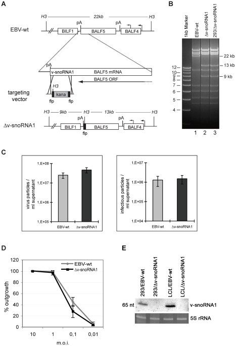
Small nucleolar RNAs (snoRNAs) are localized within the nucleolus, a sub-nuclear compartment, in which they guide ribosomal or spliceosomal RNA modifications, respectively. Up until now, snoRNAs have only been identified in eukaryal and archaeal genomes, but are notably absent in bacteria. By screening B lymphocytes for expression of non-coding RNAs (ncRNAs) induced by the Epstein-Barr virus (EBV), we here report, for the first time, the identification of a snoRNA gene within a viral genome, designated as v-snoRNA1. This genetic element displays all hallmark sequence motifs of a canonical C/D box snoRNA, namely C/C'- as well as D/D'-boxes. The nucleolar localization of v-snoRNA1 was verified by in situ hybridisation of EBV-infected cells. We also confirmed binding of the three canonical snoRNA proteins, fibrillarin, Nop56 and Nop58, to v-snoRNA1. The C-box motif of v-snoRNA1 was shown to be crucial for the stability of the viral snoRNA; its selective deletion in the viral genome led to a complete down-regulation of v-snoRNA1 expression levels within EBV-infected B cells. We further provide evidence that v-snoRNA1 might serve as a miRNA-like precursor, which is processed into 24 nt sized RNA species, designated as v-snoRNA1(24pp). A potential target site of v-snoRNA1(24pp) was identified within the 3'-UTR of BALF5 mRNA which encodes the viral DNA polymerase. V-snoRNA1 was found to be expressed in all investigated EBV-positive cell lines, including lymphoblastoid cell lines (LCL). Interestingly, induction of the lytic cycle markedly up-regulated expression levels of v-snoRNA1 up to 30-fold. By a computational approach, we identified a v-snoRNA1 homolog in the rhesus lymphocryptovirus genome. This evolutionary conservation suggests an important role of v-snoRNA1 during gamma-herpesvirus infection.


Conflict of interest statement

The authors have declared that no competing interests exist.

Figures

Figure 1
Figure 1. Sequence and expression profile of v-snoRNA1.
(A) Sequences of the newly identified 65 nt long v-snoRNA1 and the canonical box C/D from snoRNA U80 used as a control are shown here. The position of C/D boxes and C′/D′ boxes are indicated in red. (B) Northern blot analysis showing expression of v-snoRNA1 in a panel of EBV-positive (BL2-B95.8, BL41-B95.8, LCL-B95.8, Raji, Rael) and EBV-negative (BL2, BL41) cell lines. 5S rRNA served as internal loading control.
Figure 2
Figure 2. Schematic representation of the Epstein-Barr-virus genome.
The location of ncRNA genes, latent genes and the precise location of v-snoRNA1 is indicated. (A) Location and transcription of EBV ncRNA genes (black lines with blue lettering) and EBV latent genes (grey bars with black lettering). The v-snoRNA1 is indicated in red, the neighboring miRNA BART2 in orange and the viral DNA polymerase BALF5 is depicted in green (for coding region) and brown (for 3′-UTR). The promoters are shown in grey lines and lettering, the BARTs region as a grey bar and the B95.8 deletion are also indicated. (B) Close-up of v-snoRNA1 location within the 3′-UTR of the viral DNA polymerase gene. The v-snoRNA1 is located on the sense strand about 60 nt downstream of the mir-BART2 precursor transcript and complementary to the BALF5 3′UTR that is situated on the antisense strand. V-snoRNA124pp is indicated in blue, other transcripts are indicated in the same colors as described above. The black line illustrates the cleavage site of mir-BART2. Corresponding EBV coordinates refer to the EBV B95.8 deletion strain (Accession number V01555.2).
Figure 3
Figure 3. Fluorescent in situ hybridization and Co-immunoprecipitation of v-snoRNA1.
(A) The box C/D v-snoRNA1 (red) localizes in the nucleolus of EBV-positive BL2-B95.8 cells. Box C/D snoRNA U3 (green) was used as a nucleolar marker. In EBV-infected cells both v-snoRNA1 and U3 co-localize in the nucleoli. In EBV-negative cells only U3 is expressed. The nucleus was stained with DAPI for visualization of nuclei and the scale bar is 10 µm. (B) Co-immunoprecipitation of v-snoRNA1 with fibrillarin, NOP56 and NOP58 snoRNP proteins. Following immunoprecipitation employing antibodies specific to fibrillarin, NOP56 and NOP58, the v-snoRNA1 was co-precipitated and detected via northern blot analysis. Box C/D snoRNA U81 and 5.8 rRNA were used as positive and negative controls, respectively.
Figure 4
Figure 4. Expression of v-snoRNA1 during latency and lytic replication.
Expression of v-snoRNA1 was investigated in LCLs infected with either the wild type or the replication-defective ΔBZLF1 EBV strain. The expression of v-snoRNA1 in 293 cells that stably carry the EBV-wt genome was monitored before and after induction with a BZLF1 expression plasmid. 5S rRNA was used as an internal loading control.
Figure 5
Figure 5. Construction of a v-snoRNA1 null recombinant virus.
(A) Schematic map of the EBV genome segment that encompasses the v-snoRNA1 in EBV-wt before and after homologous recombination with the targeting vector carrying the kanamycin resistance gene flanked by flp recombinase recognition sites. The kanamycin cassette was excised in a second step. The restriction sites for HindIII (H3) and the expected fragment sizes after cleavage of EBV-wt and Δv-snoRNA1 genomes with this enzyme are given. pA: polyadenylation site, kana: kanamycin. (B) HindIII restriction fragment analysis of EBV-wt (lane 1) and Δv-snoRNA1 mutant genomes directly after construction in E.coli (lane 2) or after rescue from stably transfected 293 cells (293/Δv-snoRNA1) (lane 3). The result is fully consistent with the predicted restriction pattern (see A). (C) v-snoRNA1 is not required for virus production. Titres in supernatants from cells induced to produce viruses were determined either by measuring the concentration of viral genomes or by infecting the Raji B cell line in a limiting dilution assay. The concentration of viral genome equivalents and infectious particles is given for wild type and Δv-snoRNA1 viruses. Shown are mean values from three independent experiments. (D) Δv-snoRNA1 viruses show intact transforming properties. Primary B cells from three different normal donors were exposed to wild type and Δv-snoRNA1 viruses at various multiplicities of infection in a limiting dilution assay in cluster plates. The percentage of wells showing cell outgrowth is indicated. The presented results represent the average values from three experiments with the corresponding standard deviations. (E) v-snoRNA1 is expressed in cell lines infected with wild type EBV but not in cell lines infected with the Δv-snoRNA1 null-mutant. A northern blot analysis using a v-snoRNA1-specific probe was performed on 293 and B cells infected with either wild type EBV or with the Δv-snoRNA1-null mutant. 5S rRNA served as a loading control.
Figure 6
Figure 6. Sequence motifs of v-snoRNA1 antisense elements (AE) for computational target predictions.
(A) AE box D is indicated in blue and the two potential alternative AE for box D′ in green. (B) A list of the most likely potential rRNA targets of AE box D′ long on the human genome is given (for the full list see Table S1). It includes the predicted ribose methylated positions (red), alignment and score.
Figure 7
Figure 7. Expression analysis of v-snoRNA124pp.
Northern blot analysis demonstrating expression of the 24 nt long processing product v-snoRNA124pp, derived from v-snoRNA1, by employing a specific LNA oligonucleotide probe in 293/EBV-wt or in 293/Δv-snoRNA1 knock-out strain cells without or upon BZLF1-induction. Expression of full length v-snoRNA1 (65 nt) and a potential cleavage intermediate (40 nt) are also shown. 5S rRNA serves as an internal loading control.
Figure 8
Figure 8. v-snoRNA124pp-directed cleavage of BALF5 mRNA.
The sequence of the 3′-UTR target site within the BALF5 mRNA and the complementary v-snoRNA124pp derived from v-snoRNA1 are shown. The predicted v-snoRNA124pp-directed cleavage site according to cDNA sequences obtained from 5′-RACE is indicated by an arrow. cDNA sequences from 5′-RACE are shown at the bottom.
Figure 9
Figure 9. Expression of v-snoRNA1 in rLCV.
(A) Alignment of v-snoRNA1 and rLCV including their flanking regions. V-snoRNA1 and rLCV snoRNA sequences are marked in red, flanking nucleotides in black. Stars in red and black indicate conservation of the nucleotides between EBV and rLCV sequences. The boxes C/D and C′/D′ are encircled by black rectangles. (B) The putative rLCV snoRNA is expressed in simian LCLs. A northern blot analysis using a labeled rLCV snoRNA oligonucleotide was performed on the LCL8664 cell line that was generated by infection with rLCV. A panel of EBV-negative and EBV-positive human LCLs were used as controls. 5S rRNA was used as loading control.

References

    1. Murray PG, Young LS. Epstein-Barr virus infection: basis of malignancy and potential for therapy. Expert Rev Mol Med. 2001;3:1–20. - PubMed
    1. Straathof KC, Bollard CM, Rooney CM, Heslop HE. Immunotherapy for Epstein-Barr virus-associated cancers in children. Oncologist. 2003;8:83–98. - PubMed
    1. Hislop AD, Taylor GS, Sauce D, Rickinson AB. Cellular responses to viral infection in humans: lessons from Epstein-Barr virus. Annu Rev Immunol. 2007;25:587–617. - PubMed
    1. Rickinson AB, Kieff E. Epstein-Barr Virus. Fields Virology 5th edition. 2007:2655–2700.
    1. Miller G, Lipman M. Release of infectious Epstein-Barr virus by transformed marmoset leukocytes. Proc Natl Acad Sci U S A. 1973;70:190–194. - PMC - PubMed

Publication types

Associated data