. 2009 Aug;5(8):e1000548.
doi: 10.1371/journal.ppat.1000548. Epub 2009 Aug 14.

Two HIV-1 variants resistant to small molecule CCR5 inhibitors differ in how they use CCR5 for entry

Affiliations

Two HIV-1 variants resistant to small molecule CCR5 inhibitors differ in how they use CCR5 for entry

Reem Berro et al. PLoS Pathog. 2009 Aug.

Abstract

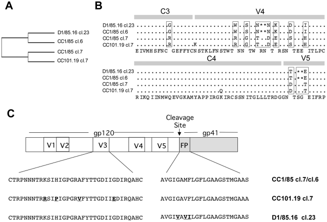
HIV-1 variants resistant to small molecule CCR5 inhibitors recognize the inhibitor-CCR5 complex, while also interacting with free CCR5. The most common genetic route to resistance involves sequence changes in the gp120 V3 region, a pathway followed when the primary isolate CC1/85 was cultured with the AD101 inhibitor in vitro, creating the CC101.19 resistant variant. However, the D1/86.16 escape mutant contains no V3 changes but has three substitutions in the gp41 fusion peptide. By using CCR5 point-mutants and gp120-targeting agents, we have investigated how infectious clonal viruses derived from the parental and both resistant isolates interact with CCR5. We conclude that the V3 sequence changes in CC101.19 cl.7 create a virus with an increased dependency on interactions with the CCR5 N-terminus. Elements of the CCR5 binding site associated with the V3 region and the CD4-induced (CD4i) epitope cluster in the gp120 bridging sheet are more exposed on the native Env complex of CC101.19 cl.7, which is sensitive to neutralization via these epitopes. However, D1/86.16 cl.23 does not have an increased dependency on the CCR5 N-terminus, and its CCR5 binding site has not become more exposed. How this virus interacts with the inhibitor-CCR5 complex remains to be understood.


Conflict of interest statement

The authors have declared that no competing interests exist.

Figures

Figure 1
Figure 1. Sequence analysis of parental and resistant viruses.
(A) Tree view of a multiple Env amino acid sequence alignment of the CC1/85-derived clones (cl.6, cl.7) and the two CCR5 inhibitor-resistant clones, CC101.19 cl.7 and D1/85.16 cl.23. (B) Alignment of gp120 sequences from residues 370–470, consisting of a segment of C3, V4, C4 and V5. The consensus amino acid sequence is given on the bottom line. Dots represent identical residues for all four clones, with gaps indicated by asterisks. Residues that are different between the two pairs (D1/85.16 cl.23 and CC1/85 cl.6 on the one hand, CC101.19 cl.7 and CC1/85 cl.7 on the other) are boxed. (C) Schematic representation of HIV-1 Env clones. The V3 and FP sequences of representative clones of CC1/85, CC101.19 and D1/85.16 are depicted. CC101.19 cl.7 contains four substitutions in the V3 region of gp120 (K305R, H308P, A316V and G321E), while D1/85.16 cl.23 contains three substitutions in the gp41 FP (G516V, M518V and F519I) ,. These sequence differences are highlighted in bold and underlined; they are necessary and sufficient to confer resistance, although there are other changes elsewhere in Env. Amino acid numbering is based on HxB2 Env.
Figure 2
Figure 2. VVC sensitivity of parental and resistant isolates and clones.
(A) The parental isolate CC1/85 (squares) and the resistant isolates CC101.19 (triangles) and D1/85.16 (circles) were tested for VVC sensitivity in an assay of HIV-1 replication in PBMCs. (B) Clones derived from the parental isolate, CC1/85 cl.7 (squares) and CC1/85 cl.6 (diamonds), or from the resistant isolates, CC101.19 cl.7 (triangles) and D1/85.16 cl.23 (circles), were used to infect PBMCs in the presence of VVC. The data in both panels A and B represent the extent of inhibition of replication (p24 antigen production) relative to that in the absence of VVC (100% replication, 0% inhibition). The data points in panels A and B are derived from a single, representative experiment.
Figure 3
Figure 3. Schematic representation of CCR5 showing positions of amino acid changes in mutant panel.
The locations of the NT and the extracellular loops are indicated. Residues altered in the various mutants are highlighted and labelled, and the positions of the critical sulfated tyrosine moieties in the NT are also shown. Bars at the Cys residues represent potential disulfide bonds between C101 and C178 and between C20 and C269, which are important for maintaining CCR5 in the correct conformation.
Figure 4
Figure 4. Sensitivity of parental and VVC-resistant clones to inhibitors of the gp120-CD4 interaction.
Chimeric molecular clones CC1/85 cl.7 (squares), CC1/85 cl.6 (diamonds), CC101.19 cl.7 (triangles) and D1/85.16 cl.23 (circles) were used to infect PBMCs in the presence of the indicated concentrations of (A) sCD4, (B) MAb b12, (C) MAb RPA-T4 or (D) BMS-806. Of these compounds, sCD4, b12 and BMS-806 bind to gp120, RPA-T4 to CD4, but each of them inhibits gp120-CD4 binding. The data represent the extent of inhibition of replication (p24 antigen production) relative to that in the absence of inhibitor (100% replication, 0% inhibition). The data points in all panels are mean values±SEM from 3 independent experiments.
Figure 5
Figure 5. Binding of CD4-IgG2 and MAb 17b to monomeric gp120.
Equal amounts of gp120 from viral lysates were captured by D7324 onto an ELISA plate and incubated with (A) CD4-IgG2; (B) CD4-IgG2 (0.4 µg/ml) plus increasing concentrations of the competitor, BMS-806; (C) MAb 17b in the absence or presence of sCD4 (300 ng/ml); (D) MAb 17b (0.4 µg/ml) plus increasing concentrations of the competitor, BMS-806. (A, C) The OD450 values shown were corrected for background binding of CD4-IgG2 and 17b, respectively, as measured in the absence of gp120. (B, D) The data shown represent the percentage inhibition of binding of CD4-IgG2 and 17b, respectively, in the presence of the indicated concentrations of BMS-806, with 0% inhibition (100% binding) occurring when BMS-806 was absent. The data points in each panel were derived from a single representative experiment.
Figure 6
Figure 6. Sensitivity of parental and VVC-resistant clones to MAbs against the CD4i epitope cluster.
Chimeric molecular clones CC1/85 cl.7 (squares), CC1/85 cl.6 (diamonds), CC101.19 cl.7 (triangles) and D1/85.16 cl.23 (circles) were used to infect PBMCs in the presence of the indicated concentrations of (A) MAb 48d or (B) MAb 17b. The data represent the extent of inhibition of replication (p24 antigen production) relative to that in the absence of inhibitor (100% replication, 0% inhibition). The data points are mean values±SEM from 3 independent experiments.
Figure 7
Figure 7. Sensitivity of parental and VVC-resistant clones to the small molecule, V3-targeted inhibitor IC9564.
Chimeric molecular clones CC1/85 cl.7 (squares), CC1/85 cl.6 (diamonds), CC101.19 cl.7 (triangles) and D1/85.16 cl.23 (circles) were used to infect PBMCs in the presence of the indicated concentrations of IC9564. The data represent the extent of inhibition of replication (p24 antigen production) relative to that in the absence of inhibitor (100% replication, 0% inhibition). The data points are mean values±SEM from 3 independent experiments.
Figure 8
Figure 8. Binding of V3 MAbs to gp120 monomers and V3 peptides.
The titration curves represent the binding of gp120 from viral lysates of CC1/85 cl.7 (squares), CC1/85 cl.6 (diamonds), CC101.19 cl.7 (triangles) and D1/85.16 cl.23 (circles) to MAbs (A) 19b; (B) 2.1e; (C) 447-52D; (D) F425-b4e8. MAb binding (OD450 values) was corrected for background binding in the absence of gp120. The data points show the results of one representative experiment. The inset panels show the binding of the same MAbs to V3 peptides derived from CC1/85 cl.7 (solid squares) and CC101.19 cl.7 (solid triangles).
Figure 9
Figure 9. Sensitivity of parental and VVC-resistant clones to V3 MAbs.
Chimeric molecular clones CC1/85 cl.7 (squares), CC1/85 cl.6 (diamonds), CC101.19 cl.7 (triangles) and D1/85.16 cl.23 (circles) were used to infect PBMCs in the presence of the indicated concentrations of (A) MAb 19b; (B) MAb 2.1e; (C) MAb 447-52D; (D) MAb F425-B4e8. The data represent the extent of inhibition of replication (p24 antigen production) relative to that in the absence of inhibitor (100% replication, 0% inhibition). The data points are mean values±SEM from 3 independent experiments.
Figure 10
Figure 10. Neutralization of parental and VVC-resistant clones by rabbit anti-V3 peptide sera.
Anti-peptide sera (day 49) from rabbits R1 (V3-CC1/85; closed squares), R2 (V3-CC1/85; closed circles), R3 (V3-CC101.19; open squares) and R4 (V3-CC101.19; open circles) were tested for their neutralizing activity against entry of Env-pseudotyped viruses (A) CC1/85 cl.7; (B) CC1/85 cl. 6; (C) CC101.19 cl.7; (D) D1/85 cl.23 into U87-CD4-CCR5 cells. The percent neutralization by immune sera was normalized to the effect of the pre-immune sera at the corresponding dilution. In most case, the effect of pre-immune sera on viral entry was negligible. The values shown are the means±SEM from two independent experiments.
Figure 11
Figure 11. Mapping sequence changes on V3 structures.
(A) The CC101.19 cl.7 and CC1/85 cl.7 gp120 structures were modelled with Swiss-Model (http://swissmodel.expasy.org) using template 1 (2B4C.pdb; red) or template 2 (2QAD.pdb; yellow) and superimposed using Pymol (http://www.pymol.org) with a root-mean-square deviation (RMSD) value of 0.892. All the V3 conformations illustrated in this figure are derived from the CD4-bound form of gp120, and differ from the conformation of the unligated form. The box in (A) encloses the V3 region, which is shown in more detail in (B). The residues that differ between the two sequences are colored red (CC1/85 cl.7) or yellow (CC101.19 cl.7) in the V3 models derived using template 1 (C and E) and template 2 (D and F). Note that, in template 2 (F), residues Arg-305 and Glu-321 are positioned close enough together to form a strand-stabilizing salt-bridge. (G) Depiction of the MAb epitopes on the V3 surface. Key contact residues are colored as follows: 447-52D: residues 312, 313 and 315 (green) ; 2.1e: residues 313, 314, 315, 316 and 317 (magenta) ; F425-B4e8: 309, 315 and 317 (yellow) ; 19b: residues 304, 307, 313, 315, 317 and 318 (red) . The V3 templates and models used were the same as shown in Fig. 11 (C,D) and are based on the CC1/85 cl.7 sequence; basing them instead on the CC101.19 cl.7 sequence was not visually informative and the outcome is not depicted.

References

    1. Kuhmann SE, Hartley O. Targeting chemokine receptors in HIV: a status report. Annu Rev Pharmacol Toxicol. 2008;48:425–461. - PubMed
    1. Ray N, Doms RW. HIV-1 coreceptors and their inhibitors. Curr Top Microbiol Immunol. 2006;303:97–120. - PubMed
    1. Kondru R, Zhang J, Ji C, Mirzadegan T, Rotstein D, et al. Molecular interactions of CCR5 with major classes of small-molecule anti-HIV CCR5 antagonists. Mol Pharmacol. 2008;73:789–800. - PubMed
    1. Dragic T, Trkola A, Thompson DA, Cormier EG, Kajumo FA, et al. A binding pocket for a small molecule inhibitor of HIV-1 entry within the transmembrane helices of CCR5. Proc Natl Acad Sci U S A. 2000;97:5639–5644. - PMC - PubMed
    1. Seibert C, Ying W, Gavrilov S, Tsamis F, Kuhmann SE, et al. Interaction of small molecule inhibitors of HIV-1 entry with CCR5. Virology. 2006;349:41–54. - PubMed

Publication types

MeSH terms