. 2009 Aug 14;4(8):e6654.
doi: 10.1371/journal.pone.0006654.

Timed somatic deletion of p53 in mice reveals age-associated differences in tumor progression

Affiliations

Timed somatic deletion of p53 in mice reveals age-associated differences in tumor progression

George Hinkal et al. PLoS One. .

Abstract

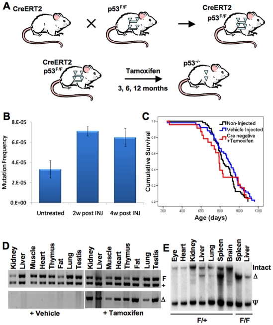
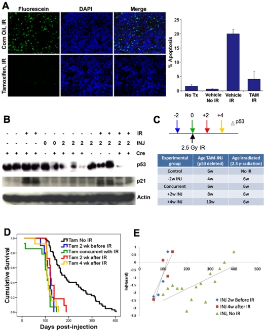
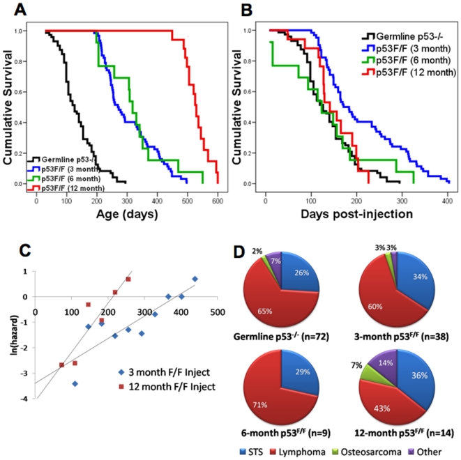
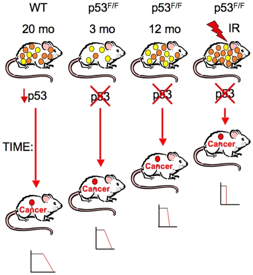
Inactivating mutations in the p53 tumor suppressor gene occur often in the progression of human cancers. p53 inhibits the outgrowth of nascent cancer cells through anti-proliferative actions (including induction of apoptosis or senescence). To test p53 tumor suppressor functions in a novel experimental context, we somatically deleted both p53 alleles in multiple tissues of mice at various ages. Mice homozygously deleted for p53 at 3 months of age showed a longer tumor latency compared to mice deleted for p53 at 6 and 12 months of age. These results are consistent with a model in which tissues accumulate oncogenically activated cells with age and these are held in check by wildtype p53. We also deleted p53 before, concurrent with, and after treatment of mice with ionizing radiation (IR). The absence or presence of p53 during IR treatment had no effect on radiation-induced lymphoma latency, confirming that the immediate p53 damage response was irrelevant for cancer prevention. Even the presence of wildtype p53 for up to four weeks post-IR provided no protection against early lymphoma incidence, indicating that long term maintenance of functional p53 is critical for preventing the emergence of a cancer. These experiments indicate that sustained p53 anti-oncogenic function acts as a final or near final line of defense preventing progression of oncogenically activated cells to malignant tumors.


Conflict of interest statement

Competing Interests: The authors have declared that no competing interests exist.

Figures

Figure 1
Figure 1. CreERT2-p53F/F and CreERT2+/F mice display efficient p53 allele excision in many tissues after tamoxifen treatment.
(A) Experimental design to generate mice that can inducibly and somatically delete p53 in many tissues. Floxed p53 alleles are represented by triangle flanked bars. Cre-excised p53 alleles are indicated by solo triangles. (B) Tamoxifen treatment of wildtype mice moderately elevates liver DNA mutation frequency. Three month C57BL/6 Big Blue Mice designed to measure mutation frequencies were treated with tamoxifen (five 1 mg daily injections) and sacrificed at 2 or 4 weeks post-injection and liver DNA subjected to the mutation frequency assay as described in the methods. Mutation frequencies are shown with or without tamoxifen injection (n = 3 for each time point, ±standard error of the mean). (C) Tamoxifen injection of mice has little or no effect on survival compared to vehicle-injected wildtype mice. Tamoxifen injection of CreERT2 negative p53F/F and p53+/F mice (red curve, n = 23) showed similar Kaplan-Meier survival curves as wildtype uninjected mice (black curve, n = 55) and vehicle injected CreERT2-p53+/F mice (blue curve, n = 50). (D) PCR assays show that vehicle-injected CreERT2-p53+/F tissues exhibit no p53 allele excision, while all tamoxifen-injected CreERT2-p53+/F tissues show evidence of p53 allele excision. Upper panels show genotyping PCR where the upper band (F) is the larger non-excised floxed allele of p53 and the lower band (+) is the non-floxed wild type p53 allele from various CreERT2-p53+/F tissues. The lower panels show PCR fragments specific for the excised p53 allele (Δ). The left set of panels contain results from vehicle (corn oil) treated tissues while the right set of panels contain PCR results from tamoxifen treated tissues. Note that in the absence of tamoxifen there is no background p53 allele excision and that in the presence of tamoxifen all tissues show evidence of p53 allele excision. (E) Tamoxifen treatment of CreERT2-p53+/F and CreERT2-p53F/F mice results in efficient p53 allele excision (Δ) in some, but not all tissues. Southern blot analysis of genomic DNA from various CreERT2-p53+/F and CreERT2-p53F/F tissues was performed as described in the methods. Note that spleen and liver show efficient excision, lung and kidney show partial excision, while eye and brain show little evidence of excision.
Figure 2
Figure 2. Tamoxifen treatment of CreERT2-p53F/F mice before, during, and after IR demonstrates the role of p53 in suppressing carcinogen-induced tumorigenesis.
(A) Deletion of p53 prior to irradiation prevents robust apoptotic response in CreERT2-p53F/F mice. Three month old CreERT2-p53F/F mice were injected with tamoxifen or vehicle (corn oil) two weeks prior to treatment with 5 Gray ionizing radiation. Five hours after IR treatment, spleens were harvested and tissue sections subjected to TUNEL assays for apoptotic cells. Note strong apoptotic response in vehicle treated mice and greatly reduced apoptotic response in tamoxifen-treated mice. Panel to the right shows percentage of TUNEL positive cells in spleens from untreated mice (No Tx), corn oil injected and non-irradiated mice (vehicle, No IR), corn oil injected and irradiated mice (vehicle, IR), and tamoxifen injected and irradiated mice (TAM, IR). (B) The p53 damage response is abrogated in tamoxifen injected CreERT2-p53F/F mice subjected to IR. Spleens were harvested from non-irradiated (-IR) and irradiated (+IR), non-injected (-INJ) and injected (+INJ 0 or 2 weeks post-injection) CreERT2-p53F/F (+Cre) and p53F/F (-Cre) mice and Western blots performed on tissue lysates using p53, p21, and actin antibodies. Note that robust p53 and p21 expression indicative of a robust p53 response occurs only in irradiated but non-Cre expressing or non-injected mice. (C) Six week old CreERT2-p53F/F mice were subjected to 2.5 Gy ionizing radiation and were injected with tamoxifen either 2 weeks prior (n = 10), simultaneous with (n = 14), 2 weeks after (n = 11), or 4 weeks after (n = 13) IR exposure. Non-irradiated tamoxifen treated mice CreERT2-p53F/F are included as controls (n = 62). (D) Kaplan-Meier tumor-free survival curves of IR-treated CreERT2-p53F/F mice show similar lymphoma latency of lymphomas post injection while non-IR treated tamoxifen-injected mice showed more delayed tumorigenesis. (E) Natural logarithm of mortality rates fitted using the T4253H smoothing algorithm comparing Gompertz variables from CreERT2-p53F/F mice injected with tamoxifen at 2 weeks prior and 4 weeks after IR treatment and non-IR treated tamoxifen-treated CreERT2-p53F/F mice (x-axis in days). Note that the IR-treated mice exhibit high mortality whether or not p53 is present at the time of radiation.
Figure 3
Figure 3. Tumor-free survival curves of CreERT2-p53F/F mice injected with tamoxifen at 3, 6, and 12 months of age.
(A) Kaplan-Meier survival curves are shown for germline p53−/− mice in black (n = 72), CreERT2-p53F/F mice injected with tamoxifen at 3 months in blue (n = 62), at 6 months in green (n = 13), and at 12 months in red (n = 18). (B) Tumor-free survival curves adjusted for time after tamoxifen injection. Line colors represent the same groups as in A. Note that the CreERT2-p53F/F mice injected with tamoxifen at 6 and 12 months of age exhibit a similar median survival as p53−/− mice and a reduced median survival compared to their 3 month injected counterparts. (C) Natural logarithm of mortality rates fitted using the T4253H smoothing algorithm comparing Gompertz variables from CreERT2-p53F/F mice injected with tamoxifen at 3, 6 and 12 months (x-axis in days) show a significantly increased mortality rate in the 12 month injected mice (p = 0.0007). (D) Tumor types observed in CreERT2-p53F/F mice treated with tamoxifen at 3 and 6 months are similar to those observed in germline p53−/− mice. In 12 month injected CreERT2-p53F/F mice, carcinomas show a modest increase and lymphomas a modest decrease.
Figure 4
Figure 4. p53 acts as a gatekeeper by preventing progression of oncogenically activated cells to full malignancy.
From left to right 20 month old wildtype p53 mice, p53F/F mice with intact p53 until 3 and 12 months of age, and 6 week old irradiated p53F/F mice are shown. All of these mice accumulate oncogenically activated clones with age, though irradiation greatly enhances the numbers of such clones. Oncogenically activated clones are represented by yellow (early stage oncogenic clones that are not yet capable of tumor formation) and orange (later stage oncogenic clones capable of tumor formation upon loss of p53) circles. These clones are held in check by wildtype p53, which prevents their progression to full malignancy. However, after the age of 20 months in wildtype mice, p53 functional activities decline, which could lead to malignant progression of one or more oncogenic clones (cancers indicated by red circle). When p53 is deleted by tamoxifen injection at 3, 6, or 12 months or following IR treatment in CreERT2-p53F/F mice, some oncogenic clones will progress to form a malignant cancer. For the p53F/F mice, the median and maximal tumor latencies following p53 deletion decrease with age or with carcinogenic insult. The relative tumor latencies are indicated by the length of the red vertical arrows. For the wildtype mice, since p53 activity is merely reduced, malignant progression is more delayed. The small graphs below each of the tumor bearing mice shows the shape of the survival curves for each cohort, indicating that tumor latencies and mortality rates depend on p53 functional status, age, or the presence of carcinogenic insult. Thus, p53 acts as a final or near-final gatekeeper keeping these oncogenic clones from progressing to full malignancy.

References

    1. Kinzler KW, Vogelstein B. Lessons from hereditary colorectal cancer. Cell. 1996;87:159–170. - PubMed
    1. Levine AJ. p53, the cellular gatekeeper for growth and division. Cell. 1997;88:323–331. - PubMed
    1. Soussi T, Wiman KG. Shaping genetic alterations in human cancer: the p53 mutation paradigm. Cancer Cell. 2007;12:303–312. - PubMed
    1. Macleod K. Tumor suppressor genes. Curr Opin Genet Dev. 2000;10:81–93. - PubMed
    1. Blondal JA, Benchimol S. The role of p53 in tumor progression. Semin Cancer Biol. 1994;5:177–186. - PubMed

Publication types