. 2009 Aug 14;4(8):e6645.
doi: 10.1371/journal.pone.0006645.

Immature and maturation-resistant human dendritic cells generated from bone marrow require two stimulations to induce T cell anergy in vitro

Affiliations

Immature and maturation-resistant human dendritic cells generated from bone marrow require two stimulations to induce T cell anergy in vitro

Thomas G Berger et al. PLoS One. .

Abstract

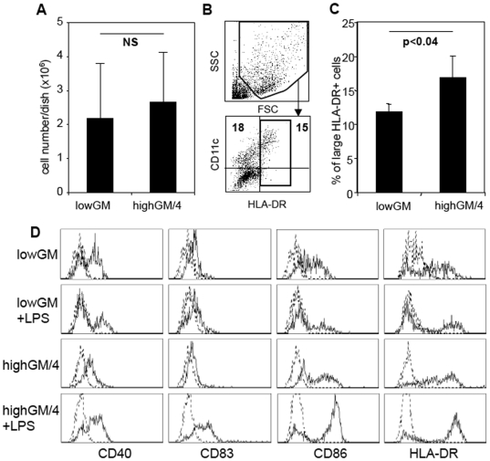
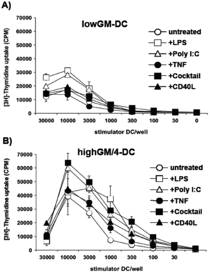
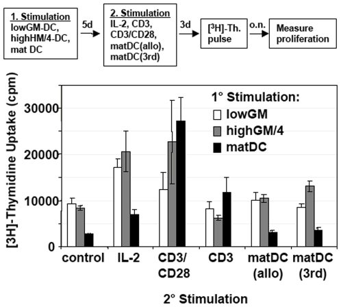
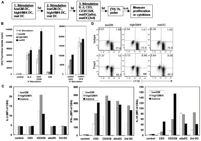
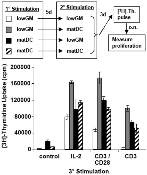
Immature dendritic cells (DC) represent potential clinical tools for tolerogenic cellular immunotherapy in both transplantation and autoimmunity. A major drawback in vivo is their potential to mature during infections or inflammation, which would convert their tolerogenicity into immunogenicity. The generation of immature DC from human bone marrow (BM) by low doses of GM-CSF (lowGM) in the absence of IL-4 under GMP conditions create DC resistant to maturation, detected by surface marker expression and primary stimulation by allogeneic T cells. This resistence could not be observed for BM-derived DC generated with high doses of GM-CSF plus IL-4 (highGM/4), although both DC types induced primary allogeneic T cell anergy in vitro. The estabishment of the anergic state requires two subsequent stimulations by immature DC. Anergy induction was more profound with lowGM-DC due to their maturation resistance. Together, we show the generation of immature, maturation-resistant lowGM-DC for potential clinical use in transplant rejection and propose a two-step-model of T cell anergy induction by immature DC.


Conflict of interest statement

Competing Interests: The authors have declared that no competing interests exist.

Figures

Figure 1
Figure 1. Human BM-derived lowGM-DC do not upregulate surface markers upon maturation.
Human BM cells were cultured under standard conditions with 800 U/ml GM-CSF plus 250 U/ml IL-4 or under lowGM conditions with 5 U/ml GM-CSF for 6 days in RPMI+1% AB plasma. A. The cells were harvested and counted under trypan blue exclusion. The mean values±standard deviations of 8 independent experiments are shown. B. The harvested cells were analysed by flow cytometry by setting gates on large cells in the FSC/SSC profile and HLA-DR+ cells. C. Analysis of 3 independent experiments as gated in B. Mean values±standard deviations are shown. D. The cells were then exposed to 100 µg/ml LPS for 24 h to induce maturation or left untreated and surface stained for the indicated markers (straight lines) or isotype controls (dotted lines). Cells were gated as in B to exclude lymphocytes and precursor cells. The data from one experiment shown are representative of 5 independent experiments.
Figure 2
Figure 2. LowGM-DC remain functionally immature after maturation.
DC were generated under low-GM (A) and standard highGM plus IL-4 conditions (B) for 6 days, stimulated with the indicated reagents or a “cocktail” consisting of TNF, IL-1β, IL-6 and PGE2 for 24 h and then added at titrated numbers to allogeneic T cells, as represented by non-adherent fraction of PBMC of an allogeneic donor. After 3 days [3H]-thymidine was added overnight to measure proliferation. The data from one experiment shown are representative of 3 independent experiments. Values represent the mean±standard deviation error bars of triplicate cultures from one experiment.
Figure 3
Figure 3. A single allogeneic T cell priming by lowGM-DC does not induce T cell anergy.
Allogeneic T cells were stimulated with either immature lowGM-DC or immature highGM+IL-4-DC or LPS-matured highGM+IL-4-DC. After 5 days the T cells were restimulated with IL-2 or the indicated antibodies or LPS-matured DC from the original allotype (matDC allo) or a third party allotype (matDC 3rd) or cells were left without stimulation (control) for another 3 days before [3H]-thymidine ([3H]-Th.) was added overnight to measure proliferation as shown in the flow chart. The data from one experiment shown are representative of 5 independent experiments. Values represent the mean±standard deviation error bars of triplicate cultures from one experiment.
Figure 4
Figure 4. Allogeneic T cell anergy induction requires two stimulations by lowGM-DC.
A. Allogeneic T cells were stimulated twice (for 5 days and then for another 3 days) with either immature lowGM-DC or immature highGM+IL-4-DC or mature highGM+IL-4-DC and then restimulated a third time for another 3 days with IL-2 or the indicated antibodies or LPS-matured DC from the original allotype (matDC allo) or a third party allotype (matDC 3rd) or cells were left without stimulation (control) for another 3 days. B. Then [3H]-thymidine ([3H]-Th.) was added overnight to measure proliferation. The data are representative of 4 independent experiments. C. Supernatants from the cultures shown in Figure 4B were tested for their content of IL-2, IFN-γ, or IL-10 by cytokine bead array (CBA). The relative mean fluorescence values of the FACS analysis are shown. The data from one experiment shown are representative for 3 independent experiments. Values in bar graphs represent the mean±standard deviation error bars of triplicate cultures from one experiment. D. CFSE-labeled T cells were restimulated by the indicated DC type for 4 days and then stained for intracellular expression of Foxp3. Cells were measured by flow cytometry. Gating was performed on CFSE+ cells and is plotted against Foxp3 or isotype. CFSElow cells represent allo-responsive proliferated cells.
Figure 5
Figure 5. Mature DC cannot substitute for immature DC at either stimulation time points.
Immature lowGM-DC or mature highGM/4-DC were generated to stimulate allogeneic naïve T cells. After 5 days the T cells were restimulated with the same type of DC or in an opposite setting as indicated in the legend. After another 3 days the T cells were restimulated with the indicated reagents for 3 days before [3H]-thymidine incorporation was detected to measure proliferation. The data from one experiment shown are representative of 3 independent experiments. Values represent the mean±standard deviation error bars of triplicate cultures from one experiment.

Similar articles

Cited by

References

    1. Banchereau J, Steinman RM. Dendritic cells and the control of immunity. Nature. 1998;392:245–252. - PubMed
    1. Steinman RM, Nussenzweig MC. Avoiding horror autotoxicus: the importance of dendritic cells in peripheral T cell tolerance. Proc Natl Acad Sci U S A. 2002;99:351–358. - PMC - PubMed
    1. Bluestone JA, Thomson AW, Shevach EM, Weiner HL. What does the future hold for cell-based tolerogenic therapy? Nat Rev Immunol. 2007;7:650–654. - PubMed
    1. Fathman CG, Lineberry NB. Molecular mechanisms of CD4+ T-cell anergy. Nat Rev Immunol. 2007;7:599–609. - PubMed
    1. Parish IA, Rao S, Smyth GK, Juelich T, Denyer GS, et al. The molecular signature of CD8+ T cells undergoing deletional tolerance. Blood. 2009;113:4575–4585. - PMC - PubMed

Publication types

MeSH terms

Substances