. 2009:2009:642502.
doi: 10.1155/2009/642502. Epub 2009 Aug 5.

Sterol Biosynthesis Pathway as Target for Anti-trypanosomatid Drugs

Affiliations

Sterol Biosynthesis Pathway as Target for Anti-trypanosomatid Drugs

Wanderley de Souza et al. Interdiscip Perspect Infect Dis. 2009.

Abstract

Sterols are constituents of the cellular membranes that are essential for their normal structure and function. In mammalian cells, cholesterol is the main sterol found in the various membranes. However, other sterols predominate in eukaryotic microorganisms such as fungi and protozoa. It is now well established that an important metabolic pathway in fungi and in members of the Trypanosomatidae family is one that produces a special class of sterols, including ergosterol, and other 24-methyl sterols, which are required for parasitic growth and viability, but are absent from mammalian host cells. Currently, there are several drugs that interfere with sterol biosynthesis (SB) that are in use to treat diseases such as high cholesterol in humans and fungal infections. In this review, we analyze the effects of drugs such as (a) statins, which act on the mevalonate pathway by inhibiting HMG-CoA reductase, (b) bisphosphonates, which interfere with the isoprenoid pathway in the step catalyzed by farnesyl diphosphate synthase, (c) zaragozic acids and quinuclidines, inhibitors of squalene synthase (SQS), which catalyzes the first committed step in sterol biosynthesis, (d) allylamines, inhibitors of squalene epoxidase, (e) azoles, which inhibit C14alpha-demethylase, and (f) azasterols, which inhibit Delta(24(25))-sterol methyltransferase (SMT). Inhibition of this last step appears to have high selectivity for fungi and trypanosomatids, since this enzyme is not found in mammalian cells. We review here the IC50 values of these various inhibitors, their effects on the growth of trypanosomatids (both in axenic cultures and in cell cultures), and their effects on protozoan structural organization (as evaluted by light and electron microscopy) and lipid composition. The results show that the mitochondrial membrane as well as the membrane lining the protozoan cell body and flagellum are the main targets. Probably as a consequence of these primary effects, other important changes take place in the organization of the kinetoplast DNA network and on the protozoan cell cycle. In addition, apoptosis-like and autophagic processes induced by several of the inhibitors tested led to parasite death.


Figures
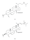
Figure 1
Figure 1
Molecular structures of cholesterol and ergosterol. The arrows indicate the parts of the molecules which have been shown to be essential for the growth of mammalian cells (cholesterol), fungi, and trypanosomatids (ergosterol and 24-methyl sterols).
Figure 2
Figure 2
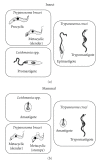
Schematic representation of main morphologies found during the life cycle of some members of the Trypanosomatidae family in the invertebrate host (insect) and vertebrate host (mammal).
Figure 3
Figure 3
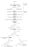
The biosynthesis of ergosterol and cholesterol showing the main steps, the enzymes involved, and the known inhibitors.
Figure 4
Figure 4
(a)-(b) Growth curves of promastigotes and (c)-(d) intracellular amastigotes of Leishmania amazonensis treated with two potent squalene synthase inhibitors, E5700 and ER-119884. The graphics are reproduced with permission from [37] American Society for Microbiology.
Figure 5
Figure 5
Ultrathin sections of L. amazonensis promastigotes control (a) and treated with azasterols, known inhibitors of the Δ24(25)-sterol methyltransferase (b)–(d). (a) General overview of an untreated-parasite showing a normal ultrastructure of the mitochondrion (M), kinetoplast (k), flagellum (F) and nucleus (N). (b)–(d) Treated-parasites presenting severe alterations in the mitochondrion structure such as a disorganization of the internal membranes ((b) and (c), arrowheads) and an intense and evident mitochondrial swelling with loss of the matrix content (b)–(d). Bars 0.5 μm.
Figure 6
Figure 6
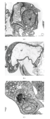
Trypanosoma cruzi epimastigotes treated with ketoconazole, an inhibitor of the C14α-demetilase (a)-(b), and L. amazonensis promastigotes treated with E5700, an inhibitor of the squalene synthase (c) showing an intense mitochondrial swelling (a)-(b) and alterations in the kinetoplast structure ((c), arrowhead). K, kinetoplast; M, mitochondrion. (a)-(b) Images are reproduced with permission from [74] American Society for Microbiology. Bars, 0.5 μm.
Figure 7
Figure 7
L. amazonensis promastigotes treated with azasterols (a)-(b), and ER-119884 (c) showing alterations in the nuclear membrane ((a), arrowhead), in the Golgi complex ((b), arrowhead), and in the endoplasmic reticulum ((c), arrowhead). In the Figures 6(b) and 6(c), the presence of a multivesicular bodies and autophagosome-like structures (stars) could be related with a remodeling process of damaged organelle by autophagy. GC, Golgi complex; M, mitochondrion; N, nucleus. Bars, 0.5 μm.
Figure 8
Figure 8
Promastigotes treated with quinuclidine and azasterol, respectively, showing the presence of structures related with autophagy such as a large vacuole containing many membrane profiles ((a), arrowheads), and a myelin-like figures involving part of the cytosol ((b), arrowhead). Star indicates the presence of a possible contractile vacuole near the flagellar pocket. A, autophagosome; F, flagellum; K, kinetoplast; M, mitochondrion; N, nucleus. (a) This image is reproduced with permission from [66] Elsevier. Bars, 0.25 μm.
Figure 9
Figure 9
(a) Diferential interference contrast (DIC) microscopy and (b) transmission electron microscopy showing the presence of a prominent contractile vacuole (arrows) near the flagellar pocket after treatment of L. amazonensis promastigotes with quinuclidine inhibitors. In the left panel it is possible to observe a rounded and swollen parasite that probably indicates osmotic changes due alterations in the plasma membrane's permeability. F, flagellum; K, kinetoplast. Bars, 5 μm and 0.5 μm, respectively.
Figure 10
Figure 10
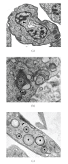
Treatment of promastigotes with ER-119884 induces the accumulation of several lipid droplets in the cytosol, sometimes appearing as an electrondense structure due to osmium tetroxide concentration ((a)-(b), small arrows), and as a classic lipid body surrounded by a phospholipid monolayer ((b), large arrows). At high magnification (stars), it is evident that the structures are completely different, probably indicating a distintic nature of the lipids that accumulate in these inclusions. M, mitochondrion; N, nucleus. All images are reproduced with permission from [37] American Society for Microbiology. Bars, 0.5 μm.
Figure 11
Figure 11
Ultrathin sections of L. amazonensis promastigotes treated with different sterol biosynthesis inhibitors showing severe alterations in the plasma membrane lining (a) the flagellar pocket, (b) the flagellum, and (c)-(d) the cell body. In (d) it is possible to observe a breakdown in the plasma membrane and release of the subpellicular microtubules (arrowhead). Star in (c) shows a classic lipid body. F, flagellum; FP, flagellar pocket; k, kinetoplast; M, mitochondrion. Images are reproduced with permission from [79] (a), and [66] (b), (d) Elsevier. Bars, 0.25 μm (a)–(c) and 0.5 μm (d).
Figure 12
Figure 12
DIC microscopy (left panel) and immunofluorescence microcospy (right panel) of L. amazonensis promastigotes control (a)-(b) and treated with ER-119884 (c)-(d). The labeling corresponds to the cytoskeleton constituted mainly by tubulin, revelead here by using of an Alexa 488-labeled secondary antibody. The black arrow in DIC image corresponds to the cell body which sometimes appeared changed and rounded as compared to the control parasites, while the white arrows point to several tubulin clusters that accumulated in the cytosol after treatment. All images are reproduced with permission from [37] American Society for Microbiology. Bars, 5 μm.
Figure 13
Figure 13
DIC microscopy ((a)-(b), left panel), fluorescence with DAPI ((a)-(b), right panel) and transmission electron microscopy (c)-(d) of treated-promastigotes to evidenciate the alterations in the cell cycle after treatment with BPQ-OH and ER-119884. (a) Control cells present a correct number of kinetoplast (white arrow), nucleus (arrowhead), and flagellum (black arrow), one of each for a unique cell. (b) After treatment, the number of these organelles is completely alterated and it is possible to find cells with one flagellum (black arrow), four kinetoplast (white arrows), and one nucleus (arrowhead). (c)-(d) Alterations in the cell cycle can also be evidenced by transmission electron microscopy with the appearance of cells with several nuclei and kinetoplasts. K, kinetoplast; N, nucleus. These images are reproduced with permission from [37] American Society for Microbiology (a), (b), (d), and from [66] Elsevier (c). Bars, 5 μm (a)-(b), and 0.5 μm (c)-(d).
Figure 14
Figure 14
Leishmania amazonensis promastigote treated with sterol biosynthesis inhibitors showing a condensation of the nuclear chromatin, a characteristic feature of the apoptosis-like cell death process. N, nucleus. Bar, 0.5 μm.

Similar articles

Cited by

References

    1. Fagundes LJM. Estudo de esteróis e lipídeos totais em Leptomonas pessoai cultivada a 28°C e 37°C. Rio de Janeiro, Brazil: Instituto de Microbiologia da Universidade Federal do Rio de Janeiro; 1974. M.S. thesis.
    1. de Souza W, Sant'anna C, Cunha-e-Silva NL. Electron microscopy and cytochemistry analysis of the endocytic pathway of pathogenic protozoa. Progress in Histochemistry and Cytochemistry. 2009;44(2):67–124. - PubMed
    1. Soares MJ, de Souza W. Endocytosis of gold-labeled proteins and LDL by Trypanosoma cruzi . Parasitology Research. 1991;77(6):461–468. - PubMed
    1. Urbina JA. Lipid biosynthesis pathways as chemotherapeutic targets in kinetoplastid parasites. Parasitology. 1997;114(supplement 1):S91–S99. - PubMed
    1. Roberts CW, McLeod R, Rice DW, Ginger M, Chance ML, Goad LJ. Fatty acid and sterol metabolism: potential antimicrobial targets in apicomplexan and trypanosomatid parasitic protozoa. Molecular and Biochemical Parasitology. 2003;126(2):129–142. - PubMed

LinkOut - more resources