Review
. 2010 Mar;39(4):541-50.
doi: 10.1007/s00249-009-0523-0. Epub 2009 Aug 13.

Viruses: incredible nanomachines. New advances with filamentous phages

Affiliations
Review

Viruses: incredible nanomachines. New advances with filamentous phages

Marcus A Hemminga et al. Eur Biophys J. 2010 Mar.

Abstract

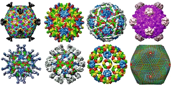
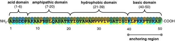
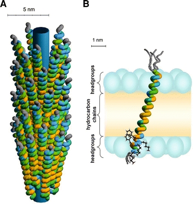
During recent decades, bacteriophages have been at the cutting edge of new developments in molecular biology, biophysics, and, more recently, bionanotechnology. In particular filamentous viruses, for example bacteriophage M13, have a virion architecture that enables precision building of ordered and defect-free two and three-dimensional structures on a nanometre scale. This could not have been possible without detailed knowledge of coat protein structure and dynamics during the virus reproduction cycle. The results of the spectroscopic studies conducted in our group compellingly demonstrate a critical role of membrane embedment of the protein both during infectious entry of the virus into the host cell and during assembly of the new virion in the host membrane. The protein is effectively embedded in the membrane by a strong C-terminal interfacial anchor, which together with a simple tilt mechanism and a subtle structural adjustment of the extreme end of its N terminus provides favourable thermodynamical association of the protein in the lipid bilayer. This basic physicochemical rule cannot be violated and any new bionanotechnology that will emerge from bacteriophage M13 should take this into account.


Figures

Fig. 1
Fig. 1
Various virus structures—a wealth of nanoshapes and nanoarchitectures for icosahedral (spherical) viruses. Courtesy: http://www.viperdb.scripps.edu/ (Carrillo-Tripp et al. 2009)
Fig. 2
Fig. 2
Primary structure of M13 major coat protein with classification of the important domains (Table 1). The colour coding is based on amino acid residue hydrophobicity scales (White and Wimley 1999) with the yellow colour corresponding to hydrophobic residues, green to neutral, and blue to charged residues. Anchoring of the protein at the membrane–water interface is provided by the C-terminal lysine residues and phenylalanines
Fig. 3
Fig. 3
a Schematic illustration of the phage-bound model for the major coat protein of bacteriophage M13. The colour coding of the amino acid residues is based on a hydrophobicity scale (Fig. 2). Unstructured protein regions are indicated in grey. The inner cylinder indicates the viral DNA. b Structure and membrane embedding of M13 coat protein in fully hydrated vesicles of 18:1PC (and mixed phospholipid systems with C18 acyl chains), based upon recent site-directed labelling spectroscopy (Koehorst et al. ; Nazarov et al. ; Stopar et al. ; Vos et al. 2005, 2007). The protein is effectively anchored with the C-terminal domain at the membrane–water interface by three “snorkelling” lysines (Lys40, Lys43, and Lys44) and two “anti-snorkelling” phenylalanines (Phe42 and Phe45). The size of the membrane regions is obtained from the literature, with the positions of the carbonyls serving as borders for the headgroup region (Ridder et al. ; White and Wimley 1999). The phospholipid headgroups are indicated with blue ellipsoids and the hydrocarbon chain region is coloured in yellow. The protein forms a mainly α-helical conformation tilted at 18° to the membrane normal. The first nine amino acid residues, which encompass the hydrophilic anchor (Table 1), are unstructured (as indicated by different possible gray structures). The membrane-bound protein structure does not differ much from the native α-helical structure of the protein in bacteriophage M13 in a. The protein survives the membrane-bound state by a simple tilt mechanism based on anchoring of its C-terminal domain at the membrane–water interface and a subtle structural adjustment at the extreme end of the N-terminal domain

References

    1. Almeida FCL, Opella SJ. Fd coat protein structure in membrane environments: structural dynamics of the loop between the hydrophobic trans-membrane helix and the amphipathic in-plane helix. J Mol Biol. 1997;270:481–495. doi: 10.1006/jmbi.1997.1114. - DOI - PubMed
    1. Bashtovyy D, Marsh D, Hemminga MA, Páli T. Constrained modeling of spin-labeled major coat protein mutants from M13 bacteriophage in a phospholipid bilayer. Protein Sci. 2001;5:979–987. doi: 10.1110/ps.43801. - DOI - PMC - PubMed
    1. Carrillo-Tripp M, Shepherd CM, Borelli IA, Venkataraman S, Lander G, Natarajan P, Johnson JE, Brooks CL, III, Reddy VS. VIPERdb2: an enhanced and web API enabled relational database for structural virology. Nucl Acids Res. 2009;37:D436–D442. doi: 10.1093/nar/gkn840. - DOI - PMC - PubMed
    1. Fernandes F, Loura LMS, Prieto M, Koehorst RBM, Spruijt RB, Hemminga MA. Dependence of M13 major coat protein oligomerization and lateral segregation on bilayer composition. Biophys J. 2003;85:2430–2441. doi: 10.1016/S0006-3495(03)74666-9. - DOI - PMC - PubMed
    1. Fischlechner M, Donath E. Viruses as building blocks for materials and devices. Angew Chem Int Edit. 2007;46:3184–3193. doi: 10.1002/anie.200603445. - DOI - PubMed

Publication types

LinkOut - more resources