. 2009 Dec;118(6):737-43.
doi: 10.1007/s00401-009-0582-4.

Evidence for antibody-mediated pathogenesis in anti-NMDAR encephalitis associated with ovarian teratoma

Affiliations

Evidence for antibody-mediated pathogenesis in anti-NMDAR encephalitis associated with ovarian teratoma

Erdem Tüzün et al. Acta Neuropathol. 2009 Dec.

Abstract

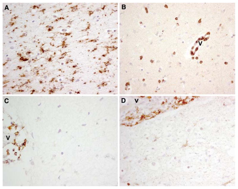
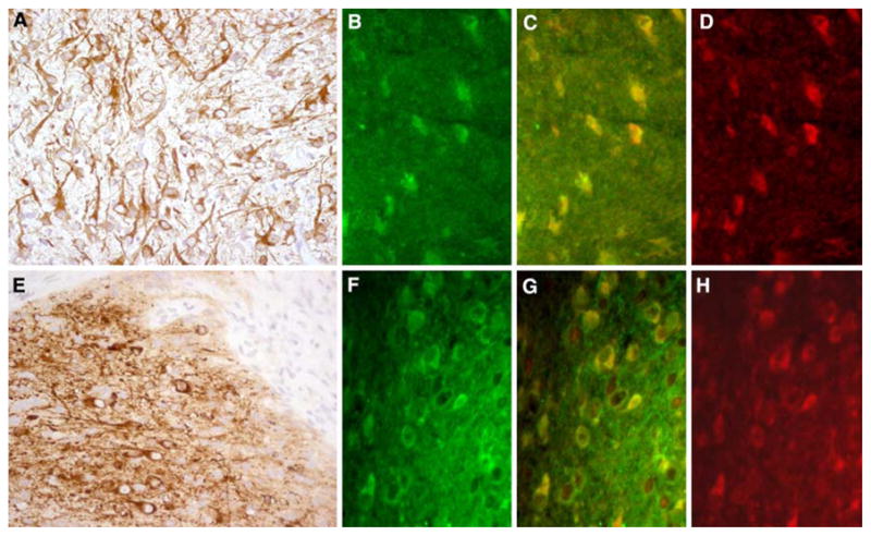
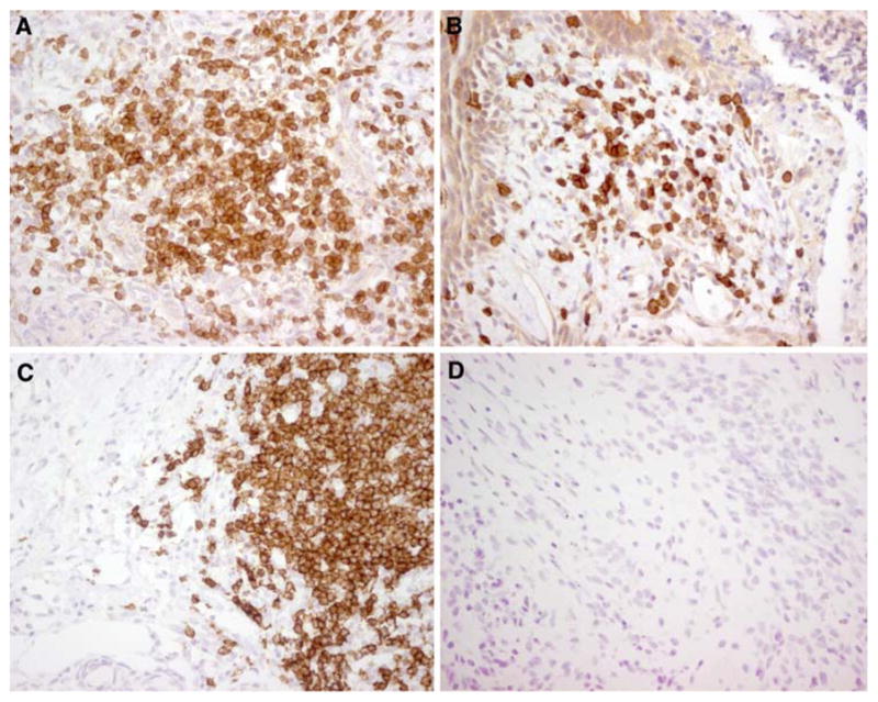
We report the immunopathological analysis of the brain and tumor of two patients who died of anti-NMDAR-associated encephalitis, and of the tumor of nine patients who recovered. Findings included prominent microgliosis and deposits of IgG with rare inflammatory infiltrates in the hippocampus, forebrain, basal ganglia, and spinal cord. Detection of cells expressing markers of cytotoxicity (TIA, granzyme B, perforin and Fas/Fas ligand) was extremely uncommon. All tumors showed NMDAR-expressing neurons and inflammatory infiltrates. All patients’ NMDAR antibodies were IgG1, IgG2, or IgG3. No complement deposits were observed in any of the central nervous system regions examined. Overall, these findings coupled with recently reported in vitro data showing that antibodies downregulate the levels of NMDA receptors suggest that the antibody immune-response is more relevant than cytotoxic T-cell mechanisms in the pathogenesis of anti-NMDAR-associated encephalitis.


Figures

Fig. 1
Fig. 1
Microgliosis and rare infiltrates of T-cells in the hippocampus and spinal cord of patient #2. a–c Paraffin embedded sections of hippocampus immunolabeled with the microglial marker CD68 (a), the T-cell marker CD3 (b) and the B cell marker CD20 (c). d Paraffin embedded spinal cord section immunolabeled with the plasma cell marker CD79a. Note the perivascular location of CD3+, CD20+ and CD79a+ cells (v blood vessel). a–d ×400, avidin–biotin–peroxidase technique with mild hematoxylin counterstaining
Fig. 2
Fig. 2
Nervous tissue in tumor sections of anti-NMDAR encephalitis and neurologically normal patients. a Ovarian teratoma of a patient with anti-NMDAR encephalitis immunolabeled with MAP-2 (brown staining), a marker specific for neurons and dendritic processes. b–d The same tumor immunolabeled with patient’s antibodies (b, green) and a specific antibody for NR2B (d, red); note that there is co-localization of reactivities (c, yellow), indicating that the patient’s antibodies react with NR2B-containing heretomers expressed in the tumor (similar findings were observed with NR1 and NR2A antibodies, not shown). e Ovarian teratoma of a patient without anti-NMDAR encephalitis immunolabeled with MAP-2 (brown staining). f–h The same tumor immunolabeled with antibodies from a patient with anti-NMDAR encephalitis (f, green) and anti-NR2B (h, red). g The co-localization of reactivities (yellow); similar findings were observed with NR1 and NR2A antibodies (not shown). a and e ×200, avidin–biotin–peroxidase technique with mild hematoxylin counterstaining, b–d and f–h ×400, immunofluorescence. All studies were performed with frozen sections
Fig. 3
Fig. 3
Inflammatory infiltrates in ovarian teratomas. a, b Ovarian teratoma of a patient with anti-NMDAR encephalitis immunolabeled for the presence of CD3+ cells (a) and CD20+ cells (b). c, d Ovarian teratoma of a patient without anti-NMDAR encephalitis immunolabeled for the presence of CD3+ cells (c) and CD20+ cells (d). While CD3+ cells (T cells) are present in both tumors, only the tumor of the patient with anti-NMDAR encephalitis shows infiltrates of CD20+ cells (B cells). ad ×400, avidin–biotin–peroxidase technique with mild hematoxylin counterstaining. All studies were performed with frozen sections

References

    1. Bernal F, Graus F, Pifarre A, Saiz A, Benyahia B, Ribalta T. Immunohistochemical analysis of anti-Hu-associated paraneoplastic encephalomyelitis. Acta Neuropathol. 2002;103:509–515. - PubMed
    1. Blumenthal DT, Salzman KL, Digre KB, Jensen RL, Dunson WA, Dalmau J. Early pathologic findings and long-term improvement in anti-Ma2-associated encephalitis. Neurology. 2006;67:146–149. - PubMed
    1. Cattoretti G, Pileri S, Parravicini C, et al. Antigen unmasking on formalin-fixed, paraffin-embedded tissue sections. J Pathol. 1993;171:83–98. - PubMed
    1. Dalmau J, Gleichman AJ, Hughes EG, et al. Anti-NMDA-receptor encephalitis: case series and analysis of the effects of antibodies. Lancet Neurol. 2008;7:1091–1098. - PMC - PubMed
    1. Dalmau J, Gultekin SH, Voltz R, et al. Ma1, a novel neuron- and testis-specific protein, is recognized by the serum of patients with paraneoplastic neurological disorders. Brain. 1999;122:27–39. - PubMed

Publication types

Substances