

Protocadherin PCDH10, involved in tumor progression, is a frequent and early target of promoter hypermethylation in cervical cancer
- PMID: 19681120
- PMCID: PMC3430375
- DOI: 10.1002/gcc.20703
Protocadherin PCDH10, involved in tumor progression, is a frequent and early target of promoter hypermethylation in cervical cancer
Abstract
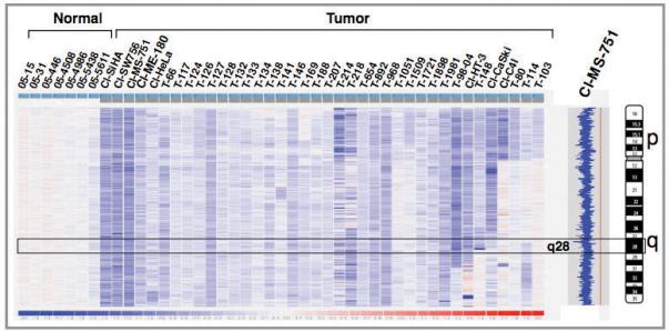
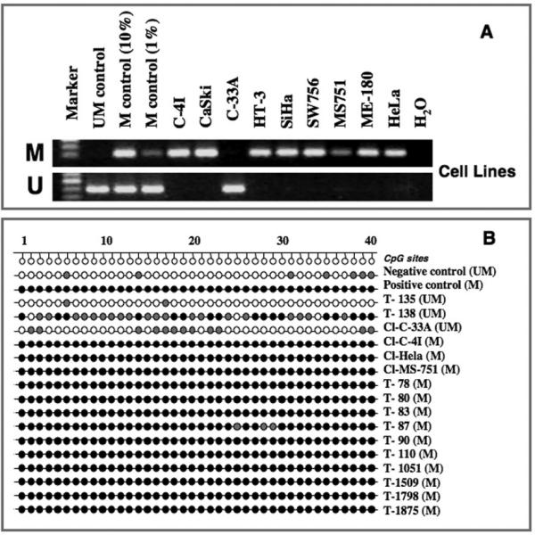
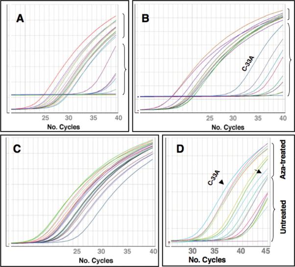
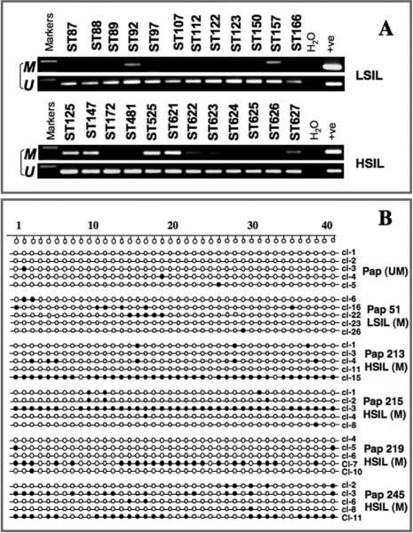
Cervical cancer (CC) is the second most common cancer in women. Currently, no tractable molecular-based therapeutic targets exist for patients with invasive CC and no predictive markers of risk assessment for progression of precancerous lesions are identified. New molecular insights into CC pathogenesis are urgently needed. Towards this goal, we first determined the copy number alterations of chromosome 4 and then examined the role of PCDH10 mapped to 4q28 as a candidate tumor suppressor gene. We identified monosomy 4 in 47% of 81 invasive CC studied by SNP array and found that 91% of 130 invasive CC harboring methylation in the promoter region of the PCDH10 gene. We then showed that aberrant promoter hypermethylation of PCDH10 is associated with downregulation of gene expression and cell lines exposed to demethylating agent resulted in profound reactivated gene expression. We also showed that the promoter methylation in the PCDH10 gene occurs at an earliest identifiable stage of low-grade squamous intraepithelial lesion. Our studies demonstrate that inactivation of PCDH10 may be a critical event in CC progression and form a potentially useful therapeutic target for CC.
Copyright 2009 Wiley-Liss, Inc.
Figures
Similar articles
-
Field methylation silencing of the protocadherin 10 gene in cervical carcinogenesis as a potential specific diagnostic test from cervical scrapings.Cancer Sci. 2009 Nov;100(11):2175-80. doi: 10.1111/j.1349-7006.2009.01285.x. Epub 2009 Jul 17. Cancer Sci. 2009. PMID: 19709077 Free PMC article.
-
Protocadherin-10 is involved in angiogenesis and methylation correlated with multiple myeloma.Int J Mol Med. 2012 Apr;29(4):704-10. doi: 10.3892/ijmm.2012.880. Epub 2012 Jan 9. Int J Mol Med. 2012. PMID: 22245948 Free PMC article.
-
Epigenetic inactivation of PCDH10 in human prostate cancer cell lines.Cell Biol Int. 2011 Jul;35(7):671-6. doi: 10.1042/CBI20100568. Cell Biol Int. 2011. PMID: 21314642
-
Aberrant DNA methylation in cervical carcinogenesis.Chin J Cancer. 2013 Jan;32(1):42-8. doi: 10.5732/cjc.012.10033. Epub 2012 Aug 28. Chin J Cancer. 2013. PMID: 22943599 Free PMC article. Review.
-
Integrative genomic approaches in cervical cancer: implications for molecular pathogenesis.Future Oncol. 2010 Oct;6(10):1643-52. doi: 10.2217/fon.10.114. Future Oncol. 2010. PMID: 21062161 Free PMC article. Review.
Cited by
-
PI3K/AKT pathway-mediated regulation of p27(Kip1) is associated with cell cycle arrest and apoptosis in cervical cancer.Cell Oncol (Dordr). 2015 Jun;38(3):215-25. doi: 10.1007/s13402-015-0224-x. Epub 2015 Mar 28. Cell Oncol (Dordr). 2015. PMID: 25821107
-
The role of lncRNAs in the development of endometrial carcinoma.Oncol Lett. 2018 Sep;16(3):3424-3429. doi: 10.3892/ol.2018.9065. Epub 2018 Jul 4. Oncol Lett. 2018. PMID: 30127944 Free PMC article. Review.
-
High methylation levels of PCDH10 predict poor prognosis in patients with pancreatic ductal adenocarcinoma.BMC Cancer. 2019 May 14;19(1):452. doi: 10.1186/s12885-019-5616-2. BMC Cancer. 2019. PMID: 31088413 Free PMC article.
-
Protocadherin 17 is a tumor suppressor and is frequently methylated in nasopharyngeal carcinoma.Cancer Manag Res. 2019 Feb 18;11:1601-1613. doi: 10.2147/CMAR.S191102. eCollection 2019. Cancer Manag Res. 2019. Retraction in: Cancer Manag Res. 2020 May 11;12:3257. doi: 10.2147/CMAR.S261450. PMID: 30863170 Free PMC article. Retracted.
-
Dysregulated expression of long noncoding RNAs in gynecologic cancers.Mol Cancer. 2017 Jun 21;16(1):107. doi: 10.1186/s12943-017-0671-2. Mol Cancer. 2017. PMID: 28637507 Free PMC article. Review.
References
-
- Backsch C, Rudolph B, Kuhne-Heid R, Kalscheuer V, Bartsch O, Jansen L, Beer K, Meyer B, Schneider A, Durst M. A region on human chromosome 4 (q35.1-->qter) induces senescence in cell hybrids and is involved in cervical carcinogenesis. Genes Chromosomes Cancer. 2005;43:260–272. - PubMed
-
- Backsch C, Wagenbach N, Nonn M, Leistritz S, Stanbridge E, Schneider A, Durst M. Microcell-mediated transfer of chromosome 4 into HeLa cells suppresses telomerase activity. Genes Chromosomes Cancer. 2001;31:196–198. - PubMed
-
- Frank M, Kemler R. Protocadherins. Curr Opin Cell Biol. 2002;14:557–562. - PubMed
-
- Hampton GM, Larson AA, Baergen RN, Sommers RL, Kern S, Cavenee WK. Simultaneous assessment of loss of heterozygosity at multiple microsatellite loci using semi-automated fluorescence-based detection: subregional mapping of chromosome 4 in cervical carcinoma. Proc Natl Acad Sci U S A. 1996;93:6704–6709. - PMC - PubMed
-
- Imoto I, Izumi H, Yokoi S, Hosoda H, Shibata T, Hosoda F, Ohki M, Hirohashi S, Inazawa J. Frequent silencing of the candidate tumor suppressor PCDH20 by epigenetic mechanism in non-small-cell lung cancers. Cancer Res. 2006;66:4617–4626. - PubMed
