. 2009 Dec;197(4):321-32.
doi: 10.1111/j.1748-1716.2009.02029.x. Epub 2009 Aug 3.

Overload-induced skeletal muscle extracellular matrix remodelling and myofibre growth in mice lacking IL-6

Affiliations

Overload-induced skeletal muscle extracellular matrix remodelling and myofibre growth in mice lacking IL-6

J P White et al. Acta Physiol (Oxf). 2009 Dec.

Abstract

Aim: Overloading healthy skeletal muscle produces myofibre hypertrophy and extracellular matrix remodelling, and these processes are thought to be interdependent for producing muscle growth. Inflammatory cytokine interleukin-6 (IL-6) gene expression is induced in overloaded skeletal muscle, and the loss of this IL-6 induction can attenuate the hypertrophic response to overload (OV). Although the OV induction of IL-6 in skeletal muscle may be an important regulator of inflammatory processes and satellite cell proliferation, less is known about its role in the regulation of extracellular matrix remodelling. The purpose of the current study was to examine if OV-induced extracellular matrix remodelling, muscle growth, and associated gene expression were altered in mice that lack IL-6, when compared with wild-type mice.

Methods: Male C57/BL6 (WT) and C57/BL6 x IL-6(-/-) (IL-6(-/-)) mice (10 weeks of age) were assigned to either a sham control or synergist ablation OV treatments for 3, 21 or 56 days.

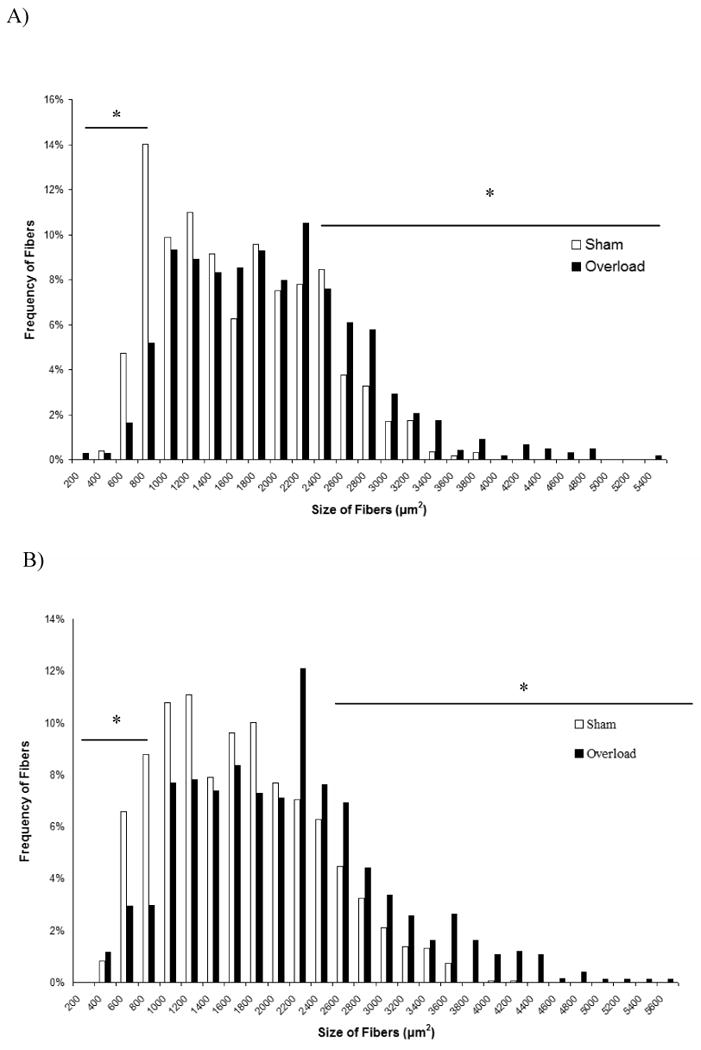
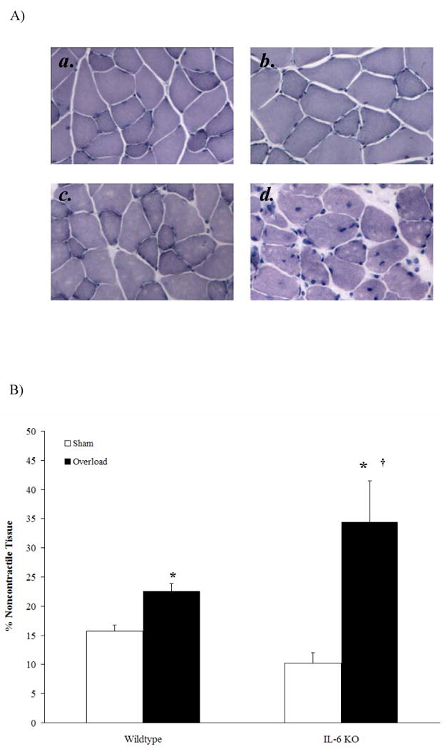
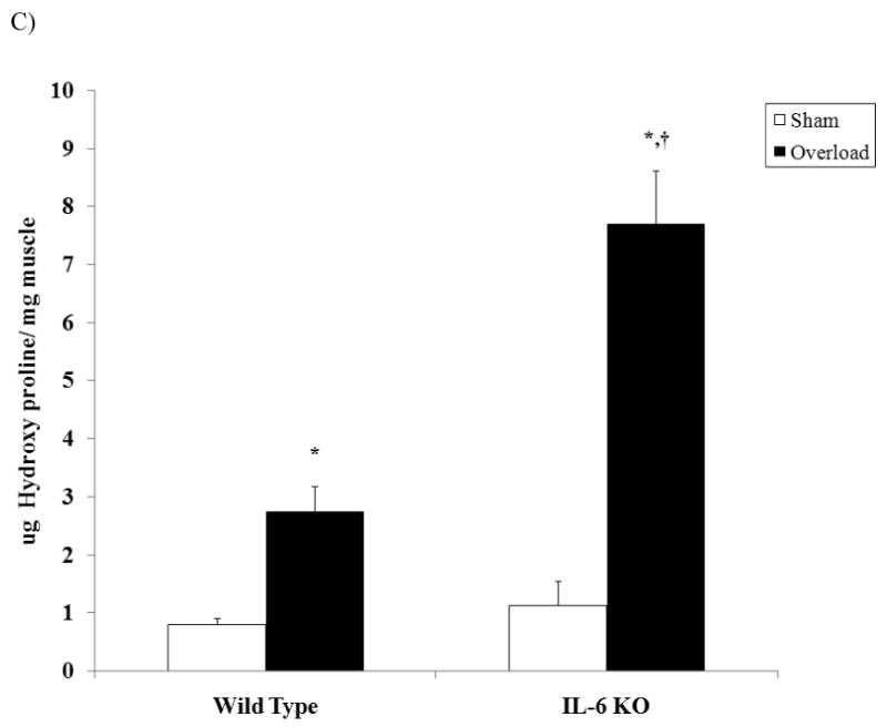
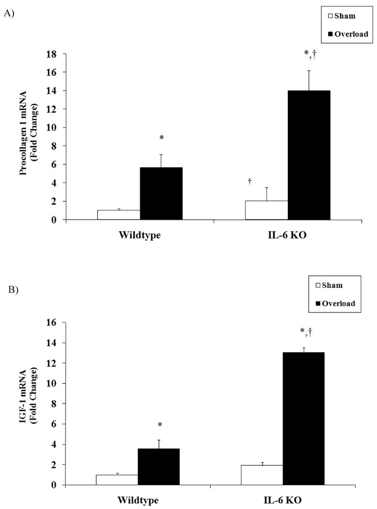
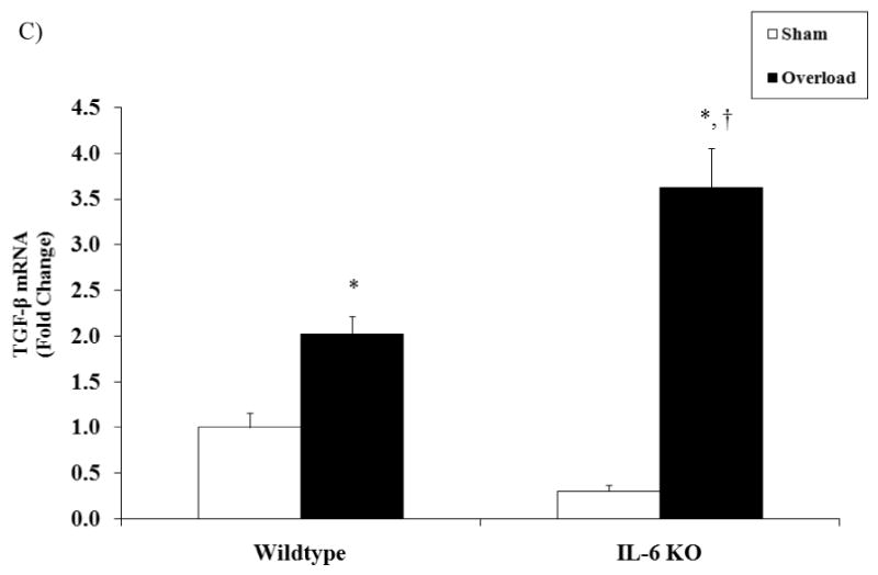
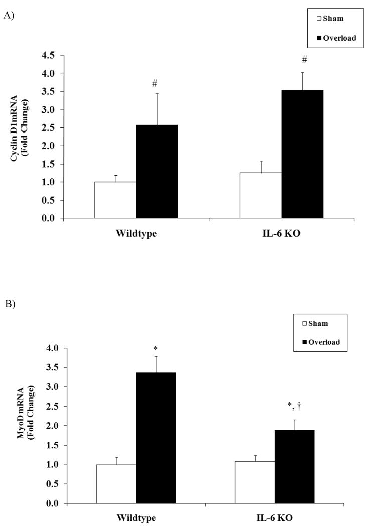
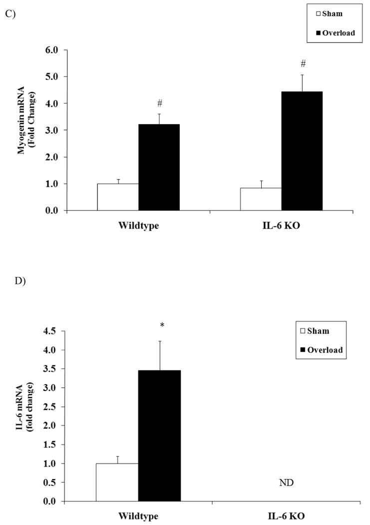
Result: Plantaris muscle mass increased 59% in WT and 116% in IL-6(-/-) mice after 21 day OV. Myofibre CSA was also increased by 21 day OV in both WT and IL-6(-/-) mice. OV induced a twofold greater increase in the volume of non-contractile tissue in IL-6(-/-) muscle compared to WT. OV also induced a significantly greater accumulation of hydroxyproline and procollagen-1 mRNA in IL-6(-/-) muscle, when compared with WT muscle after 21 day OV. Transforming growth factor-beta and insulin-like growth factor-1 mRNA expression were also induced to a greater extent in IL-6(-/-) muscle when compared with WT muscle after 21 day OV. There was no effect of IL-6 loss on the induction of myogenin, and cyclin D1 mRNA expression after 3 day OV. However, MyoD mRNA expression in 3 day OV IL-6(-/-) muscle was attenuated when compared with WT OV mice.

Conclusion: IL-6 appears to be necessary for the normal regulation of extracellular matrix remodelling during OV-induced growth.


Conflict of interest statement

Conflict of Interest: We have no conflict of interest

Figures

Figure 1
Figure 1
The effect of 21 days of functional overload on plantaris muscle myofiber size distribution after 21 days of overload. A) Wild-type plantaris muscle. B) IL-6-/- plantaris muscle. Values are means ± SE, n = 5 for each group. *Difference in the percentage of small or large fibers.
Figure 2
Figure 2
The effect of 21 days of functional overload on plantaris non-contractile tissue. A) ) H&E staining of muscle cross sections of a, wild-type sham, b, wild-type overload, c, IL-6-/- sham and d, IL-6-/- overload. B) The effect of 21 days of functional overload on volume percentage of noncontractile tissue in the plantaris muscle. C) The effect of 21 days of functional overload on hydroxyproline concentration. Values are means ± SE, n = 4-6 animals for each group. *Significantly different from same genotype control (p < 0.05). Significantly different from wild-type overload (p < 0.05).
Figure 2
Figure 2
The effect of 21 days of functional overload on plantaris non-contractile tissue. A) ) H&E staining of muscle cross sections of a, wild-type sham, b, wild-type overload, c, IL-6-/- sham and d, IL-6-/- overload. B) The effect of 21 days of functional overload on volume percentage of noncontractile tissue in the plantaris muscle. C) The effect of 21 days of functional overload on hydroxyproline concentration. Values are means ± SE, n = 4-6 animals for each group. *Significantly different from same genotype control (p < 0.05). Significantly different from wild-type overload (p < 0.05).
Figure 3
Figure 3
The effect of 21 days of functional overload on plantaris muscle procollagen-1, IGF-1, and TGF- β, and mRNA expression. A) Procollagen-1 mRNA expression. B) IGF-1 mRNA expression. C) TGF-β mRNA expression. Target gene expression corrected with 18s expression. Values are means ± SE, n = 4-6 animals per group. *Overload significantly different from same genotype control (p < 0.05). Significantly different from wild-type overload (p < 0.05).
Figure 3
Figure 3
The effect of 21 days of functional overload on plantaris muscle procollagen-1, IGF-1, and TGF- β, and mRNA expression. A) Procollagen-1 mRNA expression. B) IGF-1 mRNA expression. C) TGF-β mRNA expression. Target gene expression corrected with 18s expression. Values are means ± SE, n = 4-6 animals per group. *Overload significantly different from same genotype control (p < 0.05). Significantly different from wild-type overload (p < 0.05).
Figure 4
Figure 4
Cell cycle regulatory factors and IL-6 gene expression after 3 days of functional overload. A) Cyclin D1 mRNA expression. B) MyoD mRNA expression. C) Myogenin mRNA expression. D) IL-6 expression. Target gene expression corrected with 18s expression. Values are means ± SE, n = 4-6 for each group. *Overload significantly different from same genotype control. #Main effect of overload. (p < 0.05). ND, not detectable.
Figure 4
Figure 4
Cell cycle regulatory factors and IL-6 gene expression after 3 days of functional overload. A) Cyclin D1 mRNA expression. B) MyoD mRNA expression. C) Myogenin mRNA expression. D) IL-6 expression. Target gene expression corrected with 18s expression. Values are means ± SE, n = 4-6 for each group. *Overload significantly different from same genotype control. #Main effect of overload. (p < 0.05). ND, not detectable.

Similar articles

Cited by

References

    1. Adams GR, Haddad F, Baldwin KM. Time course of changes in markers of myogenesis in overloaded rat skeletal muscles. J Appl Physiol. 1999;87:1705–1712. - PubMed
    1. Baeza-Raja B, Munoz-Canoves P. p38 MAPK-induced nuclear factor-kappaB activity is required for skeletal muscle differentiation: role of interleukin-6. Mol Biol Cell. 2004;15:2013–2026. - PMC - PubMed
    1. Baltgalvis KA, Berger FG, Pena MM, Davis JM, Muga SJ, Carson JA. Interleukin-6 and cachexia in ApcMin/+ mice. American journal of physiology. 2008;294:R393–401. - PubMed
    1. Baltgalvis KA, Berger FG, Pena MM, Davis JM, White JP, Carson JA. Muscle wasting and interleukin-6-induced atrogin-I expression in the cachectic Apc (Min/+) mouse. Pflugers Arch. 2009;457:989–1001. - PMC - PubMed
    1. Bodell PW, Kodesh E, Haddad F, Zaldivar FP, Cooper DM, Adams GR. Skeletal muscle growth in young rats is inhibited by chronic exposure to IL-6 but preserved by concurrent voluntary endurance exercise. J Appl Physiol. 2009;106:443–453. - PMC - PubMed

Publication types

MeSH terms