. 2009 Oct;9(10):2270-80.
doi: 10.1111/j.1600-6143.2009.02755.x. Epub 2009 Jul 22.

Mast cell degranulation breaks peripheral tolerance

Affiliations

Mast cell degranulation breaks peripheral tolerance

V C de Vries et al. Am J Transplant. 2009 Oct.

Abstract

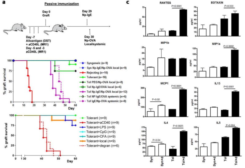
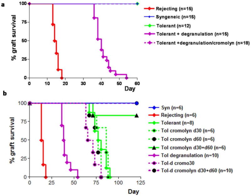
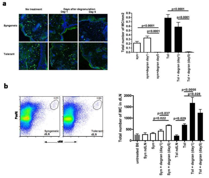
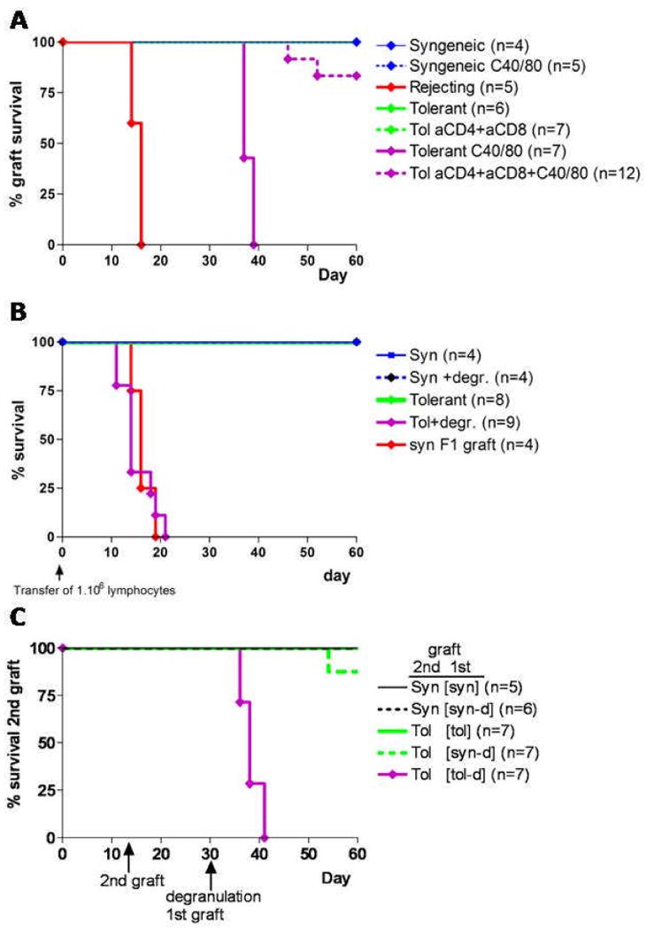
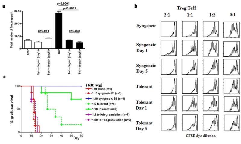
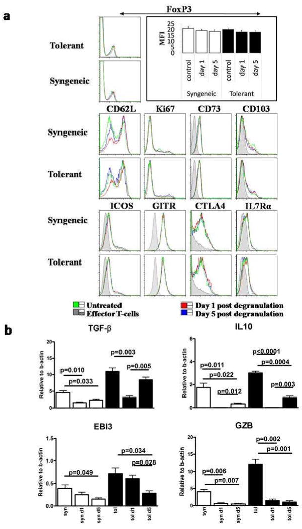
Mast cells (MC) have been shown to mediate regulatory T-cell (T(reg))-dependent, peripheral allograft tolerance in both skin and cardiac transplants. Furthermore, T(reg) have been implicated in mitigating IgE-mediated MC degranulation, establishing a dynamic, reciprocal relationship between MC and T(reg) in controlling inflammation. In an allograft tolerance model, it is now shown that intragraft or systemic MC degranulation results in the transient loss of T(reg) suppressor activities with the acute, T-cell dependent rejection of established, tolerant allografts. Upon degranulation, MC mediators can be found in the skin, T(reg) rapidly leave the graft, MC accumulate in the regional lymph node and the T(reg) are impaired in the expression of suppressor molecules. Such a dramatic reversal of T(reg) function and tissue distribution by MC degranulation underscores how allergy may causes the transient breakdown of peripheral tolerance and episodes of acute T-cell inflammation.


Figures

Figure 1
Figure 1. Degranulation of mast cell leads to acute rejection of established tolerant skin grafts
A. To induce tolerance, C57BL/6 mice were administered a regimen of donor specific transfusion (DST) of allogeneic splenocytes and anti-CD154 (200μg i.p.). Subsequently, mice received an allogeneic skin graft and survival was monitored. These mice maintain the graft for up to 100 days post-grafting, with day 0 designated as the day the mice received the graft. MCs were degranulated in mice 30 days after engraftment receiving the tolerance-inducing regiment and an allograft, in all studies described. At day 0 mice received either an CB6F1 graft (designated tolerant/tol) or a syngeneic (syn) B6 graft. Syngeneic and rejecting mice did not receive pretreatment. At day 29 mice were sensitized with 5μg of NP specific IgE (NP-IgE) and 24 hours later challenged with NP17-OVA or NP23-BSA, systemically (20ng/mouse intraperitoneal) or locally (2ng/mouse intragraft). Graft rejection was monitored for another 30 days. B. Survival of grafts for both systemic and local treatment. Controls included were either sensitized with PBS followed by local challenge with NP17-OVA or sensitized with NP-IgE followed by local challenge with 20ng of OVA. The total number of mice pooled from multiple independent experiments is shown in brackets. C. Tolerant and syngeneic with and without degranulation were collected 18 hours after degranulation and incubated in HBSS for 1h at 37°C. Supernatants were collected and tested for functionality in a proliferation assay (data not shown). Cytokine analysis was performed by multiplex and results were confirmed by ELISA. Shown are chemokines and cytokines known to be abundantly present under allergic conditions(15). D. In order to see whether degranulation induced rejection was solely the effect of inflammation various strategies were used to induce overt inflammatory responses. Mice were treated with 50μg TLR4 or TLR9 agonist, 200μl CFA or local by chemically induced inflammation. As positive controls agonistic CD40 and degranulation were included. Data shown is combined from two independent experiments.
Figure 2
Figure 2. Mast cell stabilization protects against degranulation induced rejection and prolongs graft survival in general
A. Mice were treated according figure 1A. However, 30 minutes prior to local challenge with 20ng of NP17-OVA one group received an intragraft injection of 100μl of a 39mM solution of cromolyn, a chemical known to stabilize MC thus preventing degranulation. The total number of mice pooled from three independent experiments is shown in brackets. B. Passively immunized mice were treated locally with Cromolyn at day 30 and/or day 60 post-grafting either with or without local degranulation. Grafts were monitored for a total of 120 days. The number of mice in each group, as shown in brackets is from multiple independent experiments. For both figure A and B the relevant syngeneic controls for this experiment did not show rejection for the entire duration of the experiments and are omitted from this figure for clarity.
Figure 3
Figure 3. Local IgE mediated degranulation leads to migration of MC from the graft to the draining LN
A. Grafts were degranulated locally after passive immunization. Confocal images of representative tolerant and syngeneic grafts with MC (cKit+FcεRI+) shown in green. Nuclear stain with Hoegst are shown in blue. Quantification of the number of MC in the skin was performed by counting 7 randomly chosen fields in a total of 6 individual mice. B. Flowcytometric analysis of the draining, pooled inguinal and axillary, lymph nodes. After digestion with DNAse/liberase single cell suspensions were counted and stained with cKit and FcεRI. A representative plot is showing MC in the gate after pre-enrichment with FcεRI-PE on the left. Cell counts were performed before and after enrichment to calculate the absolute number of MC (cKithighFcεRIhigh) in the dLN as shown on the right. Data is combined from 3 independent experiments with a total of 9 mice.
Figure 4
Figure 4. Break of peripheral tolerance by degranulation is T-cell dependent
A. Graft survival after depletion of CD4 and CD8 T-cells with αCD4 and αCD8 antibodies (300μl intraperitoneal and 50μl local) 2 days prior to and 5 days after local degranulation at day 30. Grafts were monitored for another 30 days after degranulation and the data shown is pooled from two independent experiments. B. RAG-/- were grafted 2 weeks prior to adoptive transfer of 1.106 lymphocytes from the inguinal and axilliary draining LN of the indicated groups. One day after degranulation lymph nodes were isolated and T-cells were purified by two rounds of negative depletion yielded over 92% purity. Graft survival measurement starts at the day of transfer of lymphocytes. Pooled data is shown from in total 3 independent experiments. C. In order to see whether degranulation could lead to rejection of a graft in a distant location, mice received two graft two weeks apart. The first graft was placed close to the base of the tail and the second graft was in the neck to assure both grafts drained to separate lymph nodes. All mice receiving an allogeneic CB6F1 graft were pretreated with DST/αCD154. At day 29 mice with two accepted grafts were included in the study. After passive immunization the first graft was locally challenged. Graft survival of a secondary graft was monitored for another 30 days. In order to control for aspecific rejection due to antigen drainage to the second graft a control was included where the first graft was syngeneic, and thus would not reject by degranulation, and the second graft was a allogeneic CB6F1 graft. Treatment and source of the first graft is indicated in between squared brackets. Data is combined from multiple experiments and the total number of mice used is shown in round brackets.
Figure 5
Figure 5. Mast cell degranulation leads to efflux of Treg from the graft and a transient block in regulatory T-cell function
A. Quantification of CD4+FoxP3+ T-cells (Treg) in the grafts of tolerized and syngeneic mice with and without local degranulation after passive immunization. Grafts were collected and individually weighted before digestion with DNAse/Dispase/Collagenase. Single cell suspension were counted and stained with CD4 before analysis by flowcytometry. Total number of Treg/mg graft tissue is shown for a total of 6 mice from 2 independent experiments. B. Following degranulation of tolerized, allografted mice, Ly-5.2+ Treg were harvested from the dLNs of Foxp3-GFP mice and FACS-purified based on GFP expression at day 1 or day 5 after local degranulationto be used in a standard suppressor assay. Briefly, naïve CD4 T-cells from wildtype, C57Bl/6 mice (Teff cells), expressing Ly5.1 were purified and labeled with CFSE. FACS-purified Ly5.2 Treg based from either degranulated or non-degranulated hosts were mixed at different ratios with Teff cells, and Treg dependent suppression was measured by CFSE dye dilution. Grafting of mice was staggered in order to be able to isolate the Treg on the same day. Cells were flow sorted and mixed with congenically (Ly5.2+) marked CFSE labeled naïve polyclonal T-cells at the indicated ratios. After 4 days of culture the Ly5.2+ cells were analyzed for CFSE dye dilution. Shown are representative histograms of multiple independent experiments. C. Treg were purified from tolerized, allografted FoxP3+-GFP mice which were untreated or degranulated with allergen. As in figure 5B, 24 hours after degranulation, Treg from tolerized controls or degranulated, tolerized groups were FACS purified based on GFP expression. Treg from each of these groups were mixed at ratios with WT, Teff (Teff: Treg) to be used in an in vivo suppressor assay. RAG-/- mice were grafted 2 weeks prior to adoptive transfer of in total 1.106 lymphocytes. Grafts were monitored for rejection for 60 after transfer of the T-cells. Data is pooled from 3 independent experiments with the total number of mice shown in brackets.
Figure 6
Figure 6. Degranulation blocks transcription of multiple important mediators used by Treg for suppression
A. Phenotypic analysis of Treg isolated from the draining lymph nodes of treated and control mice by flowcytometry. T-cells were isolated from the draining, brachial and axillary, lymph nodes of the graft at day 1 and day 5 after degranulation and grafting was staggered in order to analyze all sample on the same day. Single cell suspensions were stained with CD4 and FoxP3 in combination with the shown markers. Histograms shown are from CD4+/FoxP3+ lymphocytes except for the FoxP3 histogram where all CD4+ cells within the lymphocyte gate are shown with the mean fluorescence intensities (MFI) of FoxP3 in the bar diagram. B. RT-PCR for TGFβ, IL10, EBI3 and GzB on flow sorted Treg of the dLN of FoxP3-GFP mice one and five days after degranulation. In every experiment mice were pooled in order to get sufficient numbers of Treg. Mean and SEM are calculated from triplicate wells.

Comment in

References

    1. Dawicki W, Marshall JS. New and emerging roles for mast cells in host defence. Curr Opin Immunol. 2007;19(1):31–38. - PubMed
    1. Galli SJ, Grimbaldeston M, Tsai M. Immunomodulatory mast cell : negative, as well as positive, regulators of immunity. Nat Rev Immunol. 2008;8(6):478–486. - PMC - PubMed
    1. Sayed BA, Christy A, Quirion MR, Brown MA. The master switch: the role of mast cell in autoimmunity and tolerance. Annu Rev Immunol. 2008;26:705–739. - PubMed
    1. Grimbaldeston MA, Nakae S, Kalesnikoff J, Tsai M, Galli SJ. Mast cell-derived interleukin 10 limits skin pathology in contact dermatitis and chronic irradiation with ultraviolet B. Nat Immunol. 2007;8(10):1095–1104. - PubMed
    1. Depinay N, Hacini F, Beghdadi W, Peronet R, Mecheri S. Mast cell-dependent down-regulation of antigen-specific immune responses by mosquito bites. J Immunol. 2006;176(7):4141–4146. - PubMed

Publication types