. 2009 Oct;111(2):510-21.
doi: 10.1111/j.1471-4159.2009.06335.x. Epub 2009 Aug 13.

Docosahexaenoic acid promotes hippocampal neuronal development and synaptic function

Affiliations

Docosahexaenoic acid promotes hippocampal neuronal development and synaptic function

Dehua Cao et al. J Neurochem. 2009 Oct.

Abstract

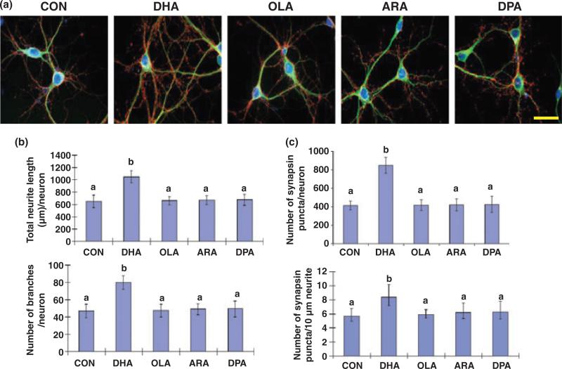
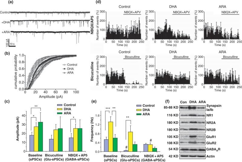
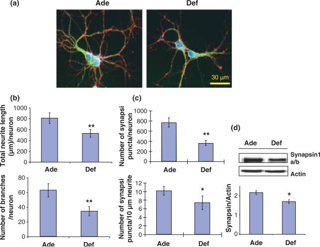
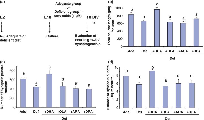
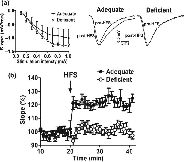
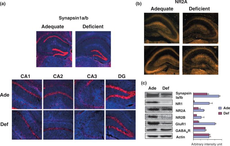
Docosahexaenoic acid (DHA, 22:6n-3), the major polyunsaturated fatty acid accumulated in the brain during development, has been implicated in learning and memory, but underlying cellular mechanisms are not clearly understood. Here, we demonstrate that DHA significantly affects hippocampal neuronal development and synaptic function in developing hippocampi. In embryonic neuronal cultures, DHA supplementation uniquely promoted neurite growth, synapsin puncta formation and synaptic protein expression, particularly synapsins and glutamate receptors. In DHA-supplemented neurons, spontaneous synaptic activity was significantly increased, mostly because of enhanced glutamatergic synaptic activity. Conversely, hippocampal neurons from DHA-depleted fetuses showed inhibited neurite growth and synaptogenesis. Furthermore, n-3 fatty acid deprivation during development resulted in marked decreases of synapsins and glutamate receptor subunits in the hippocampi of 18-day-old pups with concomitant impairment of long-term potentiation, a cellular mechanism underlying learning and memory. While levels of synapsins and NMDA receptor subunit NR2A were decreased in most hippocampal regions, NR2A expression was particularly reduced in CA3, suggesting possible role of DHA in CA3-NMDA receptor-dependent learning and memory processes. The DHA-induced neurite growth, synaptogenesis, synapsin, and glutamate receptor expression, and glutamatergic synaptic function may represent important cellular aspects supporting the hippocampus-related cognitive function improved by DHA.


Figures

Fig. 1
Fig. 1
Hippocampal neurite growth and synaptogenesis promoted uniquely by DHA supplementation. (a) Representative photomicrographs of E18 mouse hippocampal neurons after culturing 10 days with various fatty acids: microtubule-associated protein 2 (MAP2, a neuron-marker protein; green), synapsin1 (red), and DAPI for nuclei (blue). Scale bar, 30 μm. (b and c) Quantitative changes in neurite growth (b) and synapsin-positive puncta formation (c) after fatty acid treatments. Statistical analysis was performed by post hoc Tukey's HSD test at the significance level of p < 0.01. Different alphabetical letters indicate statistically significant differences.
Fig. 2
Fig. 2
Hippocampal neuronal synaptic activity enhanced by DHA supplementation. (a) Representative spontaneous synaptic current (sPSC) traces from hippocampal neurons supplemented with fatty acids for 10 days in culture. (b and c). Cumulative probability curves of the sPSC amplitude (b) for each condition, showing that sPSC amplitude is increased by the treatment with DHA and ARA (c). The acute application of bicuculline or NBQX/AP5 did not affect sPSC amplitude. The resting membrane current levels were similar among groups (–69.0 ± 9.27, –60 ± 4.59, and –67.8 ± 1.16 pA for control, DHA-, and ARA-treated neurons, respectively). (d) Frequency histograms of sPSCs, GABA-sPSCs (NBQX/AP5-treated condition), and Glu-sPSCs (bicuculline-treated condition) in cultured neurons of unsupplemented control, DHA-, and ARA-treated groups expressed as percentages of average frequency of initial condition. Bin size in the histogram is 5 s. (e) The frequency of sPSCs, Glu-sPSCs, and GABA-sPSCs in control, DHA-, and ARA-supplemented hippocampal neurons. DHA-treatment increased the frequency of sPSCs. The frequency of sPSCs from unsupplemented control neurons was significantly decreased by bicuculline while NBQX/AP5 decreased the frequency in the neurons supplemented with DHA or ARA. (f) Western blot analysis for synaptic protein expression. The expression of synapsins and subunits of glutamate receptors were elevated in the DHA-treated neuronal culture. Error bars represent SE (n = 5 for each condition). Paired t-test were performed against the baseline value of each group (#p < 0.05) or between indicated groups (*p < 0.05, **p < 0.01, and ***p < 0.001).
Fig. 3
Fig. 3
Neurite growth and synaptogenesis in cultured hippocampal neurons inhibited by prenatal depletion of DHA. (a) Representative photomicrographs of hippocampal neurons at 10 days in vitro obtained from E18 fetuses of pregnant mice fed with an n-3 fatty acid adequate (Ade) or deficient (Def) diet: MAP2 (green), synapsin 1 (red), and nucleus (blue). (b and c) Inhibition of hippocampal neurite growth and branching (b) as well as synapsin-positive puncta formation (c) by prenatal n-3 fatty acid deficiency. (d) Western blot data showing that synapsin expression was inhibited by prenatal n-3 fatty acid deprivation (n =3; *p < 0.05, **p < 0.01).
Fig. 4
Fig. 4
Restoration of hippocampal neuronal development inhibited by in vivo DHA-depletion by subsequent DHA supplementation in vitro. (a) Time line of the experimental design. E18 hippocampal neuronal cultures were obtained from pregnant mice fed adequate (Ade) or deficient (Def) diets for 16 days during pregnancy (E2–E18). The culture was grown in chemically defined medium in the presence or absence of 1 μM fatty acids for 10 days. (b–d). Total neurite length/neuron (b), number of synapsin-positive puncta/neuron (c), and number of synapsin-positive puncta/10 μm neurite (d) which were reduced by in vivo DHA-depletion but restored by subsequent DHA supplementation in vitro. Statistical analysis was performed by post hoc Tukey's HSD test at the significance level of p < 0.05. Different alphabetical letters indicate statistically significant differences.
Fig. 5
Fig. 5
Impaired LTP in young mouse hippocampi deprived of DHA during development. (a and b) fEPSPs recorded from CA1 after stimulation of Schaffer Collaterals using hippocampal slices obtained from DHA-adequate (Ade) or DHA-deficient (Def) mice at P-18. (a) Left: Input–output curves showing no difference in synaptic efficacy between the two groups. Right: Representative fEPSPs before and after high-frequency stimulation (HFS) indicating distinctive differences in the initial falling phase of fEPSPs between the two groups. (b) Inhibition of LTP by DHA-deficiency shown by the relative slope of the initial falling phase of fEPSPs plotted for 20 min before and after HFS with an averaging bin size of 1 min.
Fig. 6
Fig. 6
Localized reduction of synapsins and NR2A in young mouse hippocampi depleted with DHA during development. (a) Immunohistochemical probing of synapsin1 and NR2A in P-18 hippocampal slices, showing dramatic decreases of synapsin1 expression in CA1, CA2, and CA3 regions from DHA-depleted (Def) mice in comparison to DHA-adequate (Ade) mice; synapsin1 in red and DAPI counterstaining in blue. (b) Immunohistochemical probing of NR2A in P-18 hippo-campal slices from DHA-adequate (Ade) or DHA-depleted (Def) mice, indicating particular decrease of NR2A in the CA3 region of DHA-depleted hippocampi. (c) Western blot analysis of synapsin1, NR, GluR and GABA receptor expression in hippocampi of DHA-adequate or deficient mice at P-18.

Similar articles

Cited by

References

    1. Aarnisalo P, Kim CH, Lee JW, Perlmann T. Defining requirements for heterodimerization between the retinoid X receptor and the orphan nuclear receptor Nurr1. J. Biol. Chem. 2002;277:35118–35123. - PubMed
    1. Ahmad A, Moriguchi T, Salem N., Jr Decrease in neuron size in docosahexaenoic acid-deficient brain. Pediatr. Neurol. 2002;26:210–218. - PubMed
    1. Akbar M, Calderon F, Wen Z, Kim HY. Docosahexaenoic acid: a positive modulator of akt signaling in neuronal survival. Proc. Natl Acad. Sci. USA. 2005;102:10858–10863. - PMC - PubMed
    1. Baldelli P, Fassio A, Valtorta F, Benfenati F. Lack of synapsin I reduces the readily releasable pool of synaptic vesicles at central inhibitory synapses. J. Neurosci. 2007;27:13520–13531. - PMC - PubMed
    1. Birch EE, Garfield S, Hoffman DR, Uauy RD, Birch DG. A randomized controlled trial of early dietary supply of long-chain polyunsaturated fatty acids and mental development in term infants. Dev. Med. Child Neurol. 2000;42:174–181. - PubMed

Publication types

MeSH terms