Comparative Study
. 2009 Oct;7(10):1749-52.
doi: 10.1111/j.1538-7836.2009.03570.x. Epub 2009 Aug 11.

von Willebrand factor cleaved from endothelial cells by ADAMTS13 remains ultralarge in size

Comparative Study

von Willebrand factor cleaved from endothelial cells by ADAMTS13 remains ultralarge in size

S-Y Jin et al. J Thromb Haemost. 2009 Oct.
No abstract available


Figures

Figure 1
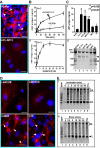
Figure 1. Cleavage of cell bound UL-vWF by recombinant ADAMTS13 and variants
Panel A. Immunofluorescent microscopic images of UL-vWF polymers on endothelial cells: HUVECs at passages 2 and 3 grown on cover slides were stimulated at 37 °C with histamine (100 μM) in phosphate buffered saline (PBS) for 2 min. After being washed with PBS, the cells were treated for 2 min at 37 °C without (A-i, buffer) or with 10 nM of recombinant full-length ADAMTS13 (A-ii, FL-AD13). After being washed with HEPES, the cells were fixed with 4% paraformaldehyde in PBS without permeablization. The fixed cells were incubated for 2 hours with mouse or rabbit anti-vWF IgG (1:100) (DAKO), followed by incubation with Cy3 -labeled IgG (1:50) (Invitrogen) for 30 min. The cell nuclei were stained with 4′, 6-diamidino-2-phenylindole (DAPI) in the mounting medium (Vector Laboratories). The images were collected under a Leica inverted fluorescent microscope (Witzlar, Germany) with SPOT Advanced Software version 4.6. The arrowheads indicate the UL-vWF strings or bundles. The bar in each figure represents 25 μm in length. Panel B. ELISA determined the vWF antigen released from endothelial cells in the conditioned medium: This panel shows the vWF antigen levels in the conditioned media of histamine-stimulated HUVECs that were treated without (dashed line, +buffer) or with (solid line, +ADAMTS13) ADAMTS13 (10 nM) for 0, 2, 5, 10, 30 and 60 min (B-i) or for 10 min with various concentrations of ADAMTS13 (0, 1, 2.5, 5, 10, 20 and 40 nM) (B-ii) in the absence of flow. Three independent experiments (N=3) were performed for each datum points and the means ±SD were shown in both sub-panels. Panel C. ELISA quantification of vWF antigen released into conditioned media of HUVECs treated with ADAMTS13 and variants. The vWF antigen levels determined by ELISA (C-i) and the proteolytic cleavage products determined by 5% SDS-polyacrylamide gel and Western blot (ii) in the conditioned media of HUVECs, after being treated for 5 min with buffer alone (buffer) or with recombinant full-length ADAMTS13 (FLA13) and various C-terminal truncated variants including delCUB, MDTCS, MDT, and M as described in Panel A. Three independent experiments were performed (N=3). The means ±SD were shown in C-i. ANOVA was performed comparing the vWF antigen levels among various groups. There was no statistically significant difference in the amount of vWF antigen released from cells treated with MDT or M compared with the buffer control. The difference between FLAD13 (or delCUB or MDTCS) and M (or MDT or buffer) was statistically significant as shown by the p values. The arrowheads in C-ii indicate the proteolytic cleavage products (176 kDa and 140 kDa) under reducing conditions. The sign ### indicates the proteolytic products of vWF by other unidentified proteases or non-specific binding of rabbit anti-vWF IgG to unknown proteins in the highly concentrated conditioned media from HUVECs treated with buffer alone, MDT and M. Panel D. Immunofluorescent microscopic imaging of UL-vWF polymers on HUVECs treated with ADAMTS13 variants. Histamine-stimulated and washed cells were treated with 10 nM of ADAMTS13 variants truncated after 8th TSP1 repeat (D-i, delCUB), after spacer domain (D-ii, MDTCS), after the first TSP1 repeat (d-iii, MDT) and after metalloprotease domain (D-iv, M) as in Panel A. The cells were fixed and stained identically as in Panel A. Three independent experiments were performed and the results were consistent. Only were the representative images shown here. The bar in each figure represents 25 μm in length. Panel E. vWF multimer distribution in the conditioned media treated with or without ADAMTS13 in the absence or presence of flow. Agarose gel (1%) electrophoresis and Western blot with anti-vWF IgG were used to determine the vWF multimer distribution in the conditioned media collected from histamine-stimulated and washed HUVECs that were treated with recombinant ADAMTS13 (10 nM) for 0 to 60 min in the absence of flow shear stress (E-i, lanes 2-8) and in the presence of flow shear stress (up to 25 dynes/cm2) (E-ii, lanes 3-7). Normal human plasma (NHP, 0.25 μl) was used as controls. In addition, vWF released from HUVECs immediate after stimulation with histamine (100 μM) was used for other controls (E-i, lane 9 and E-ii, lane 8). EDTA (20 mM) was added in the ADAMTS13-treated conditioned media to block the potential cleavage of ultra large vWF in solution. Approximately the same amount of vWF antigen was loaded into each lane (except for lane 2 in both panels where high concentrations were difficult to achieve). The ultra large vWF multimers (UL) are present in all conditioned media of HUVECs regardless of being treated with ADAMTS13 in the absence or presence of flow. However, this UL-vWF was not present in the normal human plasma. L indicates the lowest molecular weight form of vWF detected in the conditioned medium of HUVECs in culture, which is larger than the bottom two banks observed in the NHP (E-i, lanes 1 and 9; E-ii, lane 1).

References

    1. Wagner DD, Marder VJ. Biosynthesis of von Willebrand protein by human endothelial cells: processing steps and their intracellular localization. J Cell Biol. 1984;99:2123–2130. - PMC - PubMed
    1. Padilla A, Moake JL, Bernardo A, Ball C, Wang Y, Arya M, Nolasco L, Turner N, Berndt MC, Anvari B, Lopez JA, Dong JF. P-selectin anchors newly released ultra-large von Willebrand factor multimers to the endothelial cell surface. Blood. 2004;103:2150–6. - PubMed
    1. Huang J, Roth R, Heuser JE, Sadler JE. Integrin {alpha}v{beta}3 on human endothelial cells binds von Willebrand factor strings under fluid shear stress. Blood. 2008;113:1589–1697. - PMC - PubMed
    1. Sadler JE. von Willebrand factor: two sides of a coin. J Thromb Haemost. 2005;3:1702–1709. - PubMed
    1. Dong JF, Moake JL, Nolasco L, Bernardo A, Arceneaux W, Shrimpton CN, Schade AJ, McIntire LV, Fujikawa K, Lopez JA. ADAMTS-13 rapidly cleaves newly secreted ultralarge von Willebrand factor multimers on the endothelial surface under flowing conditions. Blood. 2002;100:4033–9. - PubMed

Publication types

MeSH terms