. 2009 Sep;73(5):963-74.
doi: 10.1111/j.1365-2958.2009.06825.x. Epub 2009 Aug 4.

SirA enforces diploidy by inhibiting the replication initiator DnaA during spore formation in Bacillus subtilis

Affiliations

SirA enforces diploidy by inhibiting the replication initiator DnaA during spore formation in Bacillus subtilis

Jennifer K Wagner et al. Mol Microbiol. 2009 Sep.

Abstract

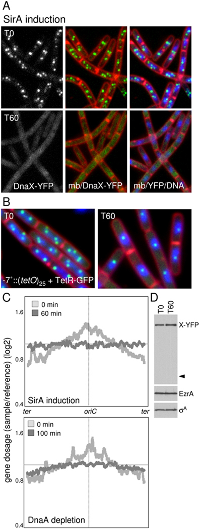
How cells maintain their ploidy is relevant to cellular development and disease. Here, we investigate the mechanism by which the bacterium Bacillus subtilis enforces diploidy as it differentiates into a dormant spore. We demonstrate that a sporulation-induced protein SirA (originally annotated YneE) blocks new rounds of replication by targeting the highly conserved replication initiation factor DnaA. We show that SirA interacts with DnaA and displaces it from the replication origin. As a result, expression of SirA during growth rapidly blocks replication and causes cell death in a DnaA-dependent manner. Finally, cells lacking SirA over-replicate during sporulation. These results support a model in which induction of SirA enforces diploidy by inhibiting replication initiation as B. subtilis cells develop into spores.


Figures

Figure 1
Figure 1
(A) The Spo0A transcription factor activates the key regulators that set the developmental pathway of sporulation into motion. These include SpoIIE, required for asymmetric cell division; RacA, responsible for reorganization of the replicated chromosomes into an axial filament; the first forespore (σF) and mother cell (σE) transcription factors and their regulators; and SirA, required to maintain diploidy. Representative sporulating cells are shown. The DNA was stained with DAPI (blue), the membranes with FM4-64 (red) or TMA-DPH (white). A forespore promoter (PspoIIQ) or a mother-cell promoter (PspoIIIA) fused to the gene encoding GFP (green) shows compartment-specific transcription factor activity. TetR-GFP (green) bound to a tetO array inserted adjacent to oriC shows that the sporulating cell has only two origins, one in each compartment (B) SirA enforces diploidy in sporulating cells by inhibiting DnaA, thereby preventing new rounds of initiation at oriC.
Figure 2
Figure 2
SirA expression is lethal to vegetatively growing cells. (A) Cells harboring an IPTG-inducible promoter (Phyperspank) fused to sirA (Phy-sirA; strain BJW38) or gfp (Phy-gfp; strain BDR1003) were streaked on LB agar plates with and without IPTG. (B) Phenotypic consequences of SirA induction (strain BJW38) and DnaA depletion (strain LAS223) in liquid CH medium. Time (in minutes) after induction or depletion is indicated. Membranes were stained with the fluorescent dye FM4-64 (red) and DNA was stained with DAPI (false-colored green). Examples of anucleate cells (white carets) and guillotined nucleoids (yellow carets) are indicated.
Figure 3
Figure 3
SirA inhibits DNA replication at the initiation step. (A) Cells were analyzed for ongoing replication using a DnaX-YFP fusion. Images show cells (strain BJW84) before and after SirA induction. Time (in min) after the addition of IPTG is indicated. Prior to SirA induction, DnaX-YFP (false-colored green) is present in replisome foci. After SirA induction, DnaX-YFP foci are lost. Membranes (mb) were stained with FM4-64 (red) and DNA was stained with DAPI (blue). (B) Cells were analyzed for origin content using TetR-GFP bound to a tetO array adjacent to oriC. Images show cells (strain BJW141) before and after SirA induction. Prior to SirA induction, most cells have 2 or 4 origin foci per DNA mass. After SirA induction, cells have a single origin focus per nucleoid. (C) Genomic DNA microarray analysis following SirA induction and DnaA depletion. The top graph shows the average gene dosage before (light grey) and after (dark gray) 60 min of SirA induction (strain BJW38). The bottom graph shows the average gene dosage before (light gray) and after (dark gray) 100 min of DnaA depletion (strain LAS223). All the probed genes in the B. subtilis chromosome arranged from −172° to +172° (ter-oriC-ter) are represented on the x-axis. The y-axis represents the gene dosage (log2) relative to a reference DNA with an oriC/ter ratio of 1 (see Experimental Procedures). The smoothed line was generated by plotting the average gene dosage of the 25 genes before and 25 genes after each gene probed. (D) Immunoblot analysis shows that DnaX-YFP remains intact after SirA induction. DnaX-YFP was analyzed using anti-GFP antibodies and the caret identifies the predicted size of free GFP. EzrA and σA were used to control for loading.
Figure 4
Figure 4
Cells that initiate replication in a DnaA-independent manner are immune to SirA induction. Cells harboring a DnaA-independent origin of replication (oriN, strain BJW173) or a DnaA-dependent origin (oriC, strain BJW174) were streaked on plates with and without IPTG. Both strains contain an IPTG-inducible promoter (Phyperspank) fused to sirA.
Figure 5
Figure 5
SirA disrupts DnaA-GFP localization. (A) Localization of DnaA-GFP (green) before and after SirA induction (strain BJW218). Time (in min) after the addition of IPTG is indicated. Membranes (mb) were stained with FM4-64 (red) and DNA was stained with DAPI (blue). Immunoblot analysis shows that DnaA-GFP remains intact after SirA induction. DnaA-GFP was analyzed using anti-GFP antibodies and the caret identifies the predicted size of free GFP. EzrA and σA were used to control for loading. The apparent septal membrane association of DnaA-GFP is due to bleed through from the membrane dye.
Figure 6
Figure 6
DnaA is required for GFP-SirA localization. (A) Localization of GFP-SirA (green) produced at low levels during vegetative growth (strain BJW196). Membranes (mb) were stained with FM4-64 (red) and DNA was stained with DAPI (blue). Yellow carets highlight nucleoid-associated GFP-SirA foci. (B) GFP-SirA localization before and after depletion of DnaA (strain BJW197). Time (in min) after the removal of IPTG (to deplete DnaA) is indicated. (C) Immunoblot analysis shows that GFP-SirA remains intact after DnaA depletion. GFP-SirA was analyzed using anti-GFP antibodies and the caret identifies the predicted size of free GFP. EzrA and σA were used to control for loading (D) SirA and DnaA interact in the yeast-two hybrid assay. A yeast strain harboring the Gal4 activation domain fused to SirA (AD-SirA) and the Gal4 DNA binding domain fused to DnaA (BD-DnaA) was able to activate transcription of the GAL2 promoter fused to the ADE2 gene. Growth on medium lacking adenine indicates a positive interaction between the hybrid proteins. Strains containing the AD-SirA fusion and the unfused DNA binding domain (BD) or the BD-DnaA fusion and the unfused activation domain (AD) did not activate transcription and were unable to grown in the absence of adenine.
Figure 7
Figure 7
Sporulating cells over-replicate in the absence of SirA (A) Cells induced to sporulate by resuspension in defined minimal medium were analyzed for ongoing replication using a DnaX-YFP fusion. The images show wild-type (strain BKM1585) and the SirA mutant (strain BJW91) at hour 1.5 of sporulation. DnaX-YFP (false-colored green) and membranes stained with TMA-DPH (false-colored red) are shown. Yellow carets highlight sporulating cells with replisome foci. (B) Cells expressing a constitutively active allele of Spo0A (Spo0Asad67) for 60 min were analyzed for ongoing replication using an SSB-GFP fusion. The images show wild-type (strain BJW271) and the SirA mutant (strain BJW275). Yellow carets highlight cells with ongoing replication as assessed by SSB-GFP foci. (C) Cells sporulated in rich medium by induction of the sensor kinase KinA were analyzed for ongoing replication using a DnaX-YFP fusion. The images show wild-type (strain BJW230) and the SirA mutant (strain BJW232) after 2 hrs of KinA induction. Yellow carets highlight sporulating cells with replisome foci. (D) Cells sporulated in rich medium by induction of the sensor kinase KinA were analyzed for origin content using TetR-GFP bound to a tetO array adjacent to oriC. The images show wild-type (strain BJW279) and the SirA mutant (strain BJW279) after 2 hrs of KinA induction. Yellow carets highlight cells with more than two TetR-GFP (green) foci. Membranes were stained with FM4-64 (red) and DNA was stained with DAPI (blue). An example of a mother cell (with two chromosomes) and twin forespores after 2.5 hrs of KinA induction is shown on the right.

Similar articles

Cited by

References

    1. Angert ER, Losick RM. Propagation by sporulation in the guinea pig symbiont Metabacterium polyspora. Proc Natl Acad Sci U S A. 1998;95:10218–10223. - PMC - PubMed
    1. Bastedo DP, Marczynski GT. CtrA response regulator binding to the Caulobacter chromosome replication origin is required during nutrient and antibiotic stress as well as during cell cycle progression. Mol Microbiol. 2009;72:139–154. - PubMed
    1. Ben-Yehuda S, Losick R. Asymmetric cell division in B. subtilis involves a spiral-like intermediate of the cytokinetic protein FtsZ. Cell. 2002;109:257–266. - PubMed
    1. Ben-Yehuda S, Rudner DZ, Losick R. RacA, a bacterial protein that anchors chromosomes to the cell poles. Science. 2003;299:532–536. - PubMed
    1. Berkmen MB, Grossman AD. Subcellular positioning of the origin region of the Bacillus subtilis chromosome is independent of sequences within oriC, the site of replication initiation, and the replication initiator DnaA. Mol Microbiol. 2007;63:150–165. - PubMed

Publication types

MeSH terms