. 2009 Sep;73(5):950-62.
doi: 10.1111/j.1365-2958.2009.06821.x. Epub 2009 Aug 4.

ESX-1 secreted virulence factors are recognized by multiple cytosolic AAA ATPases in pathogenic mycobacteria

Affiliations

ESX-1 secreted virulence factors are recognized by multiple cytosolic AAA ATPases in pathogenic mycobacteria

Patricia A DiGiuseppe Champion et al. Mol Microbiol. 2009 Sep.

Abstract

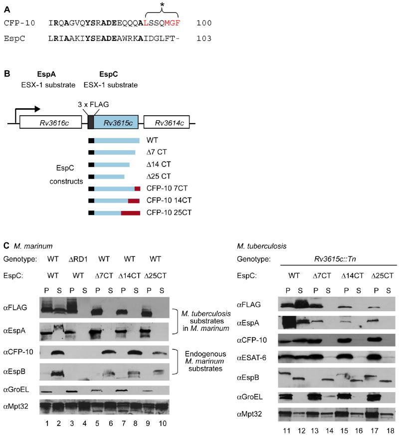
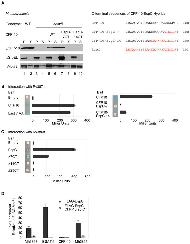
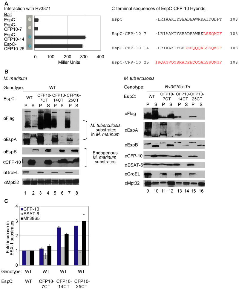
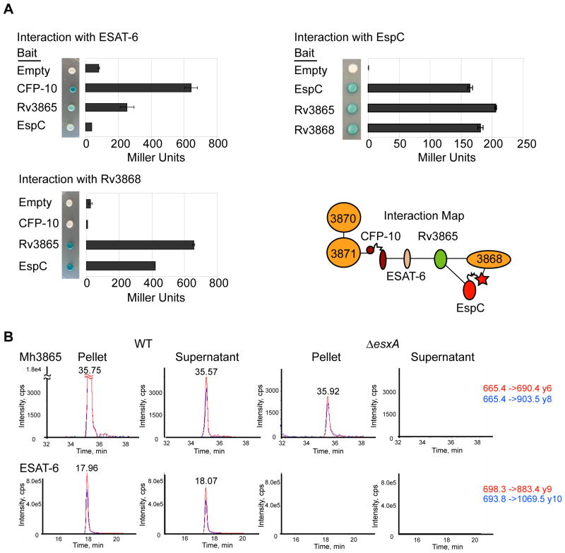
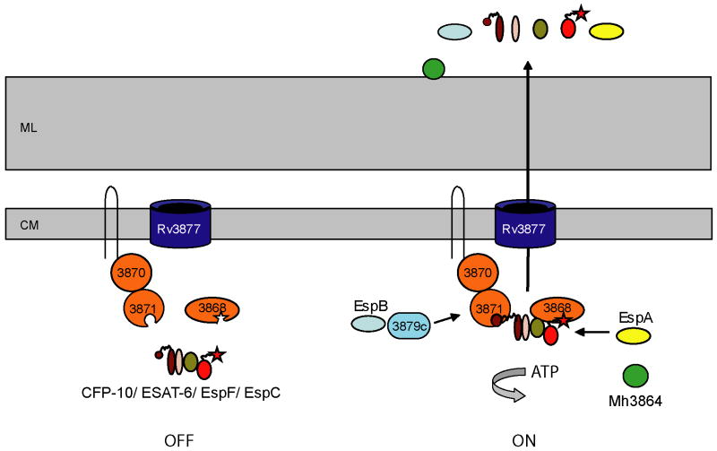
The ESX-1 secretion system of Mycobacterium tuberculosis delivers bacterial virulence factors to host cells during infection. The most abundant factor, the ESAT-6/CFP-10 dimer, is targeted for secretion via a C-terminal signal sequence on CFP-10 that is recognized by the cytosolic ATPase, Rv3871. However, the selection determinants for other ESX-1 substrates appear to be more complex. Some substrates, such as ESAT-6, are secreted despite lacking signal sequences. Furthermore, all substrates require targeting of the other ESX-1 secreted proteins, a distinguishing feature of this system. How ESX-1 substrates are selected and the basis for co-dependent secretion is unknown. Here we show that the EspC substrate interacts with Rv3868, a cytosolic AAA ATPase, through its C-terminus. Swapping the C-termini of EspC and CFP-10 revealed that these signals are functionally distinct, suggesting that the proteins are targeted via interactions with different ATPases. Surprisingly, biochemical purification experiments demonstrate that these substrates and ATPases form multi-protein complexes inside the cell and identified a new secreted substrate. By interfering with this protein interaction network, we have partially uncoupled co-dependent substrate secretion. Our results suggest that proper functioning of the ESX-1 pathway requires the interaction of multiple ESX-1 substrates and components prior to their secretion. Ultimately, understanding the details of ESX-1 targeting may allow for engineering of better vaccines to prevent tuberculosis.


Figures

Fig. 1
Fig. 1. The C-terminus of EspC is required for secretion
(A) Alignment of the C-terminal amino acids of CFP-10 and EspC. Residues required for CFP-10 signal recognition are in red. (B) Schematic of the M. tuberculosis Rv3616c operon. Rv3615c is FLAG-tagged at its N-terminus. Constructs for expression of EspC truncated or hybrid proteins are shown. (C) Immunoblot analysis of cell pellets (P) and supernatants (S) from (left) wild-type or ΔRD1 M. marinum or (right) Rv3615c∷Tn M. tuberculosis cells expressing FLAG-EspC or truncated forms of FLAG-EspC. GroEL is used as a control for autolysis, and Mpt32 is used as a loading control for all samples.
Fig. 2
Fig. 2. Rv3868 interacts with the C-terminus of EspC in vitro and in vivo
(A) Immunoblot analysis of cell pellets (P) and supernatants (S) from wild-type or ΔesxB M. tuberculosis cells harboring plasmids expressing ESAT-6 and either CFP-10 or CFP-10-EspC hybrid proteins. GroEL is used as a control for autolysis, and Mpt32 is used as a loading control for pellet and supernatant samples. The resulting C-terminal sequences of the CFP-10 EspC hybrid proteins are shown (right). (B) Yeast two-hybrid analysis of the interaction between CFP-10 or CFP-10-EspC hybrid proteins with Rv3871 (amino acids 248-591). (C) Yeast two-hybrid analysis of interaction between C-terminal truncations of EspC and Rv3868. For B and C, blue yeast colonies grown on solid media containing X-gal were positive for interaction. β-galactosidase activity is shown; error bars represent standard deviation. For all yeast two-hybrid experiments, M. tuberculosis constructs were used to study interaction. (D) Relative fold-enrichment of each protein selectively identified in the immunoprecipitation from M. marinum was assessed using quantitative mass spectrometry. The average enrichment over three injections is plotted for each protein. The fold enrichment was normalized to the average enrichment from immunoprecipitation with FLAG-Mpt64.
Fig. 3
Fig. 3. Interaction of substrates with at least two ESX-1 associated ATPases is required for secretion
(A) Yeast two-hybrid analysis of the interaction between EspC or EspC-CFP-10 hybrid proteins with Rv3868. Blue yeast colonies grown on solid media containing X-gal were positive for interaction. β-galactosidase activity is shown; error bars represent standard deviation. M. tuberculosis constructs were used to study interaction. The resulting C-terminal sequences of the EspC-CFP-10 hybrid proteins are shown (right). (B) Immunoblot analysis of cell pellets (P) and supernatants (S) of wild-type M. marinum strains (left) or M. tuberculosis bearing a transposon insertion in Rv3615c (right) expressing FLAG-EspC or FLAG-EspC-CFP-10 hybrid proteins. GroEL is used as a control for autolysis, and Mpt32 is used as a loading control for pellet and supernatant samples (C) Accumulation of M. marinum ESX-1 substrates in M. marinum due to expression of FLAG-EspC-CFP-10 hybrid proteins. Shown is the fold-change of cytosolic ESX-1 proteins in strains expressing the FLAG-EspC with the CFP-10 targeting region relative to wild-type M. marinum. 3 peptides were averaged together for CFP-10 and ESAT-6, and 5 were used for Mh3865 (EspFMm). Error bars are the %CV for the average of three measurements per peptide. Peak areas are available in the supplementary data (Table S4) and confirmatory spectra can be found in Figures S6 and S7.
Fig. 4
Fig. 4. At least three ESX-1 substrates interact for form a multi-protein complex in the cytosol
(A) Yeast two-hybrid analysis measuring the interaction of M. tuberculosis ESX-1 substrates with Rv3868. Error bars represent standard deviation. Interaction map of ESX-1 associated AAA ATPases and substrates is shown. (B) Mh3865 (EspFMm) is an ESX-1 substrate. Shown are the MRM elution profiles for EspFMm and ESAT-6Mm peptides in digested M. marinum pellets (P) or supernatants (S). Two transitions (Red&Blue) for each peptide in WT or ΔesxA M. marinum cells are shown. For Mh3865 (EspFMm) and ESAT-6Mm, the transitions are shown as parent mass (m/z) -> fragment mass (m/z), and are shown to the right in red and blue. Additional peptides, including those for GroES and confirmatory spectra are in the supplementary data (Figures S6 and S7).
Fig. 5
Fig. 5. A model for ESX-1 substrate targeting in mycobacteria
Our work supports a model in which the C-terminal regions of ESX-1 substrates function to target them to cognate ATPases, either directly or through protein interaction with other substrates. The CFP-10 signal sequence targets substrates to Rv3871, while the C-terminal amino acids of EspC targets substrates to Rv3868. One possibility is that prior to engagement of these sequences by Rv3871 and Rv3868, the secretion machine is non-functional (left). Either prior to or after the formation of a multi-substrate complex (likely including CFP-10, ESAT-6, EspF and EspC), engagement of the C-termini by the ESX-1 associated ATPases activates the machine for secretion (right). EspB likely is indirectly recognized through Rv3879c by Rv3871 (McLaughlin et al., 2007), while EspA is likely targeted through Rv3868, though the mechanism by which this occurs is unknown thus far. Mh3864, a recently identified substrate that localizes to the cell wall, is also secreted by ESX-1, but the way that it is targeted remains unknown (Carlsson et al., 2009). ML is mycolate layer, CM is cytoplasmic membrane.

References

    1. Abdallah AM, Gey van Pittius NC, Champion PA, Cox J, Luirink J, Vandenbroucke-Grauls CM, Appelmelk BJ, Bitter W. Type VII secretion--mycobacteria show the way. Nat Rev Microbiol. 2007;5:883–891. - PubMed
    1. Anderson L, Hunter CL. Quantitative mass spectrometric multiple reaction monitoring assays for major plasma proteins. Mol Cell Proteomics. 2006;5:573–588. - PubMed
    1. Bahk YY, Kim SA, Kim JS, Euh HJ, Bai GH, Cho SN, Kim YS. Antigens secreted from Mycobacterium tuberculosis: identification by proteomics approach and test for diagnostic marker. Proteomics. 2004;4:3299–3307. - PubMed
    1. Brodin P, Majlessi L, Marsollier L, de Jonge MI, Bottai D, Demangel C, Hinds J, Neyrolles O, Butcher PD, Leclerc C, Cole ST, Brosch R. Dissection of ESAT-6 system 1 of Mycobacterium tuberculosis and impact on immunogenicity and virulence. Infect Immun. 2006;74:88–98. - PMC - PubMed
    1. Burts ML, Williams WA, DeBord K, Missiakas DM. EsxA and EsxB are secreted by an ESAT-6-like system that is required for the pathogenesis of Staphylococcus aureus infections. Proc Natl Acad Sci U S A. 2005;102:1169–1174. - PMC - PubMed

Publication types

MeSH terms

Substances

LinkOut - more resources