. 2009 Sep;73(5):844-57.
doi: 10.1111/j.1365-2958.2009.06801.x. Epub 2009 Aug 6.

Decreased outer membrane permeability protects mycobacteria from killing by ubiquitin-derived peptides

Affiliations

Decreased outer membrane permeability protects mycobacteria from killing by ubiquitin-derived peptides

Georgiana E Purdy et al. Mol Microbiol. 2009 Sep.

Abstract

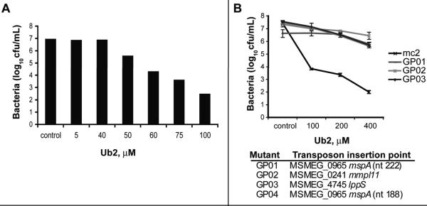
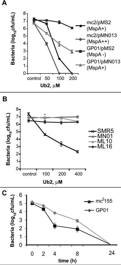
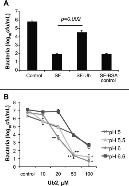
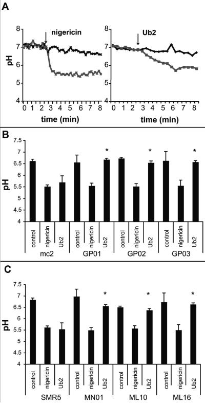
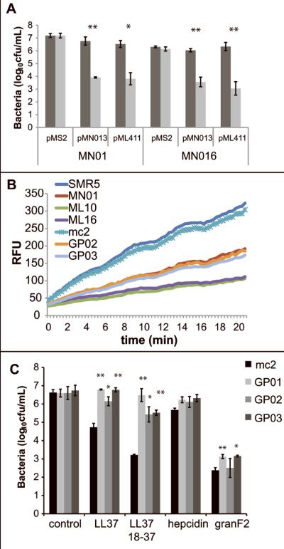
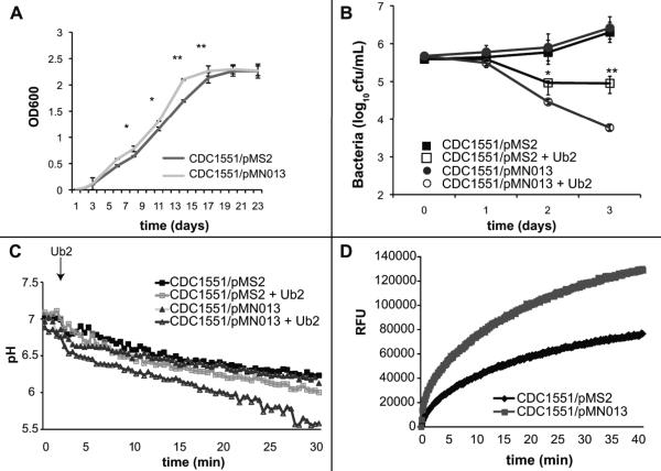
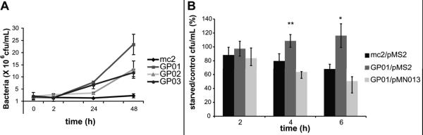
Ubiquitin-derived peptides are bactericidal in vitro and contribute to the mycobactericidal activity of the lysosome. To further define interactions of ubiquitin-derived peptides with mycobacteria, we screened for mutants with increased resistance to the bactericidal activity of the synthetic ubiquitin-derived peptide Ub2. The four Ub2-resistant Mycobacterium smegmatis mutants were also resistant to the bactericidal action of other antimicrobial peptides and macrophages. Two mutants were in the mspA gene encoding the main M. smegmatis porin. Using a translocation-deficient MspA point mutant, we showed that susceptibility of M. smegmatis to Ub2 was independent of MspA channel activity. Instead, the M. smegmatis Ub2-resistant mutants shared a common phenotype of decreased cell wall permeability compared with wild-type bacteria. Expression of mspA rendered Mycobacterium tuberculosis CDC1551 more susceptible both to ubiquitin-derived peptides in vitro and to lysosomal killing in macrophages. Finally, biochemical assays designed to assess membrane integrity indicated that Ub2 treatment impairs membrane function of M. smegmatis and M. tuberculosis cells. The M. smegmatis Ub2-resistant mutants were more resistant than wild-type M. smegmatis to this damage. We conclude that Ub2 targets mycobacterial membranes and that reduced membrane permeability provides mycobacteria intrinsic resistance against antimicrobial compounds including bactericidal ubiquitin-derived peptides.


Figures

Fig. 1
Fig. 1. Ub2 treatment of M. smegmatis hyper-resistant mutants
A. Killing of M. smegmatis with the Ub2 peptide. M. smegmatis mc2155 was subcultured to 5×10⁵ cfu/mL in 7H9 media containing the indicated concentration of Ub2 peptide. A representative experiment is shown. B. Mutant and wild type bacteria were sub-cultured to 5×10⁵ cfu/mL in 7H9 media or 7H9 containing the indicated concentration of Ub2 and incubated overnight. Bacterial viability was determined by plating serial dilutions. The average +/- standard deviation of three independent experiments is shown. The identities of the transposon mutants GP01-GP04 are shown below the graph.
Fig. 2
Fig. 2. Bacterial susceptibility to Ub2 and solubilized lysosomes (SF) correlates with the relative levels of MspA
A. Bactericidal activity of Ub2 was determined against wild type M. smegmatis (mc2155) and the mspA mutant GP01 carrying either the vector control (pMS2) or mspA expression vector (pMN013). Bacteria were incubated overnight in 7H9 media with either a buffer control or the indicated concentration of Ub2. B. Bactericidal activity of Ub2 was determined against wild type M. smegmatis (SMR5) and isogenic porin mutants MN01 (ΔmspA), ML10 (ΔmspA, ΔmspC) and ML16 (ΔmspA, ΔmspC, ΔmspD). Bacteria were incubated overnight in 7H9 media with either a buffer control or the indicated concentration of Ub2. C. Bacterial susceptibility to solubilized lysosomes (SF) was determined against wild type M. smegmatis mc2 155 (black line) and the mspA mutant GP01 (grey line). Bacteria were incubated in the presence of 50 μg/mL SF and bacterial viability determined at the indicated time points.
Fig. 3
Fig. 3. Ubiquitin contributes to the bactericidal capacity of lysosomal extracts
A. Bactericidal activity of SF upon immunodepletion of ubiquitin. M. smegmatis mc2 155 was incubated overnight with either buffer control, 50 μg/mL SF, 50 μg/mL SF pre-depleted of ubiquitin, or 50 μg/mL SF pre-depleted of BSA (negative control). Statistical significance between SF and SF-Ub was determined by Student's t test: p= 0.002. B. The bactericidal activity of Ub2 was determined in 7H9 media at pH 6.6 (standard), pH 6, pH5.5, and pH5. Statistical significance between pH 6.6 and pH 5.5 or pH 6 was determined by Student's t test: *, p<0.05; **, p<0.005. In A and B, the number of viable bacteria was determined by plating serial dilutions, and the average of three independent experiments +/- standard deviation is shown.
Fig. 4
Fig. 4. Action of Ub2 on intact M. smegmatis bacteria
A. CMFDA-labeled M. smegmatis mc2155 was resuspended in buffer (pH 5.5) and treated with either 10 mM nigericin (left) or 100 μM Ub2 (right) and fluorescence emission at 520 nm measured at excitation wavelengths of 450 nm and 490 nm. The excitation ratio was converted to pH by regression. A representative of five independent experiments is shown. B-C. CMFDA-labeled M. smegmatis strains were resuspended in buffer (pH 5.5) and treated with either nigericin or 100 μM Ub2. Fluorescence emission at 520 nm was measured at excitation wavelengths of 450 nm and 490 nm, and the excitation ratio converted to pH by regression. The average +/- standard deviations from three assays are shown. In panel B, the difference in pH observed between mc2155 and GP01, GP02, and GP03 upon treatment with Ub2 was determined to be statistically significant by a Student's t test, (*, p<0.01). Likewise, in panel C the difference in pH observed between SMR5, MN01, ML10, and ML16 was determined to be statistically significant (*, p<0.01).
Fig. 5
Fig. 5. Reduced mycobacterial cell wall permeability contributes to antimicrobial peptide resistance
A. Porin mutants MN01 (ΔmspA) and ML16 (ΔmspA, ΔmspC, ΔmspD) carrying pMS2, pMN013 (MspA), or pML411 (MspAD90L) were incubated overnight in buffer control (dark grey) or in 100 μM Ub2 (light grey). Statistical significance between bacterial viability in control and Ub2-treated samples was determined by Student's t test: *, p<0.05; **, p<0.005. B. M. smegmatis strains were incubated in the presence of 20 μM ethidium bromide. Accumulation of ethidium bromide was followed over time by monitoring emission at 590 nm upon excitation at 530 nm and expressed as Relative Fluorescence Units (RFU). C. Mutant and wild type bacteria were sub-cultured to 5×10⁵cfu/mL in 7H9 media or 7H9 containing the indicated antimicrobial peptide and incubated overnight. Statistical significance between wild type and mutant viability in each condition was determined by Student's t test: *, p<0.02; **, p<0.005. In A and C, the number of viable bacteria was determined by plating serial dilutions, and the average of three independent experiments +/- standard deviation is shown.
Fig. 6
Fig. 6. Expression of mspA in M. tuberculosis enhances growth rate, but renders bacteria hyper-susceptible to ubiquitin-derived peptides
A. Expression of mspA by CDC1551 increases growth rate in broth culture. Growth of M. tuberculosis CDC1551 carrying either pMS2 (black) or pMN013 (grey) was followed over the course of 21 days. Three independent experiments were performed. Statistical significance between CDC1551/pMS2 and CDC1551/pMN13 was determined by Student's t test: *, p<0.05; **, p<0.005. B. Bactericidal susceptibility to Ub2 killing. M. tuberculosis CDC1551 carrying either pMS2 (squares) or pMN013 (circles) was incubated in 7H9 media containing either buffer control (filled) or 100 μM Ub2 (empty). Bacterial viability was determined by plating serial dilutions at the indicated time points. The average +/- standard deviation of three replicates from a representative experiment is shown. Statistical significance between CDC1551/pMS2 and CDC1551/pMN013 upon treatment with Ub2 was determined by Student's t test: *, p<0.05; **, p<0.005. C. Bacterial susceptibility to Ub2 membrane damage. CMFDA-labeled M. tuberculosis CDC1551 carrying either pMS2 (squares) or pMN013 (triangles) was resuspended in buffer (pH 5.5) and treated with either buffer control (filled) or 100 μM Ub2 (empty) and fluorescence emission at 520 nm measured at excitation wavelengths of 450 nm and 490 nm. The excitation ratio was converted to pH by regression. A representative of three independent experiments is shown. C. Ethidium bromide accumulation of M. tuberculosis CDC1551 carrying either pMS2 (black) or pMN013 (grey). Strains were incubated in the presence of 20 μM ethidium bromide and emission at 590 nm upon excitation at 530 nm monitored over time and expressed as Relative Fluorescence Units (RFU). A representative of three independent experiments is shown.
Fig. 7
Fig. 7. Survival in macrophages is enhanced in the M. smegmatis mspA mutant
A. Bacterial survival of wild type in resting RAW 264.7 macrophages. Macrophages were infected with M. smegmatis mc2 155, the mspA mutant GP01, the mmpL11 mutant GP02, and the lppS mutant GP03. Bacterial survival over a 48 h infection was determined at the indicated time points, by harvesting the infected macrophages and the number of viable bacteria determined by plating serial dilutions. The average and standard deviations of three independent infections are shown. Statistical significance between wild type and mutants at 48 h was determined by students t test: GP01, p=0.02; GP02, p=0.01; GP03, p=0.037. B. Bacterial survival of wild type M. smegmatis mc2155/pMS2, the mspA mutant GP01/pMS2, and the complemented mspA mutant GP01/pMN013 was determined in resting and autophagic RAW264.7 macrophage-like cells. At the indicated time points, the infected macrophages were harvested and the number of viable bacteria determined by plating serial dilutions. The average and standard deviations of three independent experiments are shown. Statistical significance between survival of mc2155/pMS2 and GP01/pMS2 was determined by Students t test: *, p<0.05; **, p<0.01.
Fig. 8
Fig. 8. Expression of mspA in M. tuberculosis renders bacteria more susceptible to killing by autophagic macrophages and impairs long-term intracellular survival
A. RAW 264.7 were infected with CDC1551/pMS2 (black bars) or CDC1551/pMN013 (grey bars). Bacterial survival in resting macrophages and autophagic macrophages was determined and compared to resting control macrophages over a two-hour time course. Statistical significance between CDC1551/pMS2 and CDC1551/pMN13 upon starvation or rapamycin treatment was determined by Student's t test: *, p<0.001; **, p<0.01. B. RAW 264.7 macrophages were infected with CDC1551/pMS2 (black squares), CDC1551/pMN013 (grey triangles) and CDC1551/pML411 (grey circles) and bacterial survival monitored over ten days. The difference between CDC1551/pMS2 and CDC1551/pMN13 was determined to be significant by a Student's t test: *, p=0.012; **, p=0.015. C. RAW 264.7 seeded onto coverslips were infected with CMFDA-stained CDC1551/pMS2 (black bars) or CDC1551/pMN013 (grey bars). At 2 hours, 2 days and 4 days, the coverslips were fixed, pearmeabilized and probed with primary antibody against the lysosomal marker LAMP1 and Texas Red-conjugated secondary antibody. Co-localization of bacteria with LAMP1-positive vacuoles was scored using confocal microscopy. Over 30 bacteria-containing phagosomes were scored for each time point. Each bar represents the average of three experiments, +/- standard deviation.

References

    1. Adams LB, Dinauer MC, Morgenstern DE, Krahenbuhl JL. Comparison of the roles of reactive oxygen and nitrogen intermediates in the host response to Mycobacterium tuberculosis using transgenic mice. Tuber Lung Dis. 1997;78:237–246. - PubMed
    1. Alonso S, Pethe K, Russell DG, Purdy GE. Lysosomal killing of Mycobacterium mediated by ubiquitin-derived peptides is enhanced by autophagy. Proc Natl Acad Sci U S A. 2007;104:6031–6036. - PMC - PubMed
    1. Andreu D, Carreno C, Linde C, Boman HG, Andersson M. Identification of an anti-mycobacterial domain in NK-lysin and granulysin. Biochem J. 1999;344(Pt 3):845–849. - PMC - PubMed
    1. Barry C.E.r. Interpreting cell wall 'virulence factors' of Mycobacterium tuberculosis. Trends Microbiol. 2001;9:237–241. - PubMed
    1. Brogden KA. Antimicrobial peptides: pore formers or metabolic inhibitors in bacteria? Nat Rev Microbiol. 2005;3:238–250. - PubMed

Publication types

MeSH terms