. 2009 Oct;10(10):1458-70.
doi: 10.1111/j.1600-0854.2009.00958.x. Epub 2009 Jun 22.

A helical membrane-binding domain targets the Toxoplasma ROP2 family to the parasitophorous vacuole

Affiliations

A helical membrane-binding domain targets the Toxoplasma ROP2 family to the parasitophorous vacuole

Michael L Reese et al. Traffic. 2009 Oct.

Abstract

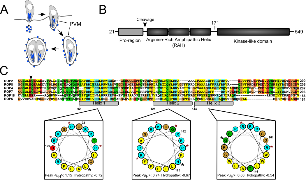
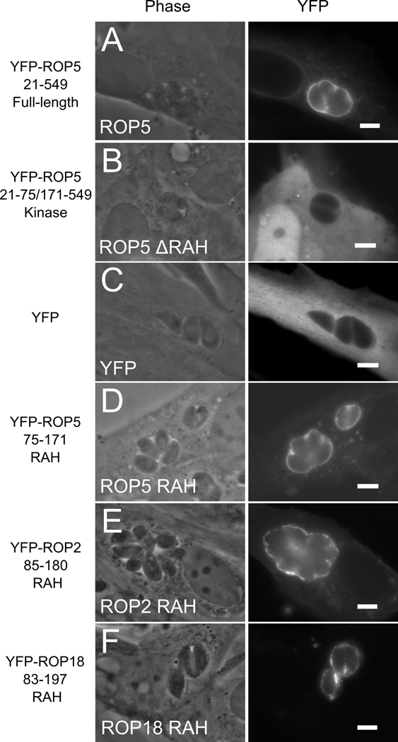
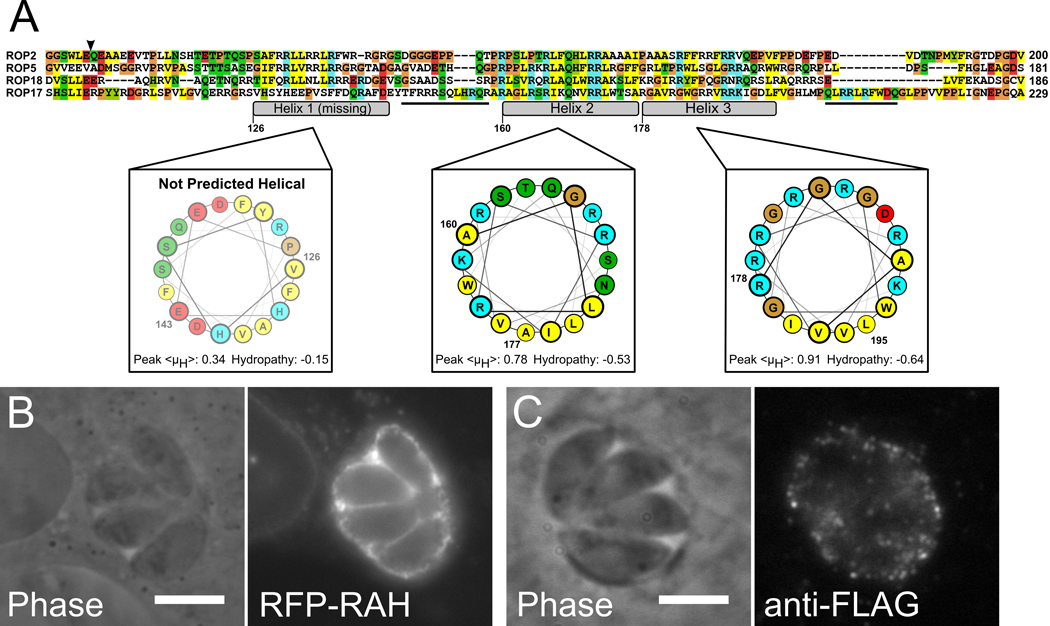
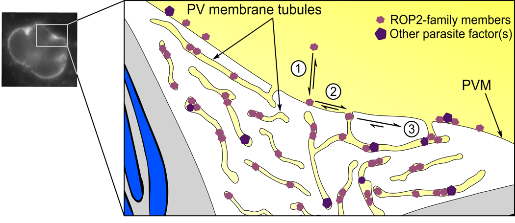
During invasion, the obligate intracellular pathogen, Toxoplasma gondii, secretes into its host cell a variety of effector molecules, several of which have been implicated in strain-specific variation in disease. The largest family of these effectors, defined by the canonical member ROP2, quickly associates with the nascent parasitophorous vacuole membrane (PVM) after secretion. Here we demonstrate that the NH(2)-terminal domain of the ROP2 family contains a series of amphipathic helices that are necessary and sufficient for membrane association. While each of the amphipathic helices is individually competent to bind cellular membranes, together they act to bind the PVM preferentially, possibly through sensing its strong negative curvature. This previously uncharacterized helical domain is an evolutionarily robust and energetically efficient design for membrane association.


Figures

Figure 1
Figure 1. The ROP2-family domain architecture
A) During Toxoplasma invasion of a host cell, its rhoptries (R) secrete their contents of effector molecules (blue) into the host cytosol. Many of these molecules associate with the PVM, which expands as the parasite replicates inside. B) Members of the ROP2-family have three domains following their signal peptides: a pro-region that is proteolytically processed in all members except for ROP5, a series of three arginine-rich amphipathic helices (RAH domain) and a protein kinase-like domain that appears to be catalytically inactive in all family members except for ROP18. Numbering is according to ROP5. The schematic is not drawn to scale. C) The RAH domain is the region of highest identity among the various family members. The putative processing site that precedes the RAH is marked with an arrowhead. The three amphipathic helices are marked in the alignment and numbered according to the ROP5 sequence. The ROP5 helices are projected on helical wheels and the average hydropathy and peak hydrophobic moment, <μH>, of each is shown, calculated according to the Eisenberg consensus scale (27). Blue shading indicates basic residues, red indicates acidic residues, yellow indicates hydrophobic residues, green indicates polar, and glycines and prolines are shaded tan. Residues that have been mutated to glutamate in the present study are marked with a red star. Residues that have been mutated to proline are marked with a black star.
Figure 2
Figure 2. The N-terminal RAH domain of ROP2-family is necessary and sufficient for membrane association
ROP2, ROP5, and ROP18 were fused to yellow fluorescent protein (YFP) and heterologously expressed in human fibroblasts. The localization of the protein after Toxoplasma infection was visualized by fluorescence. (A) Full-length mature YFP-ROP5 localizes strongly to the PVM. (B) YFP-ROP5 protein lacking the RAH domain fails to associate with host membranes and the PVM; note the diffuse localization identical to (C) YFP alone. (D) The RAH of ROP5 is sufficient to target YFP to the PVM. (E) YFP-ROP2RAH also shows strong and specific PVM association, as does (E) YFP-ROP18RAH.
Figure 3
Figure 3. The RAH domain associates weakly with various membranous structures in uninfected cells
1µm slices of confocal micrographs of uninfected cells expressing either YFP-ROP2RAH (A, B) or YFP-ROP5RAH (C, D) exhibit distributed, punctate fluorescence. Little YFP fluorescence specifically colocalizes with host mitochondrial (A, C; MitoRFP; red), ER (A, C; anti-KDEL; blue), or trans-Golgi (B, D; CFP-GalTase; red) membranes is apparent. However, some YFP-fluorescence colocalizes with host nuclear envelope (B, D; anti-lamin; blue). Scale bar represents 10µm.
Figure 4
Figure 4. The RAH domain associates specifically with the PVM
1 µm slices of confocal micrographs of Toxoplasma-infected cells expressing YFP-ROP2RAH. YFP-RAH (green) localizes strongly to the PVM of infected cells, and does not colocalize with (A) the mitochondrial-targeted RFP (red) and the endoplasmic reticular marker KDEL (blue) or (B) the trans-Golgi membrane (CFP-GalTase; red) and the nuclear envelope marker lamin A (blue). (C,D) The same is true for infected cells expressing YFP-ROP5RAH. (E) Even mitochondria associated with the PVM (arrows) do not colocalize with YFP-RAH, nor does YFP-RAH signal intensity correspond to proximity to mitochondria. YFP-RAH localizes not just to the cytosolic-facing PVM, but also to regions reminiscent of the tubular vesicular network of the PV, which is seen as the complex fluorescence “between” parasites. (F) In cells infected with Δgra2 parasites, which have a severely attenuated tubular network, YFP-RAH localizes only to the perimeter of the PV, not to the internal spaces between parasites. Scale bar for (A–D) represents 10 µm. Scale bar for (E–F) represents 5 µm.
Figure 5
Figure 5. Endogenous ROP2-family proteins appear resident in the PVM tubular network
1µm slice through the center of a confocal stack of a Toxoplasma PV. The Toxoplasma strain is an otherwise wild-type strain that transgenically expresses RFP (red) as a cytosolic morphology marker and ROP5 C-terminally tagged with the 3xFLAG epitope (green). Note the typical punctate ROP2-family staining on the PVM, which, like the YFP-RAH, is apparent both on the perimeter of the PVM and in what appears to be the PVM tubular network (arrowheads). Scale bar represents 5µm.
Figure 6
Figure 6. The RAH helices act in concert to bind membrane
Deletion mutants of mCherry(mRFP)-ROP5RAH were expressed in infected HFF concurrently with soluble YFP as a cytosolic morphology marker. Constructs containing (A) the first helix of ROP5RAH or (B) the third helix are insufficient for strong PVM association; note the high level of cytosolic punctate fluorescence as compared to that localized to the PVM. (C) The second helix of ROP5RAH is sufficient to strongly target the fusion protein to the PVM. Similarly, constructs containing (D) both ROP5RAH helices 1 & 2 or (E) both ROP5RAH helices 2 & 3 show strong localization to the PVM. (F) mRFP fused to ROP5RAH helices 1 & 3 (Δhelix 2) exhibits intermediate specificity for the PVM. (G) Membrane association was assessed biochemically by fractionation. Individual mRFP-ROP5RAH constructs were expressed in uninfected fibroblasts and the soluble fraction was compared to the amount of protein extracted from membrane either with treatment with Triton-x100 or with sodium carbonate. While mCherry (mRFP) is found entirely in the soluble fraction, mRFP-ROP5RAH strongly associates with the membrane. Detergent extracts RAH from membrane, but carbonate does not. Each of the individual RAH helices show similar carbonate-resistant membrane association.
Figure 7
Figure 7. The polar surface of RAH helix 2 encodes PVM-specific binding
(A) Overview of mutant constructs. The net charge of each construct and a summary of the quality of PVM association are listed. Mutants of mRFP-ROP5RAH were expressed in infected fibroblasts concurrently with soluble YFP as a cellular morphology marker. (B) Protein containing mutations in the first helix (R99E, R100E, R102E) that neutralize the positive charge of the polar face displays wild-type specificity for the PVM. (C) Similar mutations in the third helix (R146E, R153E, R154E) also have no effect on PVM specificity. Mutations in the second helix (D) K127E, Q131E or (E) R126E, R128E, that halve the net charge, attenuate the RAH PVM specificity.
Figure 8
Figure 8. An RAH motif correctly predicts PVM association
A) Alignment of ROP17, a more divergent member of the ROP2-superfamily, with ROP2, ROP5, and ROP18. ROP17 has a divergent RAH that lacks the first amphipathic helix (nor is that region of ROP17 sequence predicted to be helical), but has additional, short arginine-rich segments (underlined sequences) outside of the conserved amphipathic helices. Numbering is according to ROP17 sequence, coloring is as in Figure 1. B) mRFP-ROP17RAH (residues 104–223) expressed in infected host cells strongly localizes to the PVM. C) Cells infected with Toxoplasma that stably express ROP17-3xFLAG show staining for ROP17-3xFLAG on the surface of the PVM.
Figure 9
Figure 9. Model for ROP2-family interaction with the PVM tubular network
A schematic of a Toxoplasma PV with the parasites in grey, their unsecreted rhoptries blue, and the host cytosolic space yellow. Note that though the PV tubular network spreads through the vacuole, it is topologically cytosolic. 1. RAH-containing proteins (pink hexagons), due to their amphipathic nature, can transition between soluble and membrane associated states. 2. However, they appear to preferentially associate with negatively curved membranes, such as the PV membrane tubules. 3. Once recruited to the tubular network, they may associate with other proteins (purple pentagons), reducing their off-rate from the membrane. In addition, the narrow tubules may limit the diffusion of their resident proteins, helping retain them.

Similar articles

Cited by

References

    1. Baekkeskov S, Kanaani J. Palmitoylation cycles and regulation of protein function (Review) Mol Membr Biol. 2009:1–13. - PubMed
    1. Lemmon MA. Membrane recognition by phospholipid-binding domains. Nat Rev Mol Cell Biol. 2008;9(2):99–111. - PubMed
    1. Charron AJ, Sibley LD. Molecular partitioning during host cell penetration by Toxoplasma gondii. Traffic. 2004;5(11):855–867. - PubMed
    1. Boothroyd JC, Dubremetz JF. Kiss and spit: the dual roles of Toxoplasma rhoptries. Nat Rev Microbiol. 2008;6(1):79–88. - PubMed
    1. Mercier C, Dubremetz JF, Rauscher B, Lecordier L, Sibley LD, Cesbron-Delauw MF. Biogenesis of nanotubular network in Toxoplasma parasitophorous vacuole induced by parasite proteins. Mol Biol Cell. 2002;13(7):2397–2409. - PMC - PubMed

Publication types

MeSH terms

LinkOut - more resources