. 2009 Dec;49(12):2686-99.
doi: 10.1111/j.1537-2995.2009.02333.x. Epub 2009 Aug 4.

Understanding loss of donor white blood cell immunogenicity after pathogen reduction: mechanisms of action in ultraviolet illumination and riboflavin treatment

Affiliations

Understanding loss of donor white blood cell immunogenicity after pathogen reduction: mechanisms of action in ultraviolet illumination and riboflavin treatment

Rachael P Jackman et al. Transfusion. 2009 Dec.

Abstract

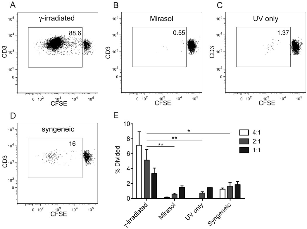
Background: Donor white blood cells (WBCs) present in transfusion products can lead to immune sequelae such as production of HLA antibodies or graft-versus-host disease in susceptible transfusion recipients. Eliminating the immunogenicity of blood products may prove to be of clinical benefit, particularly in patients requiring multiple transfusions in whom allosensitization is common. This study examines a method of pathogen reduction based on ultraviolet light illumination in the presence of riboflavin. In addition to pathogens, WBCs treated with this system are affected and fail to stimulate proliferation of allogeneic peripheral blood mononuclear cells (PBMNCs) in vitro.

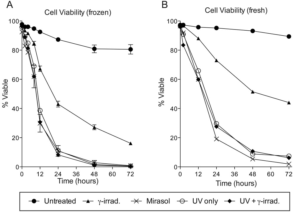
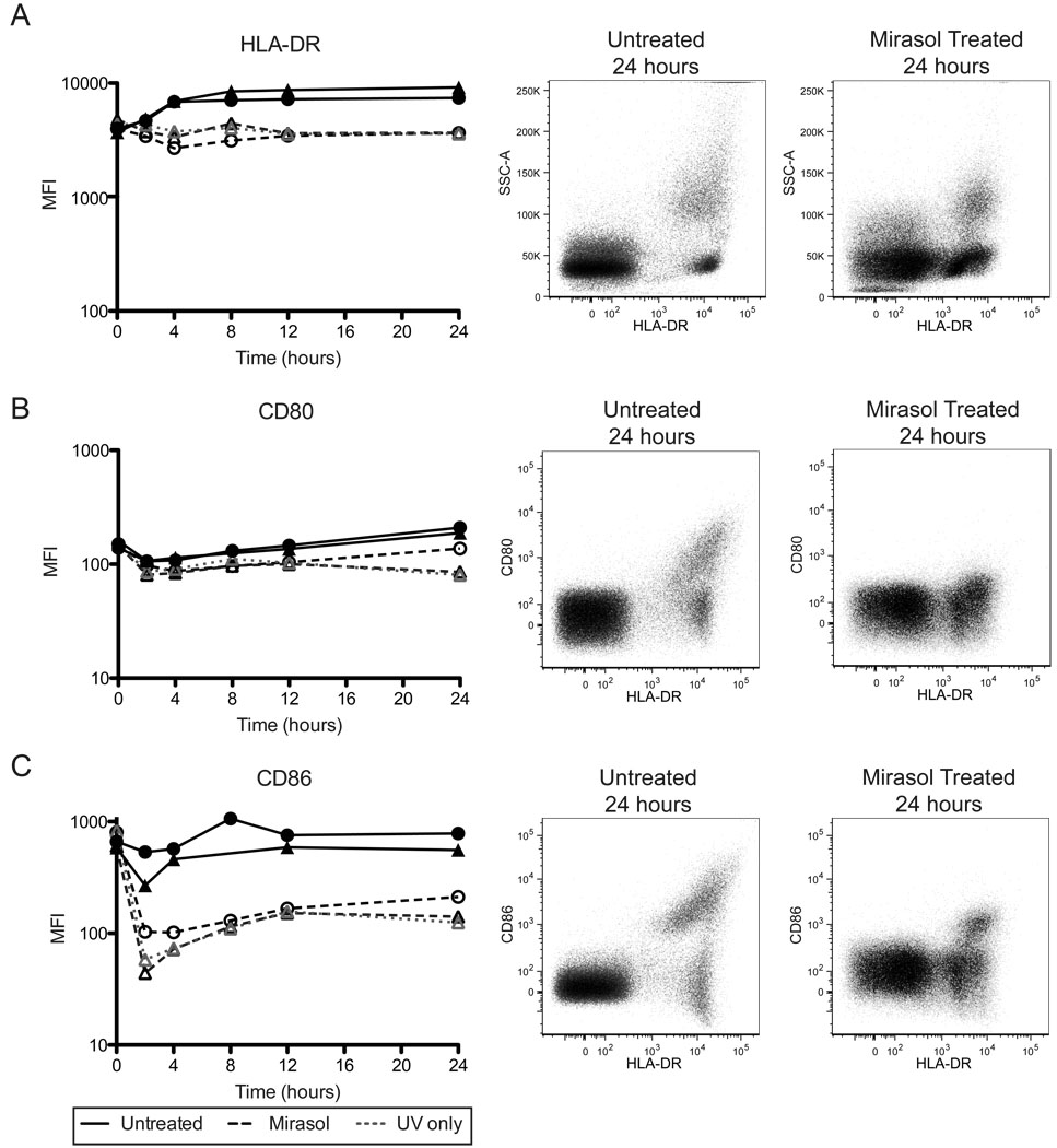
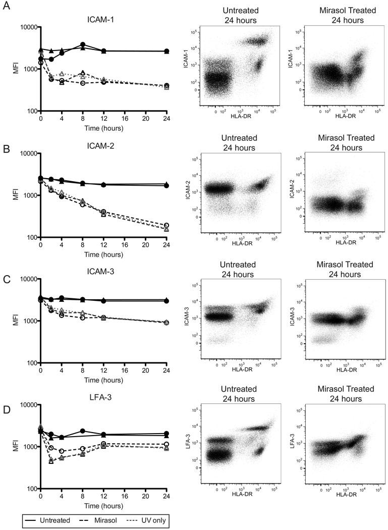
Study design and methods: This study sought to determine the mechanisms regulating this loss of immunogenicity. Treated cells were examined for surface expression of a number of molecules involved in activation and adhesion, viability, cell-cell conjugation, and ability to stimulate immune responses in allogeneic PBMNCs.

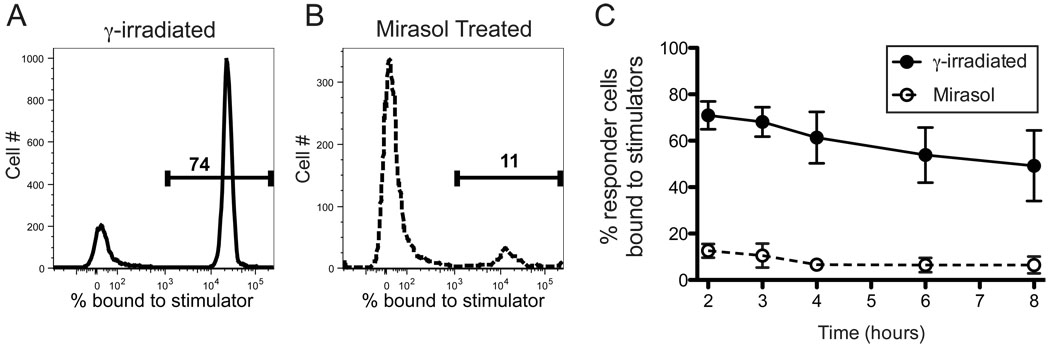
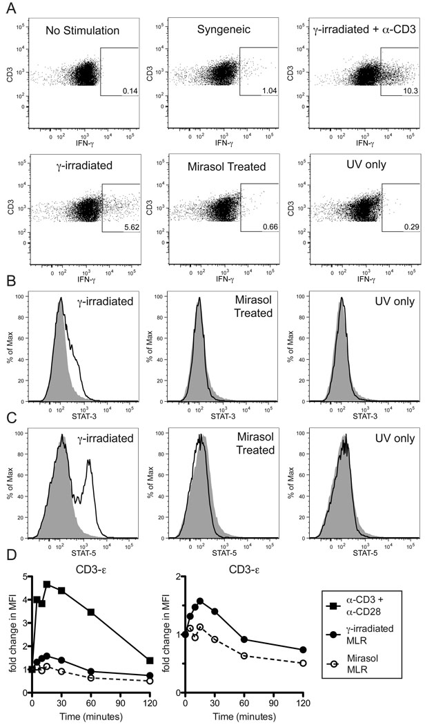
Results: Compared with untreated controls, ultraviolet (UV)-irradiated antigen-presenting cells showed slightly reduced surface expression of HLA Class II and costimulatory molecules and had more significant reductions in surface expression of a number of adhesion molecules. Furthermore, treated cells had a severe defect in cell-cell conjugation. The observed loss of immunogenicity was nearly complete, with UV-irradiated cells stimulating barely measurable interferon-gamma production and no detectable STAT-3, STAT-5, or CD3-epsilon phosphorylation in allospecific primed T cells.

Conclusion: These results suggest that defective cell-cell adhesion prevents UV-irradiated cells from inducing T-cell activation.


Conflict of interest statement

Conflict of Interest: Rachael P. Jackman and John W. Heitman have no conflict of interest. Susanne Marschner and Raymond P. Goodrich are employed by CaridianBCT Biotechnologies, Philip J. Norris has a consulting relationship with and received research funding for this project from CaridianBCT Biotechnologies.

Figures

Figure 1
Figure 1. Mirasol treated cells fail to stimulate allogeneic MLR
PBMCs were labeled with CFSE and stimulated for one week with allogeneic PBMCs that were γ-irradiated (A), Mirasol treated (B), or given a modified treatment without riboflavin (UV only) (C). γ-irradiated syngeneic stimulator cells were used as a negative control (D). A–D are representative plots of live T cells from each of these groups at day 7. The % of the initial responder population that has divided by day 7 was back calculated in FlowJo and is shown for each stimulation (E). γ-irradiated and Mirasol treated cells from three different donors were used for each experiment and the experiment was run two times (each time with a different responder population) with the results plotted as mean and standard error. Stimulations were done in 96-well plates with a stimulator:responder ratio of 4:1 (white), 2:1 (light grey), or 1:1 (dark grey). Stimulators that did not get full Mirasol treatment were γ-irradiated to prevent proliferation. 10,000 T cells were collected for each sample where possible, for samples with little to no cell division, fewer cells were available and a minimum of 2,400 cells were collected for these groups. Differences between each treatment group were evaluated including the data from all three stimulator:responder ratios. * p < 0.05, ** p < 0.01.
Figure 2
Figure 2. Viability of Mirasol treated cells
Cells that were untreated (n=3), γ-irradiated (n=3), Mirasol treated (n=2), or given a modified treatment without riboflavin (UV only) with or without γ-irradiation (n=1 for each), were cultured for 0, 2, 4, 8, 12, 24, 48, or 72 hours, then stained with aqua amine-reactive dye to determine viability. Means with standard error are displayed for groups with n>1 (A). One set of fresh cells (not frozen) was processed with each treatment and cultured for 0, 12, 24, 48, or 72 hours, then stained with aqua amine-reactive dye (B).
Figure 3
Figure 3. Mirasol treated APCs have reduced surface HLA-DR and costimulatory molecule expression
Cells that were untreated, Mirasol treated, or given a modified treatment without riboflavin (UV only), were cultured for 0, 2, 4, 8, 12, or 24 hours, then stained for surface expression of HLA-DR (A), CD80 (B), and CD86 (C). Left panels plot median fluorescent intensity for HLA-DR+ cells (APCs) over time. ▲ and ● are two different donors. The center panels are representative plots of an untreated sample at 24 hours, and the right panels are representative plots of Mirasol treated samples at 24 hours. Both the center and right panels are ungated populations.
Figure 4
Figure 4. Mirasol treated APCs have reduced surface adhesion molecule expression
Cells that were untreated, Mirasol treated, or given a modified treatment without riboflavin (UV only), were cultured for 0, 2, 4, 8, 12, or 24 hours, then stained for surface expression of ICAM-1 (A), ICAM-2 (B), ICAM-3 (C), and LFA-3 (D). Left panels plot median fluorescent intensity for HLA-DR+ cells (APCs) over time. ▲ and ● are two different donors. The center panels are representative plots of an untreated sample at 24 hours, and the right panels are representative plots of Mirasol treated samples at 24 hours. Both the center and right panels are ungated populations.
Figure 5
Figure 5. Cell conjugation is defective in Mirasol treated cells
Normal PBMCs were primed with γ-irradiated cells to enrich for allo-specific cells. These responder cells were labeled with PKH red, and cultured with stimulators stained with PKH green. Stimulators were either γ-irradiated or Mirasol treated cells from the same donor as the priming cells. Representative sample plots (A–B) from t=2 hours show cells gated on red responder events, with the green stimulator events plotted as histograms. The markers denote the percentage of responders bound to stimulators. Four replicate samples were tested longitudinally from 2 – 8 hours (C). A minimum of 4,000 red events were collected for each sample. Error bars represent 95% confidence intervals.
Figure 6
Figure 6. Mirasol treated are cells unable to activate untreated T cells
Normal PBMCs were primed with γ-irradiated cells to enrich for allo-specific cells. These primed cells were labeled with CFSE, then restimulated overnight with γ-irradiated, Mirasol treated, or UV only treated allogeneic cells from the same donor as the priming cells. Additional groups that were either unstimulated, or stimulated with syngeneic cells or γ-irradiated allogeneic cells + α-CD3 antibodies were included as controls. CFSE+CD3+ gates were used to identify responder T cells (A). Gates are drawn around IFN-γ+ T cells based on unstimulated control and confirmed with isotype control stains. A minimum of 8,000 T cells were collected for each sample. This experiment was repeated three times with representative data displayed. In B–C the primed cells were restimulated with labeled cells (from the same donor as the priming cells) that were either γ-irradiated, Mirasol treated, or UV only treated for two hours, then stained intracellularly for STAT-3, STAT-5, and CD3. Stains of responder T cells for STAT-3 (B) and STAT-5 (C) are plotted with the unstimulated control stain shown in grey for comparison. In D, normal PBMCs were stimulated with anti-CD3 + anti-CD28 antibodies ■, and primed cells were restimulated with γ-irradiated cells ●, or Mirasol treated cells ○ (from the same donor as the priming cells), for 0, 5, 10, 15, 30, 60, or 120 minutes, lysed, and examined for the phosphorylation of CD3-ε using a multiplex bead-based kit. Fold change over t=0 is plotted over time. The left panel shows all three stimulation conditions, the right panel shows only the two MLR stimulations. Experiments in B–D were repeated twice with representative data displayed.

Similar articles

Cited by

References

    1. Goodrich RP, Edrich RA, Li J, Seghatchian J. The Mirasol PRT system for pathogen reduction of platelets and plasma: an overview of current status and future trends. Transfus Apher Sci. 2006;35:5–17. - PubMed
    1. Cardo LJ, Rentas FJ, Ketchum L, Salata J, Harman R, Melvin W, Weina PJ, Mendez J, Reddy H, Goodrich R. Pathogen inactivation of Leishmania donovani infantum in plasma and platelet concentrates using riboflavin and ultraviolet light. Vox Sang. 2006;90:85–91. - PubMed
    1. Cardo LJ, Salata J, Mendez J, Reddy H, Goodrich R. Pathogen inactivation of Trypanosoma cruzi in plasma and platelet concentrates using riboflavin and ultraviolet light. Transfus Apher Sci. 2007;37:131–137. - PubMed
    1. Rentas F, Harman R, Gomez C, Salata J, Childs J, Silva T, Lippert L, Montgomery J, Richards A, Chan C, Jiang J, Reddy H, Li J, Goodrich R. Inactivation of Orientia tsutsugamushi in red blood cells, plasma, and platelets with riboflavin and light, as demonstrated in an animal model. Transfusion. 2007;47:240–247. - PubMed
    1. Fast LD, Dileone G, Li J, Goodrich R. Functional inactivation of white blood cells by Mirasol treatment. Transfusion. 2006;46:642–648. - PubMed

Publication types

MeSH terms