Review
. 2010 Mar-Apr;1799(3-4):257-65.
doi: 10.1016/j.bbagrm.2009.08.003. Epub 2009 Aug 12.

Control of alpha-herpesvirus IE gene expression by HCF-1 coupled chromatin modification activities

Affiliations
Review

Control of alpha-herpesvirus IE gene expression by HCF-1 coupled chromatin modification activities

Thomas M Kristie et al. Biochim Biophys Acta. 2010 Mar-Apr.

Abstract

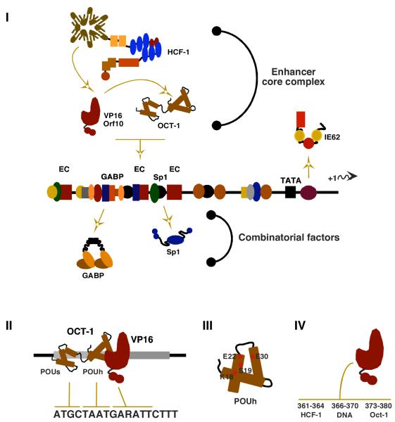
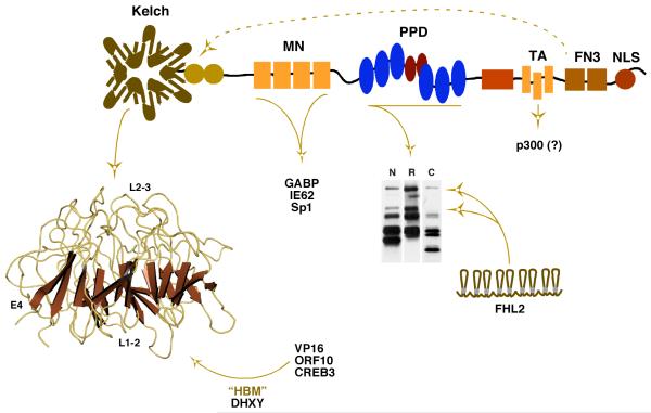
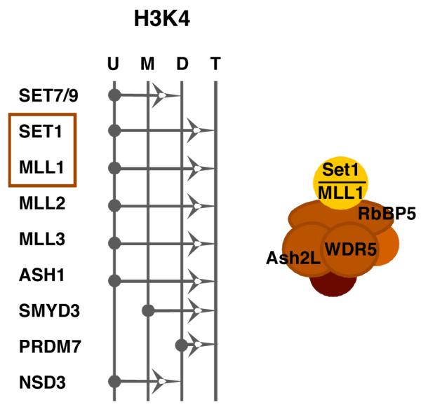
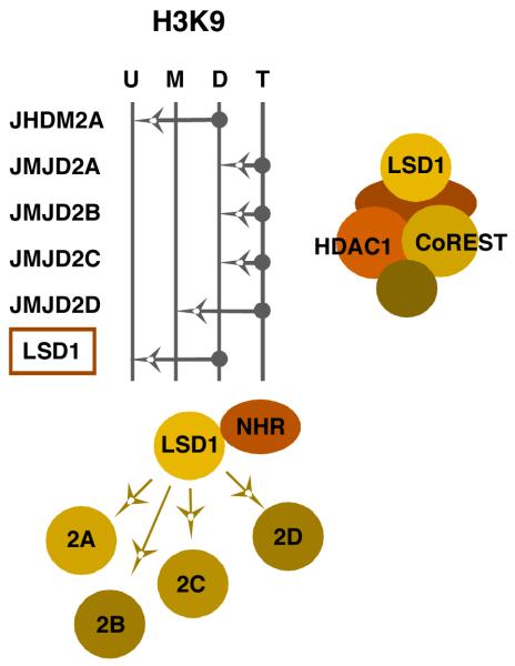
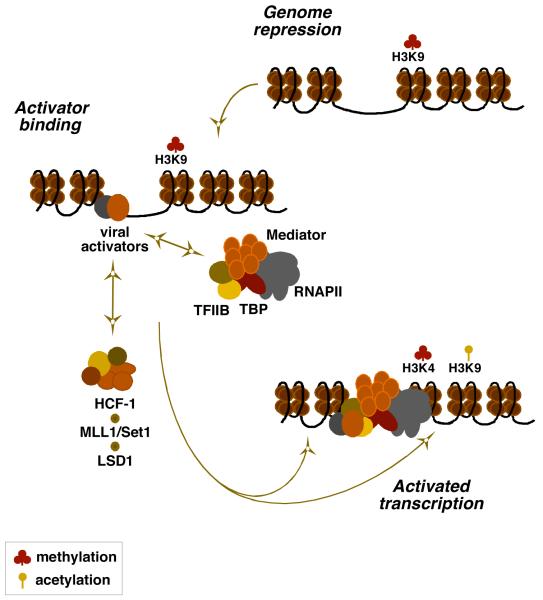
The immediate early genes of the alpha-herpesviruses HSV and VZV are transcriptionally regulated by viral and cellular factors in a complex combinatorial manner. Despite this complexity and the apparent redundancy of activators, the expression of the viral IE genes is critically dependent upon the cellular transcriptional coactivator HCF-1. Although the role of HCF-1 had remained elusive, recent studies have demonstrated that the protein is a component of multiple chromatin modification complexes including the Set1/MLL1 histone H3K4 methyltransferases. Studies using model viral promoter-reporter systems as well as analyses of components recruited to the viral genome during the initiation of infection have elucidated the significance of HCF-1 chromatin modification complexes in contributing to the final state of modified histones assembled on the viral IE promoters. Strikingly, the absence of HCF-1 results in the accumulation of nucleosomes bearing repressive marks on the viral IE promoters and silencing of viral gene expression.


Figures

Figure 1
Figure 1. Components regulating the expression of the α-herpesvirus IE genes
(I) The IE genes of HSV and VZV enhancer-promoter domains are complex and contain binding sites for multiple factors functioning synergistically or cooperatively. Viral IE activators (VP16 for HSV; ORF10 for VZV) interact with Oct-1 and HCF-1 to form the stable enhanceosome complex. A second VZV IE activator IE62 stimulates expression via its own recognition elements. Cellular factors such as GABP and Sp1 amplify the enhancer core (EC, TAATGARAT) mediated expression of the IE genes but may also function independent of the EC complex to stimulate IE gene expression. (II) Oct-1 recognizes the EC element via a bipartite DNA binding domain consisting of POU-specific (s) and POU-homeo (h) domains. (III) The viral activator, VP16, recognizes the surface of the Oct-1 POU-homeo domain via specific residues in helix 1 and 2 and provides specificity by recognition of the 3′ sequences of the EC element. (IV) Clustered residues in the carboxyterminal region of VP16 mediate interactions with HCF-1, DNA, and Oct-1.
Figure 2
Figure 2. The transcriptional coactivator HCF-1 and interactions regulating the expression of the α-herpesvirus IE genes
The essential coactivator HCF-1 interacts with numerous factors that impact the expression of the IE genes. An amino-terminal kelch domain is a barrel structure consisting of reiterated units of 4 antiparallel β-sheets connected by flexible loops. The structure presents several protein interaction surfaces composed of: (i) loops connecting sheets 2 and 3 (top surface, L2-3); (ii) loops connecting sheets 1 and 2 (bottom surface, L1-2); and (iii) the 4th β-sheet of each reiterated unit (circumference, E4). Viral and cellular activators (i.e. VP16, ORF10, CREB3/Luman) and coactivators containing an HCF-1 Binding Motif (HBM, D/EHXY) interact with this domain, presumably in a non-exclusive manner. The mid-aminoterminus (MN) binds GABP, IE62, and Sp1. The central proteolytic processing domain (PPD) consists of a series of 20 amino acid reiterations (blue/red ovals) that are the sites of specific proteolytic processing that generates the family of HCF-1 polypeptides shown in the western blot (N, R, C represent antiserum specific to the aminoterminus, repeat, and carboxyterminus, respectively). The coactivator FHL2 interacts with the PPD and this interaction is regulated by HCF-1 processing. A transactivation domain (TA) is required for cooperative stimulation by factors such as VP16 and may function to recruit the coactivator p300/CBP. Two fibronectin type III repeats (FN3) mediate the amino- and carboxyterminal subunit association of HCF-1 as indicated by the dashed arrow. NLS; nuclear localization signal.
Figure 3
Figure 3. The histone H3-lysine 4 methyltransferase family
The specificities of the members of the histone H3-lysine 4 (H3K4) family of methyltransferases are shown. The arrows represent the ability of each member to catalyze the addition of a methyl group to an unmodified (U), monomethyl (M), or dimethyl (D) lysine 4 residue. The Set1 and MLL1 methyltransferase complexes contain a set of common core proteins including Ash2L, WDR5, and RbBP5 in addition to the catalytic subunit (Set1 or MLL1). Both hSet1 and MLL1 (boxed) interact with HCF-1 and play a role in HSV and VZV IE gene expression, likely via installation of activating H3K4 methylation at the IE promoters.
Figure 4
Figure 4. The histone H3-lysine 9 demethylase family: positive and negative LSD1 complexes
The specificities of the members of the histone H3-lysine 9 (H3K9) family of demethylases are shown. The arrows represent the ability of each member to catalyze the removal of a methyl group from a trimethyl (T), dimethyl (D), or monomethyl (M) lysine 9 residue. LSD1 has a dual specificity; removing positive H3K4 methylation when in the CoREST complex or removing repressive H3K9 methylation when complexed with other components such as nuclear hormone receptors (NHR). Removal of repressive H3K9 trimethylation requires the cooperative activity of one of the other members of the family with the complimenting activity (JMJD2A, B, C, or D). LSD1 is highlighted (boxed) to emphasize its role in viral IE gene expression.
Figure 5
Figure 5. An HCF-1 complex couples methyltransferase and demethylase activities in the initiation of IE gene expression
A model for the role of the HCF-1 coactivator complex in the regulation of the α-herpesvirus IE genes is shown. Upon infection, the viral genome is subject to the accumulation of chromatin bearing repressive marks (H3K9-methylation). The viral IE activators (VP16 or ORF10 and IE62) bind to the IE regulatory domains and recruit components of the RNAPII complex to the promoter. Transcriptional activation requires the recruitment of HCF-1 dependent chromatin modification activities (H3K4 methyltransferase Set1 or MLL1 and H3K9 demethylase LSD1) that ultimately results in a decrease in repressive marks and an increase in positive marks to promote IE gene transcription. Coordinated H3K4 methylation and H3K9 acetylation is proposed although the specific acetyltransferase complexes involved have not yet been determined.
Figure 6
Figure 6. Specific sequestering and transport of HCF-1 in sensory neurons: a role for HCF-1 in regulation of α-herpesvirus reactivation from latency
(I) HCF-1 is ubiquitously expressed and localized in the nucleus of most cell types (Liver and Kidney are examples shown) but is specifically sequestered in the cytoplasm of unstimulated sensory neurons (Trigeminal Ganglia). (II) Stimuli that result in viral reactivation cause rapid transport and accumulation of HCF-1 in the nucleus of sensory neurons in a mouse trigeminal ganglia explant model system. The percentage of neurons exhibiting HCF-1 nuclear localization post explant in both mock infected and HSV latently infected trigeminal ganglia is graphically represented as a function of time.

References

    1. Kristie TM. Early events pre-initiation of viral gene expression. In: Arvin A, Campadielli-Fiume G, Roizman B, Whitley R, Yamanishi K, editors. Human Herpesviruses: Biology, Therapy, and Immunoprophylaxis. Cambridge University Press; Cambridge: 2006. pp. 395–452.
    1. Roizman B, Sears AE. Herpes simplex viruses and their replication. In: Fields BN, Knipe DM, Howley PM, editors. Fundamental Virology. Lippincott-Raven Publishers; Philadelphia: 1996. pp. 1043–1107.
    1. Kristie TM, Sharp PA. Interactions of the Oct-1 POU subdomains with specific DNA sequences and with the HSV alpha-trans-activator protein. Genes Dev. 1990;4:2383–2396. - PubMed
    1. Sturm RA, Herr W. The POU domain is a bipartite DNA-binding structure. Nature. 1988;336:601–604. - PubMed
    1. Verrijzer CP, Kal AJ, van der Vliet PC. The oct-1 homeo domain contacts only part of the octamer sequence and full oct-1 DNA-binding activity requires the POU-specific domain. Genes Dev. 1990;4:1964–1974. - PubMed

Publication types

MeSH terms

LinkOut - more resources