. 2009 Sep 15;19(17):1498-502.
doi: 10.1016/j.cub.2009.07.034. Epub 2009 Aug 13.

Centriole age underlies asynchronous primary cilium growth in mammalian cells

Affiliations

Centriole age underlies asynchronous primary cilium growth in mammalian cells

Charles T Anderson et al. Curr Biol. .

Abstract

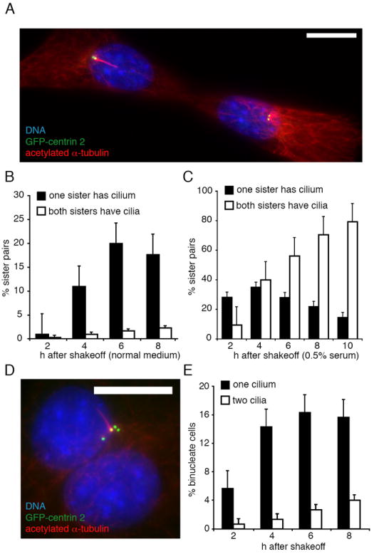
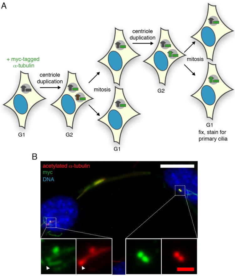
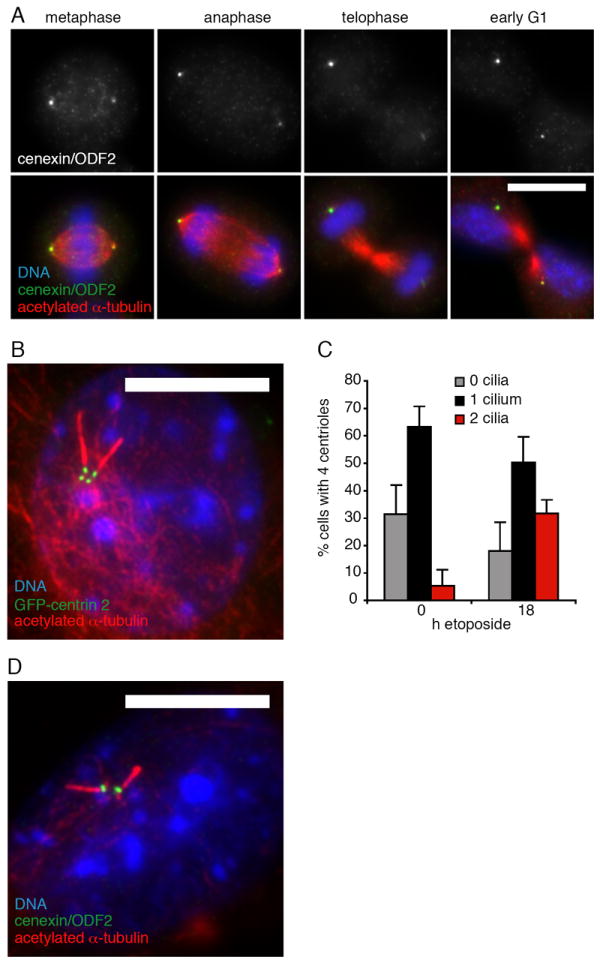
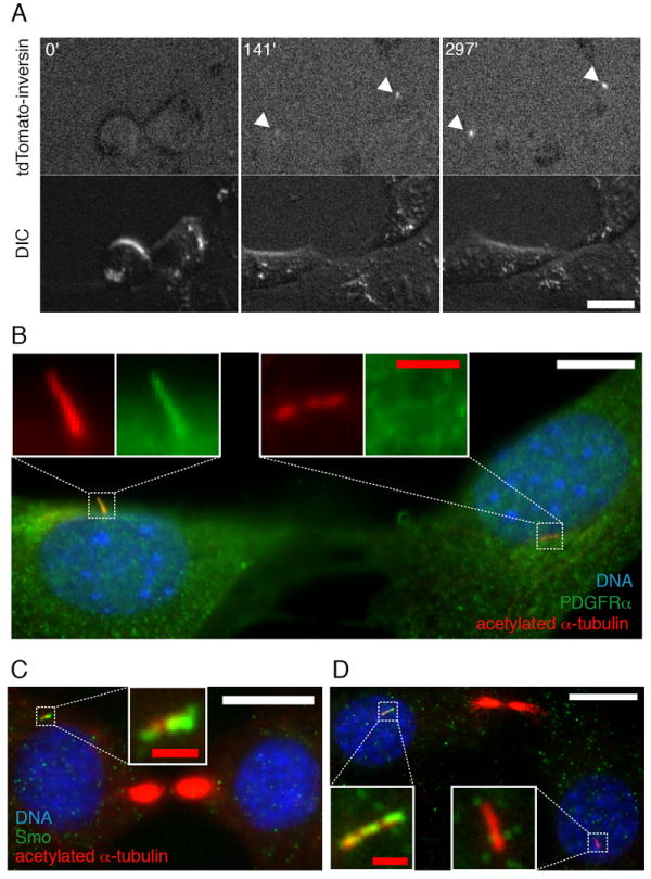
Primary cilia are microtubule-based sensory organelles that play important roles in development and disease . They are required for Sonic hedgehog (Shh) and platelet-derived growth factor (PDGF) signaling. Primary cilia grow from the older of the two centrioles of the centrosome, referred to as the mother centriole. In cycling cells, the cilium typically grows in G1 and is lost before mitosis, but the regulation of its growth is poorly understood. Centriole duplication at G1/S results in two centrosomes, one with an older mother centriole and one with a new mother centriole, that are segregated in mitosis. Here we report that primary cilia grow asynchronously in sister cells resulting from a mitotic division and that the sister cell receiving the older mother centriole usually grows a primary cilium first. We also show that the signaling proteins inversin and PDGFRalpha localize asynchronously to sister cell primary cilia and that sister cells respond asymmetrically to Shh. These results suggest that the segregation of differently aged mother centrioles, an asymmetry inherent to every animal cell division, can influence the ability of sister cells to respond to environmental signals, potentially altering the behavior or fate of one or both sister cells.


Conflict of interest statement

The authors declare that they have no conflicts of interest.

Figures

Figure 1
Figure 1. Primary cilium growth is asynchronous in sister cells, and independent of cytoplasmic differences
(A) Sister pair of NIH/3T3 cells 6 h after mitotic shake-off in which the left cell has a cilium but the right cell does not; acetylated α-tubulin is red, GFP-centrin 2 is green, and DNA is blue. Bar, 10 μm. (B,C) Quantification of ciliogenesis in sister pairs in normal (B) or reduced-serum (0.5%) (C) media. N=300 cells for each timepoint; error bars, s.e.m. from 3 experiments. (D) Binucleate cell 8 h after mitotic shake-off, in which one centriole has a cilium; acetylated α-tubulin is red, GFP-centrin 2 is green, and DNA is blue. Bar, 10 μm. (E) Quantification of ciliogenesis in binucleate NIH/3T3 cells. n=300 cells for each timepoint; error bars, s.e.m. from 3 experiments.
Figure 2
Figure 2. Asynchronous cilium growth is dependent on centriole age
(A) Schematic of centriole pulse-labeling. After transfection, myc-tagged α-tubulin incorporates into new centrioles such that in G2 each of the two centrosomes has an unlabeled mother centriole (gray) and a labeled daughter centriole (green). After duplication and segregation in the second mitosis, there are sister cell pairs in which the mother centriole of one cell is unlabeled (the older mother centriole) and the mother centriole of the other cell is labeled (the newer mother centriole). (B) Sister pair of NIH/3T3 cells (connected by a midbody remnant, arrowhead) 68 h after transfection with myc-α-tubulin. The cell on the left has a myc- (older) mother centriole, and a cilium (left insets); the cell on the right has two myc+ (newer) centrioles, and lacks a cilium (right insets). Acetylated α-tubulin labeling of centrioles and cilia is red, myc-α-tubulin is green, and DNA is blue. In 33 of 35 such pairs in which one primary cilium was present, the cell with the unlabeled (oldest) mother centriole had the primary cilium. White bar, 10 μm; red bar, 1 μm.
Figure 3
Figure 3. The timing of new mother centriole maturation correlates with asynchronous cilium growth
(A) NIH/3T3 cells in mitosis and early G1 in which one mother centriole (left side of each panel) stains more brightly for cenexin/ODF2 than the other. Cenexin/ODF2 is shown alone in the top panels; in the bottom panels, acetylated α-tubulin is red, cenexin/ODF2 is green, and DNA is blue. Bar, 10 μm. In all panels, both centrosomes were in the same focal plane. (B,D) Cells after 18 h etoposide treatment in which both mother centrioles possess primary cilia; acetylated α-tubulin is red, GFP-centrin 2 is green in (B), cenexin/ODF2 is green in (D), and DNA is blue. Bars, 10 μm. (C) Quantitation of the fraction of cells with four centrioles that have 0, 1 or 2 cilia before and after the 18 h etoposide treatment.
Figure 4
Figure 4. Inversin and PDGFRα localize to primary cilia asynchronously, and sister cells respond asymmetrically to Shh
(A) Images from a time lapse recording (Video S1) of sister NIH/3T3 cells expressing tdTomato-inversin. Time shown in minutes; tdTomato-inversin images are above and DIC images below. At time 0’, immediately after mitosis, there is no visible tdTomato-inversin; at 141’ the cell on the right has a pronounced focus of tdTomato-inversin (arrowheads); and by 297’ both cells have a focus of tdTomato-inversin. Bar, 25 μm. (B) Sister pair of NIH/3T3 cells 24 h after mitotic shake-off into 0.5% serum medium in which PDGFRα is present in the primary cilium of one cell (left cell and insets) but not the other (right cell and insets). This asymmetry was found in 39/48 pairs in which both sister cells had primary cilia. Acetylated α-tubulin is red, PDGFRα is green, and DNA is blue. White bar, 10 μm; red bar, 1 μm. (C) Sister pairs of NIH/3T3 cells 24 h after exposure to ShhN conditioned medium. In the left panel (one cilium), the left cell has a Smo+ cilium (see inset) and the right cell lacks a cilium. This asymmetry was observed in 46 of 63 such cell pairs. In the right panel (two cilia), the left cell has a Smo+ cilium (left inset) and the right cell has a Smo- cilium (right inset). This asymmetry was observed in 33 of 96 such cell pairs. This Acetylated α-tubulin is red, Smo is green, and DNA is blue. White bars, 10 μm; red bars, 1 μm.

Similar articles

Cited by

References

    1. Fliegauf M, Benzing T, Omran H. When cilia go bad: cilia defects and ciliopathies. Nat Rev Mol Cell Biol. 2007;8:880–893. - PubMed
    1. Haycraft CJ, Banizs B, Aydin-Son Y, Zhang Q, Michaud EJ, Yoder BK. Gli2 and Gli3 localize to cilia and require the intraflagellar transport protein polaris for processing and function. PLoS Genet. 2005;1:e53. - PMC - PubMed
    1. Corbit KC, Aanstad P, Singla V, Norman AR, Stainier DY, Reiter JF. Vertebrate Smoothened functions at the primary cilium. Nature. 2005;437:1018–1021. - PubMed
    1. Rohatgi R, Milenkovic L, Scott MP. Patched1 regulates hedgehog signaling at the primary cilium. Science. 2007;317:372–376. - PubMed
    1. Schneider L, Clement CA, Teilmann SC, Pazour GJ, Hoffmann EK, Satir P, Christensen ST. PDGFRalphaalpha signaling is regulated through the primary cilium in fibroblasts. Curr Biol. 2005;15:1861–1866. - PubMed

Publication types