. 2009 Oct;30(31):6202-12.
doi: 10.1016/j.biomaterials.2009.07.063. Epub 2009 Aug 15.

Dynamic in vivo biocompatibility of angiogenic peptide amphiphile nanofibers

Affiliations

Dynamic in vivo biocompatibility of angiogenic peptide amphiphile nanofibers

Shahram Ghanaati et al. Biomaterials. 2009 Oct.

Abstract

Biomaterials that promote angiogenesis have great potential in regenerative medicine for rapid revascularization of damaged tissue, survival of transplanted cells, and healing of chronic wounds. Supramolecular nanofibers formed by self-assembly of a heparin-binding peptide amphiphile and heparan sulfate-like glycosaminoglycans were evaluated here using a dorsal skinfold chamber model to dynamically monitor the interaction between the nanofiber gel and the microcirculation, representing a novel application of this model. We paired this model with a conventional subcutaneous implantation model for static histological assessment of the interactions between the gel and host tissue. In the static analysis, the heparan sulfate-containing nanofiber gels were found to persist in the tissue for up to 30 days and revealed excellent biocompatibility. Strikingly, as the nanofiber gel biodegraded, we observed the formation of a de novo vascularized connective tissue. In the dynamic experiments using the dorsal skinfold chamber, the material again demonstrated good biocompatibility, with minimal dilation of the microcirculation and only a few adherent leukocytes, monitored through intravital fluorescence microscopy. The new application of the dorsal skinfold model corroborated our findings from the traditional static histology, demonstrating the potential use of this technique to dynamically evaluate the biocompatibility of materials. The observed biocompatibility and development of new vascularized tissue using both techniques demonstrates the potential of these angiogenesis-promoting materials for a host of regenerative strategies.


Figures

Figure 1
Figure 1
Chemical structures of heparin-binding peptide amphiphile (A) and a fluorescein-conjugated peptide amphiphile (B) used in this study. Additionally, the general chemical structure of heparan sulfate showing the three sites of variable sulfation (C).
Figure 2
Figure 2
Computerized scanning of an entire histological slide produced from an assembly of many high-resolution (100x) single images. This technique allows an for quantitative analysis of the entire implantation bed in a single image. Here, the tissue and material is stained with hematoxylin and eosin, showing that the nanofiber gel structure is maintained at 3 days and demonstrating a favorable peri-implant response.
Figure 3
Figure 3
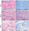
Representative histology for HBPA-heparan sulfate nanofiber gels implanted subcutaneously. The implant bed and surrounding tissue were excised at 3 days, 10 days and 30 days stained with H&E (left) and Azan (right). At 3 days (A and B), the nanofiber gel was primarily homogenous and not infiltrated by immune cells. By 10 days (C and D) the nanofiber gel showed early signs of degradation and converted into a vascularized connective tissue from the periphery, with representative blood vessels indicated by arrows and a macrophage ingesting the nanofiber gel circled in red. At 30 days (E and F) the implantation bed was a well-vascularized connective tissue, with representative blood vessel indicated by arrows. The staining and magnification for each image is shown and all scale bars represent 100 μm.
Figure 4
Figure 4
Quantification of the degradation (A) and vascularization (B) of subcutaneously implanted HBPA-heparan sulfate nanofiber gels, determined through analysis of histological images.
Figure 5
Figure 5
Representative histology for HBPA-phosphate nanofiber gels implanted subcutaneously. The implant bed and surrounding tissue was excised at 3 days (A and B), 10 days (C and D). The nanofiber gel appears as a mosaic of aggregates and has already been infiltrated with immune cells at day 3, and by day 10 shows significant degradation. There is no evidence of new vasculature within the implant bed. The staining and magnification for each image is shown and all scale bars represent 100 μm.
Figure 6
Figure 6
Intravital microscopy to track the vasculature (A, left) and fluorescent nanofiber gel (A, right) of the fluorescent HBPA-Heparan Sulfate nanofiber gel implanted within a dorsal skinfold chamber at 2, 6, and 10 days following implantation. Scale bars represent 500 μm. The same region of microcirculation in an awake mouse was examined at each day, as shown, and the diameter of vessels within the dorsal skinfold chamber adjacent to the HBPA-heparan sulfate nanofiber gel were measured and expressed as a percent of the day 0 diameter for the 4 animals (B). The material is readily apparent within the dorsal skinfold chamber (C), appearing yellow due to the presence of the FITC, with an arrow showing the area of the vasculature displayed in panel A.
Figure 7
Figure 7
Representative intravital microscopy showing the green fluorescent HBPA-Heparan nanofiber gel (A), rhodamine-illuminated leukocytes (B), and brightfield showing the microcirculation (C). In this way, leukocytes adhered to the endothelium in the vicinity of the gel could be tracked to observe the nature of the acute immune response to the nanofiber gel. Scale bars represent 200 μm.
Figure 8
Figure 8
(A) Representative histology from the endpoint of the dorsal skinfold chamber model, displaying the implantation bed stained with H&E. As shown, the HBPA-HS nanofiber gel is enveloped within a newly formed vascularized connective tissue, with arrows indicating representative vessels. (B) High magnification fluorescent image from dorsal HBPA-HS nanofiber gel immediately before sacrifice, with arrows pointing to dark regions indicating blood vessels within the nanofiber gel. Scale bars represent 100 μm (left) and 500 μm (right).

References

    1. Stupp SI. Biomaterials for regenerative medicine. Mrs Bulletin. 2005;30(7):546–553.
    1. Stupp SI, Donners JJJM, Li LS, Mata A. Expanding frontiers in biomaterials. Mrs Bulletin. 2005;30(11):864–873.
    1. Hartgerink JD, Beniash E, Stupp SI. Self-assembly and mineralization of peptide-amphiphile nanofibers. Science. 2001;294(5547):1684–1688. - PubMed
    1. Hartgerink JD, Beniash E, Stupp SI. Peptide-amphiphile nanofibers: a versatile scaffold for the preparation of self-assembling materials. Proc Natl Acad Sci USA. 2002;99(8):5133–5138. - PMC - PubMed
    1. Velichko YS, Stupp SI, de la Cruz MO. Molecular simulation study of peptide amphiphile self-assembly. J Phys Chem B. 2008:2326–2334. - PubMed

Publication types