. 2009 Aug 14;35(3):352-64.
doi: 10.1016/j.molcel.2009.06.015.

Gcn5 and SAGA regulate shelterin protein turnover and telomere maintenance

Affiliations

Gcn5 and SAGA regulate shelterin protein turnover and telomere maintenance

Boyko S Atanassov et al. Mol Cell. .

Abstract

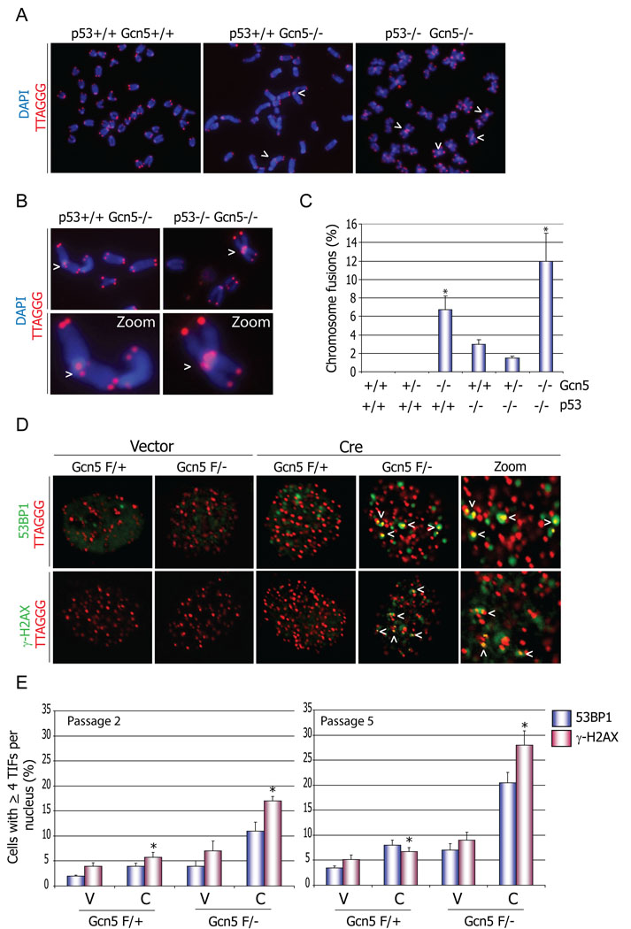
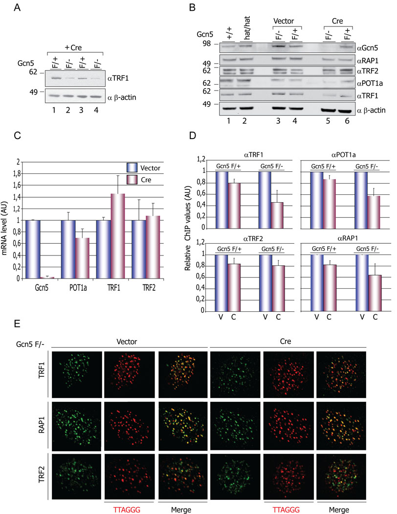
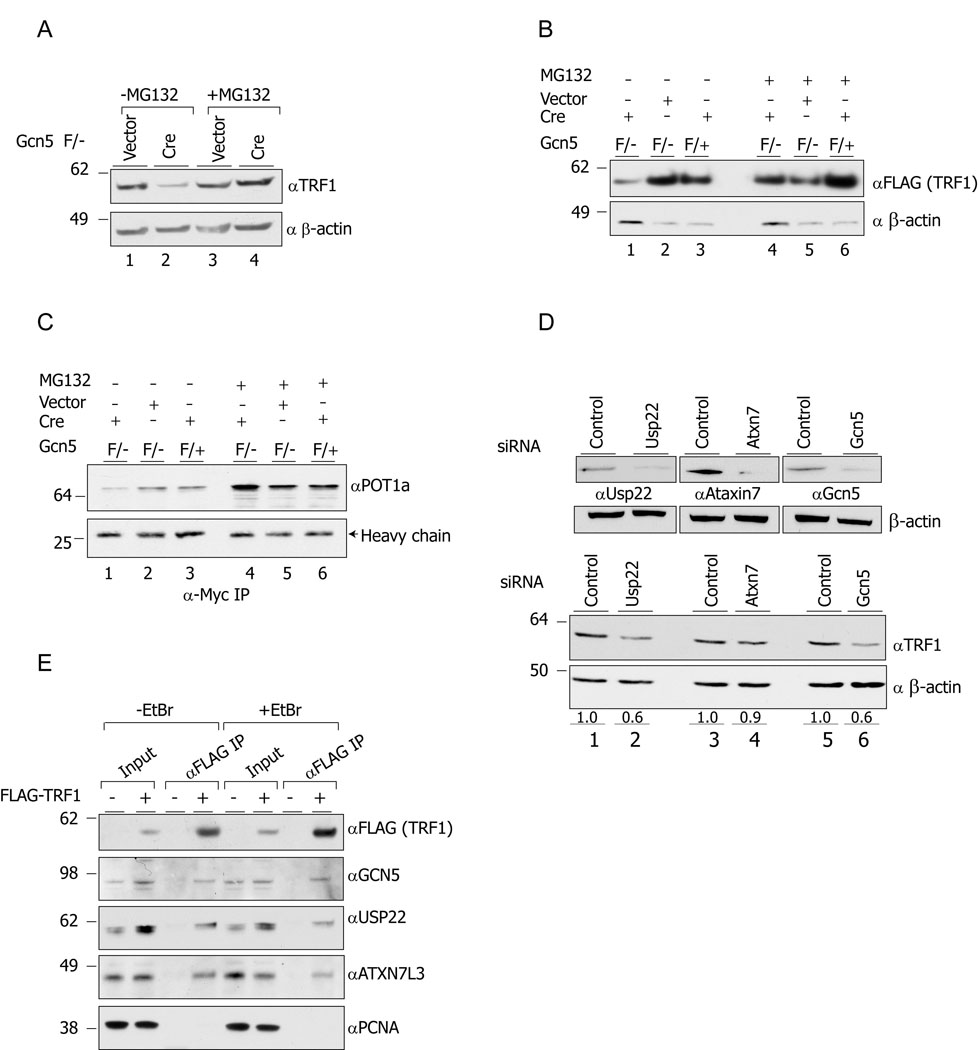
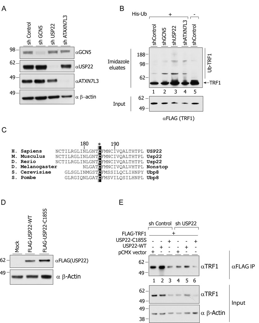
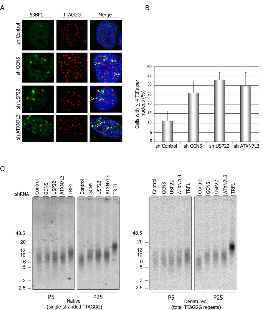
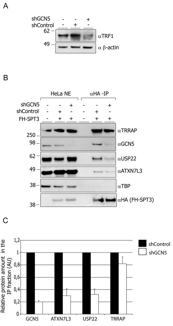
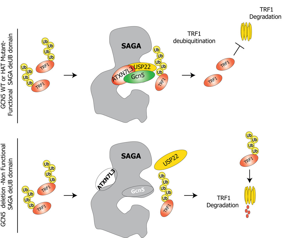
Histone acetyltransferases (HATs) play important roles in gene regulation and DNA repair by influencing the accessibility of chromatin to transcription factors and repair proteins. Here, we show that deletion of Gcn5 leads to telomere dysfunction in mouse and human cells. Biochemical studies reveal that depletion of Gcn5 or ubiquitin-specific protease 22 (Usp22), which is another bona fide component of the Gcn5-containing SAGA complex, increases ubiquitination and turnover of TRF1, a primary component of the telomeric shelterin complex. Inhibition of the proteasome or overexpression of USP22 opposes this effect. The USP22 deubiquitinating module requires association with SAGA complexes for activity, and we find that depletion of Gcn5 compromises this association in mammalian cells. Thus, our results indicate that Gcn5 regulates TRF1 levels through effects on Usp22 activity and SAGA integrity.


Figures

Figure 1
Figure 1. Loss of Gcn5 impairs proper telomere maintenance
(A) Loss of Gcn5 leads to telomere end-to-end fusions in mouse embryonic cells. Cells were isolated from E8.5 embryos with indicated genotypes, and metaphase spreads were subjected to FISH analysis. Chromosomal DNA was stained with DAPI (blue) and telomeres were labeled with a TRITC-conjugated telomere specific probe (TTAGGG). Arrow heads indicate chromosomal end-to-end fusions in cells isolated from Gcn5 null embryos. (B) Close up view of chromosome fusions represented in (A). (C) Quantification of the data represented in A and B. 30 non-apoptotic nuclei from each embryo genotype (n=2) were examined, and the number of chromosome fusions in each metaphase was scored. * p values <0.001 relative to the wild type sample, as determined by students two-tailed t-test (D) Depletion of Gcn5 induces a DNA damage response on the telomeres in MEF cells. Telomere localization of the DNA damage signal, indicated with the arrows, was verified after in situ hybridization with TRITC-conjugated telomere specific probe as above. (E) Quantification of data represented in (D). Approximately one hundred cells from each genotype were counted two and five passages after the initial Vector or Cre transfection. Cells with more than 4 TIFs per nucleus were scored. (error bars in (C) and (E) indicate S.E.M., asterisk, p<0.05 based on a two tailed Student’s t-test). Cells from three independent embryo isolates were used. V = vector and C = Cre.
Figure 2
Figure 2. Decreased levels of Shelterin components in Gcn5 null cells
(A) Decreased levels of TRF1 protein in Gcn5 null MEFs. Protein levels of TRF1 are decreased in Gcn5F/− (lanes 2 and 4) compared to Gcn5F/+ (lanes 1 and 3) MEFs following Cre treatment. (B) Immunoblot data showed decreased TRF1 and POT la protein levels in Gcn5F/− (after Cre treatment) but not in Gcn5F/+ or Gcn5hat/hat cells (lane 5 compared to lanes 1, 2, 3, 4, and 6). Levels of the other shelterin components monitored do not show significant variations in any of the genotypes examined. (C) mRNA level of shelterin components tested by real-time RT-PCR. Experiments were performed with 2 individual MEF cell lines. mRNA level data were normalized by that of GAPDH and vector-treated group values were set as 1. Error bars represent standard deviation from the mean, (D) Decreased amounts on TRF1 and POT la on telomeres. Chromatin immunoprecipitations (ChIPs) were performed using the indicated antibodies and slot blots hybridized with a γ32P-ATP end-labeled TTAGGG probe. The membranes were exposed to Phospholmager screens and the signals quantified with ImageQuant software. Error bars - S.E.M, n=3 independent ChIP experiments. V=vector, C=Cre. (E) Proper localization of TRF1, TRF2 and RAP1 on telomeres. Vector or Cre transfected Gcn5F/− MEFs were immunostained with the indicated antibodies and telomere localization of TRF1, TRF2 and RAP1 was monitored after hybridization with TRITC-conjugated TTAGGG probe.
Figure 3
Figure 3. Gcn5 loss alters TRF1 and POT1a protein turnover
(A) Treatment of the cells with the proteasome inhibitor MG132 stabilizes TRF1 protein levels in Gcn5 null cells (lanes 2 and 4). Lysates from Gcn5 null and Gcn5 heterozygous MEFs, treated with MG132 or DMSO only, were resolved by SDS-PAGE, and TRF1 protein levels were monitored by immunoblot. (B and C) Decreased levels of exogenous FLAG-mTRFl and POT1a-Myc in Gcn5 depleted cells. MEFs with the indicated genotypes were transfected with mTRFl-FLAG expressing vector or infected with POT1a-Myc expressing retroviral vectors. After MG132 or DMSO treatment, lysates were prepared and the exogenous TRF1-FLAG levels were monitored by using anti-FLAG antibody. For monitoring POT1a-Myc protein levels, anti-Myc IP was performed. (D) Depletion of Usp22 has a similar impact on TRF1 protein levels as does Gcn5 depletion (compare lanes 1 and 2 to lanes5 and 6). (E) TRF1 co-immunoprecipitates with GCN5, USP22 and ATXN7L3.
Figure 4
Figure 4. USP22 deUB activity regulates the ubiquitination and steady state levels of TRF1
(A) Efficient depletion of the indicated proteins in 293T cells. (B) Increased ubiquitination of TRF1 upon depletion of GCN5, USP22 or ATXN7L3. Cells were transfected with 6xHis-ubiquitin and FLAG-TRF1 expression vectors and the ubiquitinated species were precipitated by Ni-NTA agarose from nuclear extracts. (C) Schematic representation of Cysteine 185 residue conservation among the USP22 orthologs in different species. (D) Expression of wt -USP22 and C185S–USP22 in HeLa cells. (E) Expression of wt-USP22 restores TRF1 protein levels in USP22 depleted cells, while over expression of C185S–USP22 decreases TRF1 protein levels.
Figure 5
Figure 5. Depletion of hSAGA deUB module induces TIFs and telomere elongation
(A) GCN5, USP22 or ATXN7L3 depleted cells, as well as control shRNA treated cells, were subjected to immunostaining with anti-53BP1 antibody (green) followed by PNA-FISH (red). 100 cells of each sample were counted, and cells with ≥4 TIFs per nucleus (yellow signals indicated with arrowheads) were scored. (B) Quantification of the data represented in (A). Error bars represent standard deviation of the mean (n=2) (C) Telomere elongation after depletion of GCN5, USP22 and ATXN7L3 in HeLA cells. Cells stably expressing denoted shRNAs were harvested at the indicated passages and used for telomere restriction fragment Southern analysis. In-gel hybridizations were done using 32P labeled (GGGTTA)4 or (CCCTAA)4 probes. The hybridized gels were exposed to phosphoimager screens.
Figure 6
Figure 6. Depletion of GCN5 impacts hSAGA integrity
(A) Depletion of GCN5 in HeLa S3 cells by shRNA leads to lowered TRF1 protein levels. (B) Depletion of GCN5 compromises association of USP22 and ATXN7L3 with SAGA. SAGA was purified from nuclear extracts prepared from FLAG-HA-SPT3 expressing HeLa S3 cells. Immunoprecipitated fractions as well as nuclear extracts were resolved by SDS-PAGE and blotted with the indicated antibodies. (C) Quantification of the data from multiple experiments like that shown in (B). X-ray films were scanned and the images were quantified using ImageQuant software. Error bars represent standard deviations of the mean (n=4)
Figure 7
Figure 7
A proposed model for the role of GCN5 and SAGA in shelterin protein turnover. SAGA deUb module stabilizes TRF1 protein levels by deubiquitination, thereby inhibiting degradation. Depletion of GCN5 or ATXN7L3 leads to destabilization of the deubiquitination module, which in turn leads to altered steady state levels of TRF1.

Comment in

Similar articles

Cited by

References

    1. Allis CD, Berger SL, Cote J, Dent S, Jenuwien T, Kouzarides T, Pillus L, Reinberg D, Shi Y, Shiekhattar R, et al. New nomenclature for chromatin-modifying enzymes. Cell. 2007;131:633–636. - PubMed
    1. Bonnet J, Romier C, Tora L, Devys D. Zinc-finger UBPs: regulators of deubiquitylation. Trends Biochem Sci. 2008;33:369–375. - PubMed
    1. Brownell JE, Zhou J, Ranalli T, Kobayashi R, Edmondson DG, Roth SY, Allis CD. Tetrahymena histone acetyltransferase A: a homolog to yeast Gcn5p linking histone acetylation to gene activation. Cell. 1996;84:843–851. - PubMed
    1. Bu P, Evrard YA, Lozano G, Dent SY. Loss of Gcn5 acetyltransferase activity leads to neural tube closure defects and exencephaly in mouse embryos. Mol Cell Biol. 2007;27:3405–3416. - PMC - PubMed
    1. Candau R, Berger SL. Structural and functional analysis of yeast putative adaptors. Evidence for an adaptor complex in vivo. J Biol Chem. 1996;271:5237–5245. - PubMed

Publication types

MeSH terms