. 2009 Oct 23;393(2):435-47.
doi: 10.1016/j.jmb.2009.08.021. Epub 2009 Aug 14.

The 1.9 a structure of human alpha-N-acetylgalactosaminidase: The molecular basis of Schindler and Kanzaki diseases

Affiliations

The 1.9 a structure of human alpha-N-acetylgalactosaminidase: The molecular basis of Schindler and Kanzaki diseases

Nathaniel E Clark et al. J Mol Biol. .

Abstract

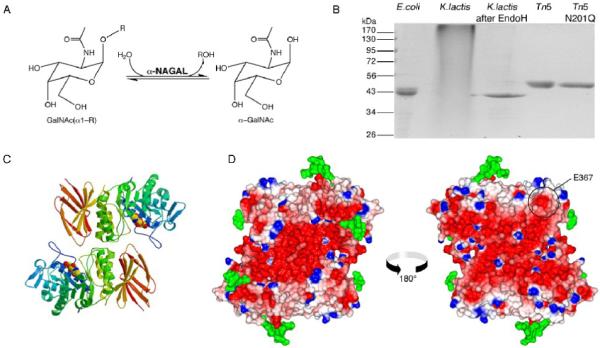
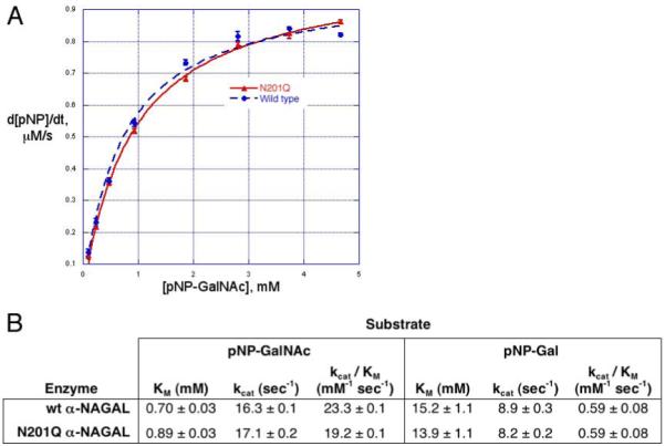
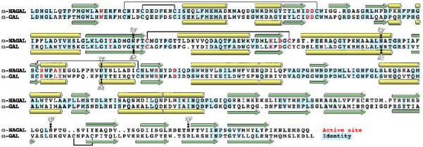
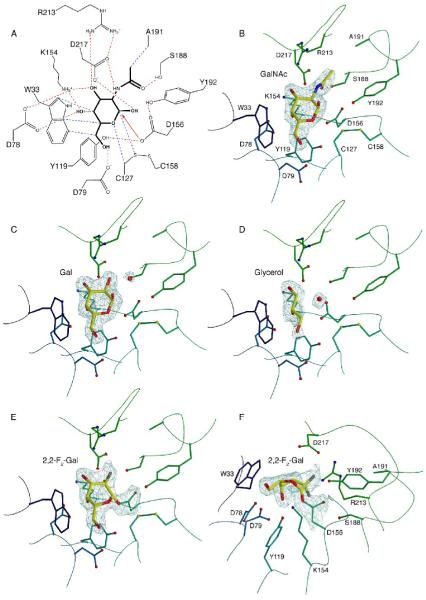
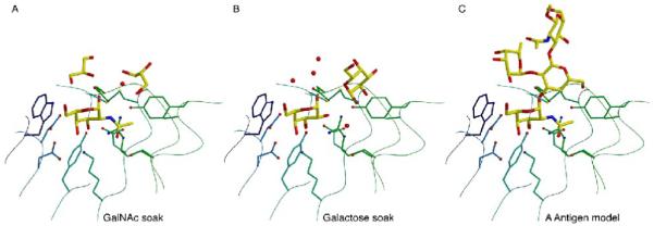
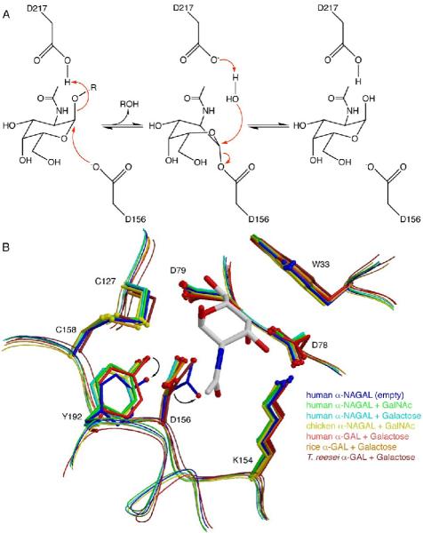
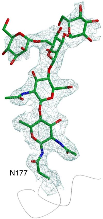
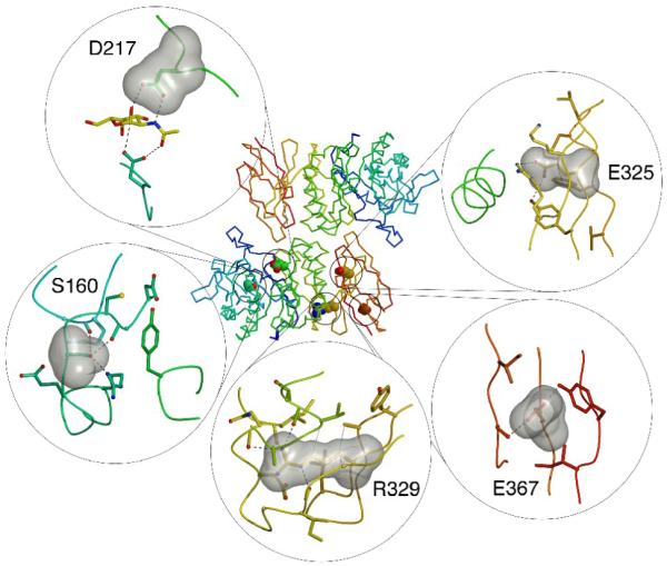
alpha-N-acetylgalactosaminidase (alpha-NAGAL; E.C. 3.2.1.49) is a lysosomal exoglycosidase that cleaves terminal alpha-N-acetylgalactosamine residues from glycopeptides and glycolipids. In humans, a deficiency of alpha-NAGAL activity results in the lysosomal storage disorders Schindler disease and Kanzaki disease. To better understand the molecular defects in the diseases, we determined the crystal structure of human alpha-NAGAL after expressing wild-type and glycosylation-deficient glycoproteins in recombinant insect cell expression systems. We measured the enzymatic parameters of our purified wild-type and mutant enzymes, establishing their enzymatic equivalence. To investigate the binding specificity and catalytic mechanism of the human alpha-NAGAL enzyme, we determined three crystallographic complexes with different catalytic products bound in the active site of the enzyme. To better understand how individual defects in the alpha-NAGAL glycoprotein lead to Schindler disease, we analyzed the effect of disease-causing mutations on the three-dimensional structure.


Figures

Figure 1
Figure 1
α-NAGAL reaction and overall structure A. The reaction catalyzed by α-NAGAL. Both substrate and product are in the α anomeric configuration. B. Expression and purification of recombinant human α-NAGAL from different sources. Lane1: E. coli expressed α-NAGAL, Ni-affinity purified from inclusion bodies. Lanes 2 and 3: K. lactis expressed α-NAGAL before and after deglycosylation with Endo H. Lane 4 and 5: Tn5 expression of wild type α-NAGAL and N201Q α-NAGAL (with the 3rd carbohydrate site removed). C. A ribbon diagram of the human α-NAGAL dimer with the enzymatic product α-GalNAc in the active sites. D. An electrostatic map of the dimer showing contoured from -10 kT/e (red) to +10 kT/e (blue). Carbohydrates are shown in green. The left image is in the same orientation as in C, and the right image is rotated 180° about a vertical axis. The surface exposed residue E367 is circled.
Figure 2
Figure 2
Enzyme kinetics of wild type and N201Q A. Michaelis-Menten plot of wild type and N201Q human α-NAGAL. Production of pNP from pNP-α-GalNAc was monitored by OD at 400nm. B. Summary of kinetic data on human α-NAGAL. The wild type and N201Q mutant glycoproteins have similar kinetic parameters. The specificity constant kcat/KM for each protein is 30-40 fold greater for the GalNAc substrate compared to the galactose substrate.
Figure 3
Figure 3
Structural alignment of human α-NAGAL and human α-GAL The structurally derived sequence alignment shows identities (blue), α helices (yellow), β strands (green), active site residues (red), disulfide bonds (black lines), and N-linked carbohydrates (branched groups).
Figure 4
Figure 4
Active site interactions and ligand binding A. Active site interactions with the GalNAc ligand. Hydrogen bonds are shown in red, van der Waals interactions in blue, with the initial nucleophilic attack shown as a red arrow. B. Active site residues with the GalNAc ligand. Residues are colored as in Figure 2C, and the ligand is shown with σA-weighted 2Fo-Fc electron density contoured at 2σ. C. The galactose ligand with σA-weighted 2Fo-Fc electron density contoured at 1.5σ. D. The glycerol ligand with σA-weighted 2Fo-Fc electron density contoured at 1.2σ. E and F. Two view of the electron density for the 2,2-difluoro-galactose ligand covalently attached to the catalytic nucleophile D156. The map is a σA-weighted 2Fo-Fc electron density contoured at 2.0σ.
Figure 5
Figure 5
Model of blood group A antigen binding A. Glycerol molecules cluster around the active site of the GalNAc ligand. B. Galactose and glycerol molecules bound near the active site when galactose is soaked into the active site. C. A docked model of the blood group A antigen bound to the active site, where the locations of the atoms in the model mimic the position of the small molecules bound around the active site of the crystal structures in panels A and B.
Figure 6
Figure 6
Reaction mechanism and inactive conformation A. The double displacement reaction mechanism in human α-NAGAL. D156 acts as the nucleophile and D217 acts as the acid/base. The ligand is bent into a high energy 1S3 skew boat conformation during the reaction. B. Human α-NAGAL without a soaked ligand shows a different conformation from all of the previous structures in the family. The residues D156 and Y192 shift into conformations where the nucleophilic lone pair rotates into a catalytically inactive location.
Figure 7
Figure 7
Glycosylation of residue N177 The entire paucimannose carbohydrate is visible in the electron density (a σA-weighted 2Fo-Fc electron density contoured at 1.2σ).
Figure 8
Figure 8
Schindler disease mutations Five residues are shown in the center panel showing the location of the residue in the polypeptide fold. The panels show a surface representation of the ligand as well as the surrounding residues. Hydrogen bonds are shown as black dashed lines and van der Waals interactions as grey dotted lines.

References

    1. van Diggelen OP, Schindler D, Kleijer WJ, Huijmans JM, Galjaard H, Linden HU, Peter-Katalinic J, Egge H, Dabrowski U, Cantz M. Lysosomal α-N-acetylgalactosaminidase deficiency: a new inherited metabolic disease. Lancet. 1987;2:804. - PubMed
    1. Schindler D, Bishop DF, Wolfe DE, Wang AM, Egge H, Lemieux RU, Desnick RJ. Neuroaxonal dystrophy due to lysosomal α-N-acetylgalactosaminidase deficiency. N. Engl. J. Med. 1989;320:1735–40. - PubMed
    1. Desnick RJ, Schindler D. α-N-Acetylgalactosaminidase Deficiency: Schindler Disease. In: Scriver CR, Beaudet AL, Sly WS, Valle D, editors. The Metabolic and Molecular Bases of Inherited Disease. 8th edit. McGraw-Hill; New York: 2001. pp. 3483–3505.
    1. van Diggelen OP, Schindler D, Willemsen R, Boer M, Kleijer WJ, Huijmans JG, Blom W, Galjaard H. α-N-acetylgalactosaminidase deficiency, a new lysosomal storage disorder. J. Inherit. Metab. Dis. 1988;11:349–57. - PubMed
    1. Wang AM, Schindler D, Desnick R. Schindler disease: the molecular lesion in the α-N-acetylgalactosaminidase gene that causes an infantile neuroaxonal dystrophy. J. Clin. Invest. 1990;86:1752–6. - PMC - PubMed

Publication types

Substances