

Cerebellum development during childhood and adolescence: a longitudinal morphometric MRI study
- PMID: 19683586
- PMCID: PMC2775156
- DOI: 10.1016/j.neuroimage.2009.08.016
Cerebellum development during childhood and adolescence: a longitudinal morphometric MRI study
Abstract
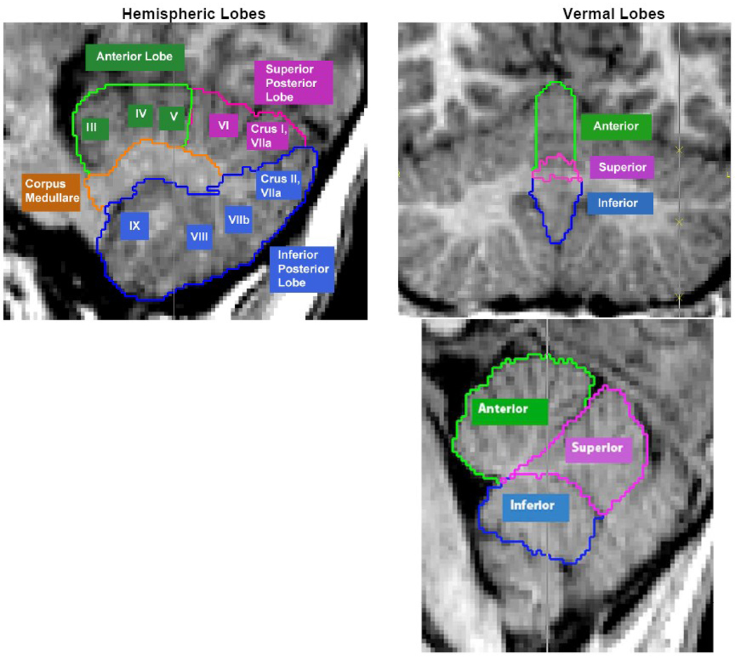
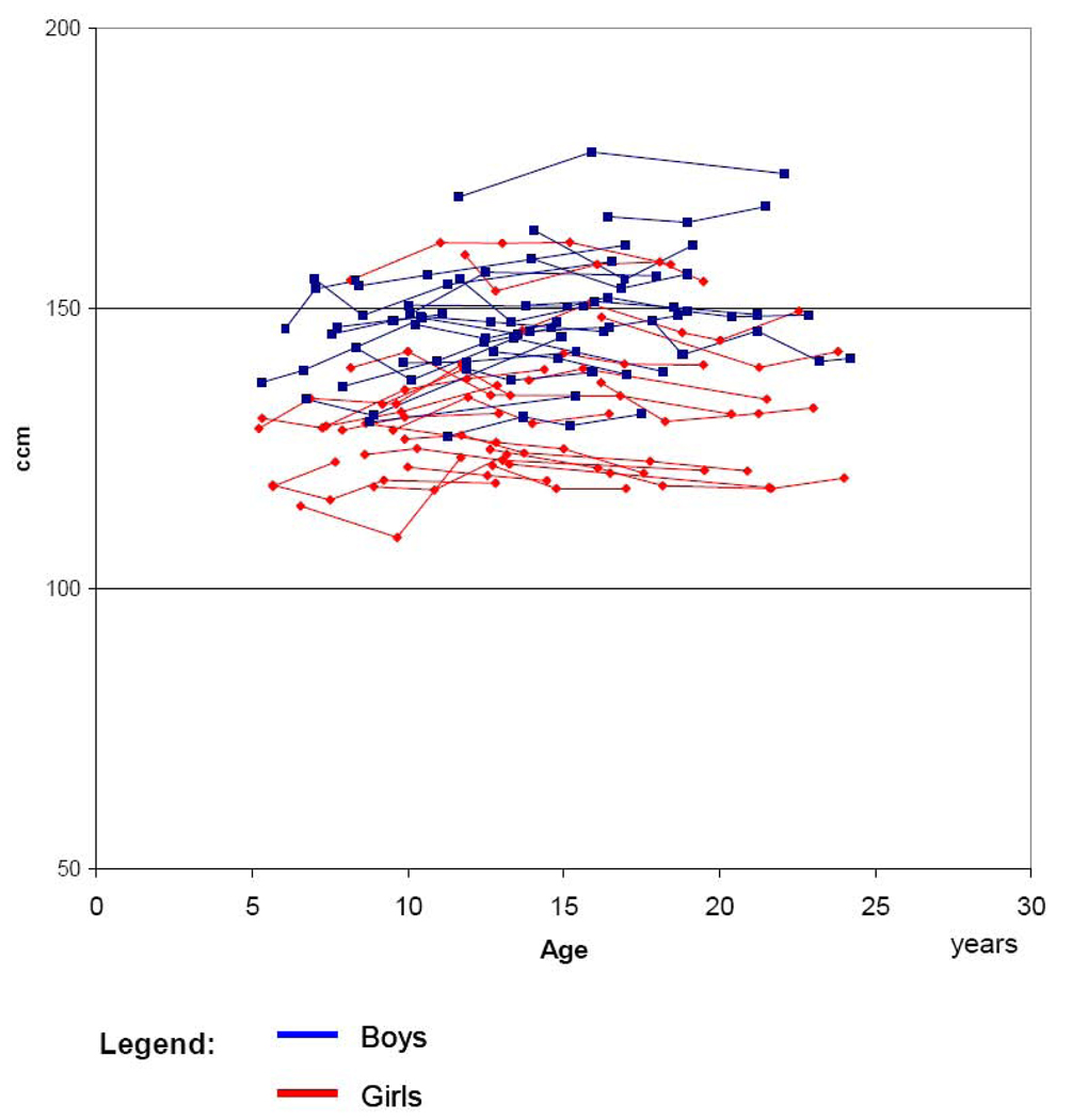
In addition to its well-established role in balance, coordination, and other motor skills, the cerebellum is increasingly recognized as a prominent contributor to a wide array of cognitive and emotional functions. Many of these capacities undergo dramatic changes during childhood and adolescence. However, accurate characterization of co-occurring anatomical changes has been hindered by lack of longitudinal data and methodologic challenges in quantifying subdivisions of the cerebellum. In this study we apply an innovative image analysis technique to quantify total cerebellar volume and 11 subdivisions (i.e. anterior, superior posterior, and inferior posterior lobes, corpus medullare, and three vermal regions) from anatomic brain MRI scans from 25 healthy females and 25 healthy males aged 5-24 years, each of whom was scanned at least three times at approximately 2-year intervals. Total cerebellum volume followed an inverted U shaped developmental trajectory peaking at age 11.8 years in females and 15.6 years in males. Cerebellar volume was 10% to 13% larger in males depending on the age of comparison and the sexual dimorphism remained significant after covarying for total brain volume. Subdivisions of the cerebellum had distinctive developmental trajectories with more phylogenetically recent regions maturing particularly late. The cerebellum's unique protracted developmental trajectories, sexual dimorphism, preferential vulnerability to environmental influences, and frequent implication in childhood onset disorders such as autism and ADHD make it a prime target for pediatric neuroimaging investigations.
Figures
 Boys
Boys  Girls Observations from one individual are connected by lines
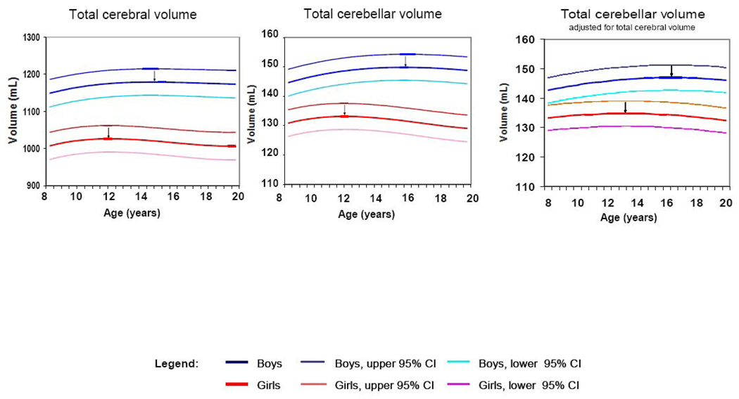
 Boys —— Boys, upper 95% CI
  Boys, lower 95% CI Girls
  Girls, upper 95% CI
  Girls, lower 95% CI
  Indicates age of peak volume
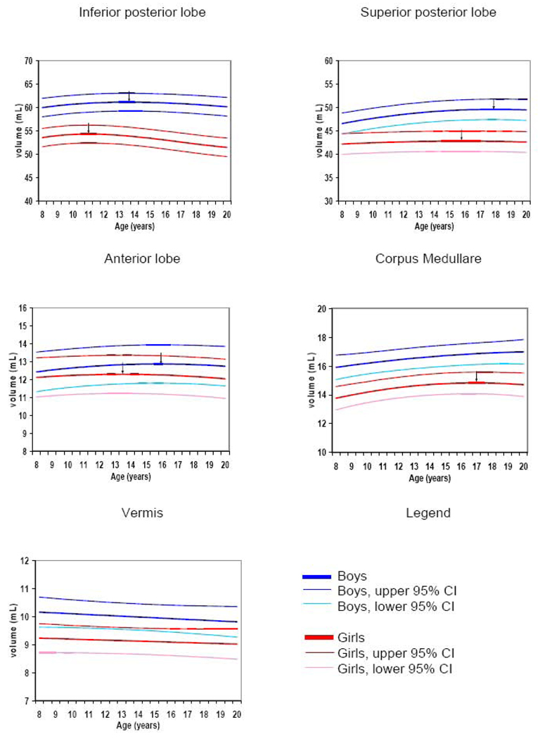 Boys —— Boys, upper 95% CI Boys, lower 95% CI Girls Girls, upper 95% CI Girls, lower 95% CI Indicates age of peak volume
References
-
- Achenbach TM, Edelbrock CS. Manual for child behavior checklist and revised behavior profile. Burlington, VT: Department of Psychiatry, University of Vermont; 1983.
-
- Bishop DV. Cerebellar abnormalities in developmental dyslexia: cause, correlate or consequence? Cortex. 2002;38:491–498. - PubMed
-
- Caviness VS, Jr, Kennedy DN, Richelme C, Rademacher J, Filipek PA. The human brain age 7–11 years, A volumetric analysis based on Magnetic Resonance Images. Cerebral Cortex. 1996;6:726–736. - PubMed
-
- Chechik G, Meilijson I, Ruppin E. Neuronal regulation: A mechanism for synaptic pruning during brain maturation. Neural Comput. 1999;11:2061–2080. - PubMed
Publication types
MeSH terms
Grants and funding
LinkOut - more resources
Full Text Sources
