. 2009 Sep-Oct;78(2-3):116-23.
doi: 10.1016/j.diff.2009.07.001. Epub 2009 Aug 15.

Generation of primordial germ cells from pluripotent stem cells

Affiliations

Generation of primordial germ cells from pluripotent stem cells

Cristina Eguizabal et al. Differentiation. 2009 Sep-Oct.

Abstract

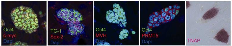
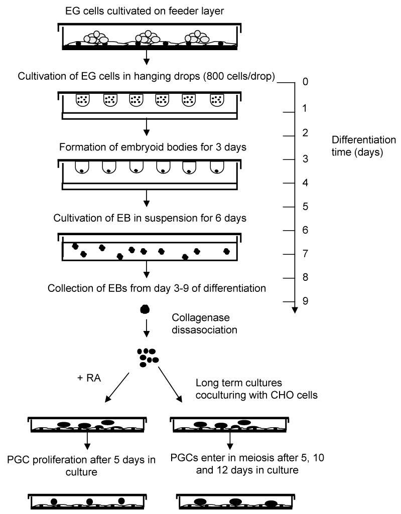
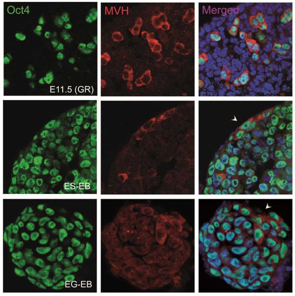
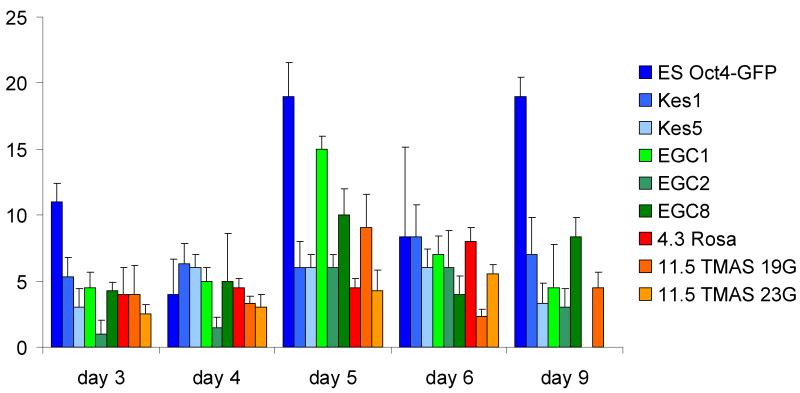
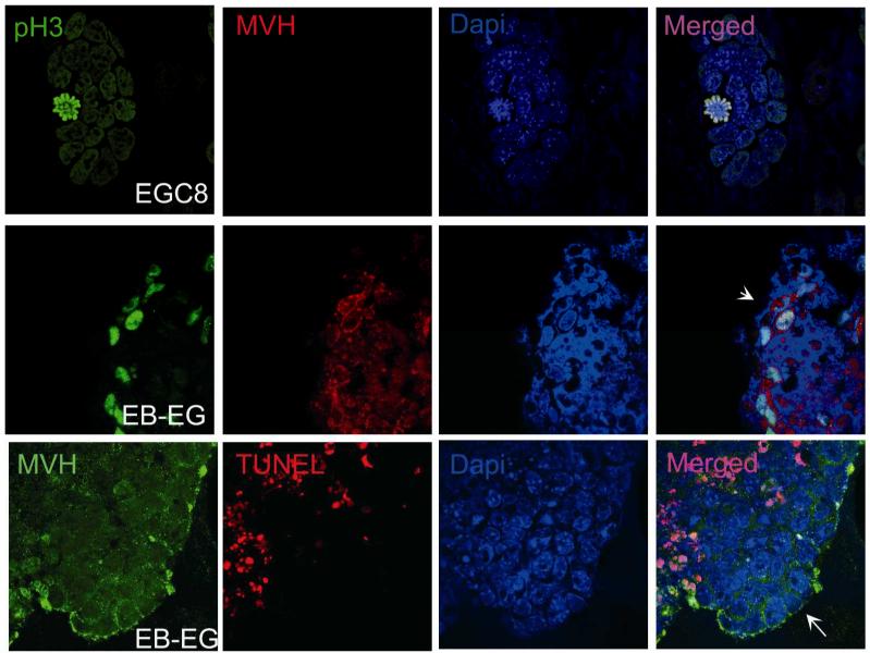
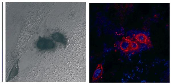
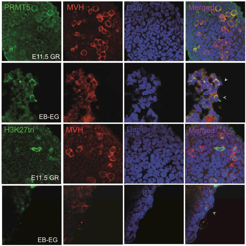
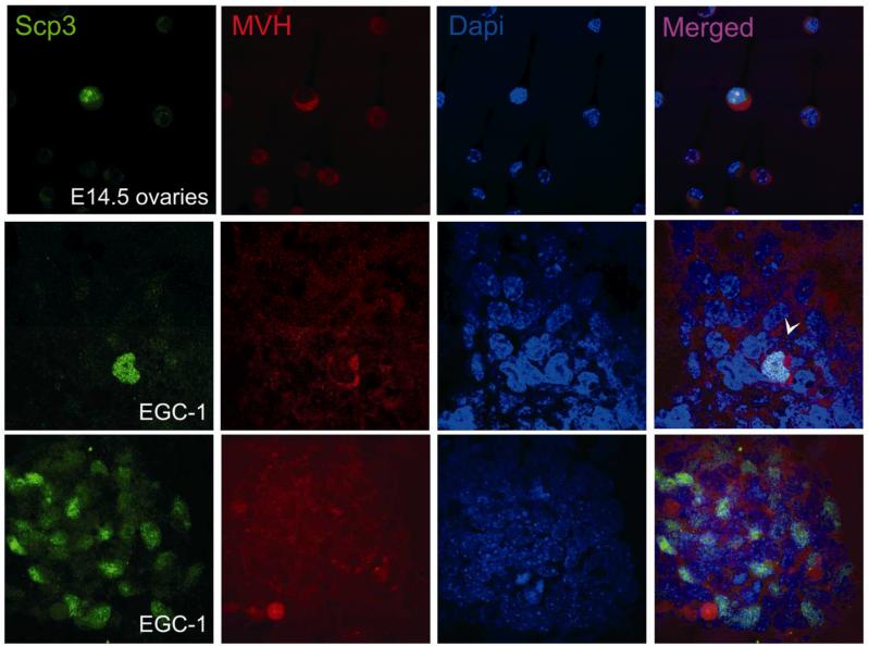
Embryonic stem (ES) cells, derived from pre-implantation embryo, embryonic germ (EG) cells, derived from embryonic precursors of gametes, primordial germ cells (PGCs), can differentiate into any cell type in the body. Moreover, ES cells have the capacity to differentiate into PGCs in vitro. In the present study we have shown the differentiation capacity of six EG cell lines to form PGCs in vitro, in comparison to ES cells. Cell lines were differentiated via embryoid body (EB) formation using the co-expression of mouse vasa homolog (Mvh) and Oct-4 to identify newly formed PGCs in vitro. We found an increase of PGC numbers in almost all analysed cell lines in 5-day-old EBs, thus suggesting that EG and ES cells have similar efficiency to generate PGCs. The addition of retinoic acid confirmed that the cultures had attained a PGC-like identity and continued to proliferate. Furthermore we have shown that the expression pattern of Prmt5 and H3K27me3 in newly formed PGCs is similar to that observed in embryonic day E11.5 PGCs in vivo. By co-culturing EBs with Chinese hamster ovary (CHO) cells some of the PGCs entered into meiosis, as judged by Scp3 expression. The derivation of germ cells from pluripotent stem cells in vitro could provide an invaluable model system to study both the genetic and epigenetic programming of germ cell development in vivo.


Figures

Figure 1
Figure 1. Characterisation of EG cell lines
Representative immunofluorescence analysis of EG cell lines grown on a feeder layer. Clear expression of EG surface marker SSEA-1, nuclear transcription factors Oct4, Sox-2 as well as c-myc and Prmt5 proteins were observed. Also we observed all colonies positives for TNAP. By contrast all colonies were negative for Mvh.
Figure 2A
Figure 2A. Schematic illustrating the in vitro differentiation of EG cells
Figure 2B
Figure 2B. Identification of PGCs in 5 days old bodies
EBs derivated from ES and EG cells were cryosectioned and stained for Oct4 (green) as a pluripotency marker and Mvh (red) to detect a specific germ cell marker. We used genital ridges (GR) from 11.5 dpc as a positive control. Arrowheads indicate some PGCs positives for both markers in EBs generated from EG and ES cells.
Figure 3A
Figure 3A. Development of PGCs at several time points by differentiating EBs made from early and late EG cell lines
Bars represent the number of Mvh positive cells per EB. We used several ES, 8.5-11.5 EG cells.
Figure 3B
Figure 3B. Proliferation and apoptosis in newly formed PGCs from EG cells
To detect proliferation in EBs derivated from EG cells were cryosectioned and stained with phosphorilated Histone 3 (PHH3) (green) as a mitotic marker and Mvh (red) to detect a specific germ cell marker. We used EG cell line as a positive control. Then we used TUNEL reaction to detect apoptosis (red) in combination with Mvh (green). Arrowhead shows proliferating PGCs and the arrow indicates non apoptotic cells during the in vitro PGC formation.
Figure 4
Figure 4. Generation of PGCs after culturing 5 days old bodies with RA treatment for 5 days
We cultured EBs formed from EG cells with RA during 5 days. Then, we combined AP staining with immunofluoresce technique, using Mvh (red) marker to demonstrate both typical markers of PGCs.
Figure 5
Figure 5. Epigenetic status of in vitro PGCs
Expression of several histone modification markers of late PGCs, as Prmt5 and H3K27-tri (green) in combination with another specific germ cell marker, Mvh (red). We used genital ridges (GR) from 11.5 dpc as a positive control. Arrowheads indicate the expression of both histone modifications in these newly formed PGCs are similar to in vivo 11.5 PGCs.
Figure 6
Figure 6. Expression of a meiotic marker in long-term cultures of EBs
Immunofluorescence of 5 days old bodies derived from EG and ES cells co-cultured with CHO cells for 5 days. We observed positive cells for Scp3 (green) and Mvh (red) markers indicating enter into meiosis (arrowhead). We used single cell suspension of fetal ovaries from 14.5 dpc as a Scp3 and Mvh controls.

Similar articles

Cited by

References

    1. Ancelin K, Lange UC, Hajkova P, Schneider R, Bannister AJ, Kouzarides T, Surani MA. Blimp1 associates with Prmt5 and directs histone arginine methylation in mouse germ cells. Nat Cell Biol. 2006;8:623–630. - PubMed
    1. Beddington RS, Robertson EJ. An assessment off the developmental potential of embryonic stem cells in the midgestation mouse embryos. Development. 1989;105:733–737. - PubMed
    1. Chuva de Sousa Lopes SM, Hayashi K, Shovlin TC, Mifsud W, Surani MA, McLaren A. X Chromosome activity in Mouse XX primordial germ cells. PLoS Genetics. 2008;4(2):30–37. - PMC - PubMed
    1. De Felici M, Scaldaferri ML, Lobascio M, Iona S, Nazzicone V, Klinger FG, Farini D. Experimental approaches to the study of primordial germ cell lineage and proliferation. Hum Reprod Update. 2004;10(3):197–206. - PubMed
    1. Donovan PJ, De Miguel MP. Turning germ cells into stem cells. Curr Opin Genet Dev. 2003;13(5):463–471. - PubMed

Publication types

LinkOut - more resources