Review
. 2009 Aug;19(4):395-401.
doi: 10.1016/j.conb.2009.07.010. Epub 2009 Aug 15.

Development of form and function in the mammalian cochlea

Affiliations
Review

Development of form and function in the mammalian cochlea

Michael C Kelly et al. Curr Opin Neurobiol. 2009 Aug.

Abstract

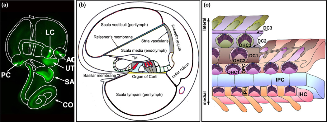
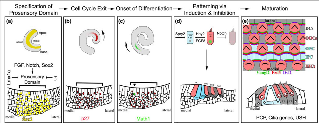
The cochlea possesses specialized features to receive sound signals and to resolve and convert the frequency and intensity components within each signal for auditory perception. It consists of precisely patterned and polarized sensory cells adorned with a highly specialized mechanotransduction apparatus for sensitivity and adaptation, and discrete nonsensory cellular networks for biochemical and mechanical support to drive an integrated cellular response and mechanotransduction. This review summarizes recent discoveries about the roles of FGF, Notch, and Hedgehog signaling and transcriptional factors in the differentiation and patterning of the auditory sensory organ, the Usher complex, and the planar cell polarity pathway in the formation and polarization of mechanotransduction component hair bundles, and the contribution of nonsensory cell networks in the stria vascularis and the sensory region toward the maturation of the mammalian cochlea.


Figures

Figure 1
Figure 1
The cochlea. (A) The inner ear consists of the cochlea (CO) and the vestibule. The cochlea has one sensory organ, the organ of Corti, which is marked by green fluorescent protein (green). The vestibule has five sensory organs: the maculae of the saccule (SA), the utricle (UT), and three cristae (AC, PC, and LC) that are also marked by green fluorescent protein (green). (B) A diagram of the cross-section of the cochlea, illustrating its three chambers and partitioning of the endolymphatic and perilymphatic fluids. The scala vestibule and scala tympani are filled with the perilymph while the scala media is filled with the endolymph. The stria vascularis contributes significantly to the unusual ion content of the endolymph. TM: tectorial membrane. The sensory hair cells are shown in red. (C) A schematic diagram of the whole-mount organ of Corti. In the organ of Corti, the inner (IHCs) and outer hair cells (OHCs) are interdigitated with several types of distinct nonsensory supporting cells: the inner phalangeal cells (IPHs), inner pillar cells (IPCs), outer pillar cells (OPCs), and three rows of Deiters’ cells (DC1–DC3s). A single kinocilium (blue) and numerous stereocilia (purple) constitute a ‘V’-shaped hair bundle on the apical surface of each nascent hair cell. All of the ‘V’-shaped hair bundles are uniformly aligned, showing a distinctive PCP.
Figure 2
Figure 2
The development of the auditory sense organ. (A) The cochlear duct begins as an expansion of the ventral-most portion of the otocyst. Initial specifications of prosensory domain most likely begin as soon as the cochlear duct is recognizable. The prosensory domain is further restricted to a group of cells on the floor of the cochlea duct (shown with bracket) by the action of Sox2, Notch and Hedgehog (HH) signaling, and Lmx1a. Diagrams for the whole-mount cochlea (top) and the cross-section of the cochlea at this stage highlight the expression of Sox2 (yellow) in the prosensory domain. (B) The sensory precursor cells become postmitotic under the control of several cyclin-dependent kinase inhibitors, including p27. Cell cycle exit is initiated in the cells in the apical region and progresses toward the basal region, coinciding with the gradient of p27 onset in the cochlea. The arrow indicates the direction of onset of p27 and the gradient of cell cycle withdrawal along the longitudinal axis of the cochlear duct. (C) The first sign of differentiation within the postmitotic prosensory domain is the expression of the transcription factor Math1, which starts in the midbasal region and progresses toward both the apex and the base of the cochlea. There is also a second gradient along the medial-lateral axis of the cochlea from the inner to outer hair cells. The arrow indicates the longitudinal gradient of Math1 onset and hair cell differentiation. (D) Inductive and inhibitory signaling creates the correct cellular patterning of the organ of Corti. Much of this appears to be mediated by Notch signaling, which inhibits hair cell neighbors from adopting the same fate. Furthermore, the initial differentiation of the inner hair cells appears to direct the differentiation of other cells types, such as the Pillar cells (blue) through FGF8 and Hey2 in a Notch-independent manner. Sprouty 2 (SPY2) further restricts the differentiation of pillar cells. (E) During terminal differentiation and maturation, all cells in the organ of Corti coordinate their cellular morphologies under the regulation of the planar cell polarity (PCP) signaling pathway, which, in part, involves the asymmetric distribution of a core set of proteins (some examples are shown). The result of PCP signaling in the ear can be most clearly observed by the uniform orientation of the ‘V’-shaped hair bundles on the apical surfaces of hair cells. In mice, late embryonic and early postnatal hair cell and supporting cell types all undergo morphological and maturational changes that ultimately result in a highly sensitive sensory structure that is functional by two weeks after birth.

References

    1. Dallos P, Popper AN, Fay RR, editors. Springer Handbook of Auditory Research. Vol. 8. New York: Springer; 1996.
    1. Rida PC, Chen P. Line up and listen: planar cell polarity regulation in the mammalian inner ear. Semin Cell Dev Biol. 2009 - PMC - PubMed
    1. Lelli A, Asai Y, Forge A, Holt JR, Geleoc GS. Tonotopic gradient in the developmental acquisition of sensory transduction in outer hair cells of the mouse cochlea. J Neurophysiol. 2009;101:2961–2973. - PMC - PubMed
    1. Muller U. Cadherins and mechanotransduction by hair cells. Curr Opin Cell Biol. 2008;20:557–566. - PMC - PubMed
    1. Hudspeth AJ. Making an effort to listen: mechanical amplification in the ear. Neuron. 2008;59:530–545. - PMC - PubMed

Publication types