. 2009 Oct 9;284(41):28104-28114.
doi: 10.1074/jbc.M109.044925. Epub 2009 Aug 14.

Human copper transporter 1 lacking O-linked glycosylation is proteolytically cleaved in a Rab9-positive endosomal compartment

Affiliations

Human copper transporter 1 lacking O-linked glycosylation is proteolytically cleaved in a Rab9-positive endosomal compartment

Edward B Maryon et al. J Biol Chem. .

Abstract

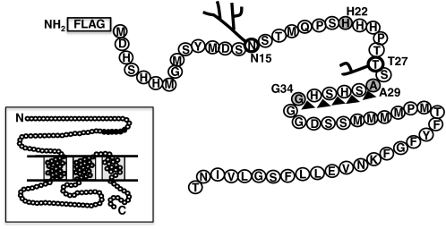
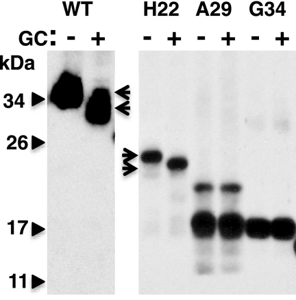
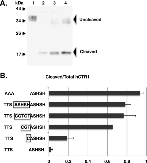
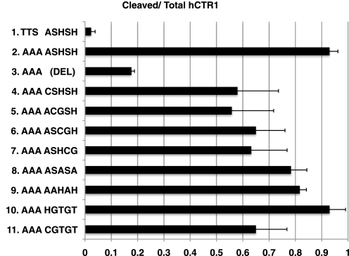
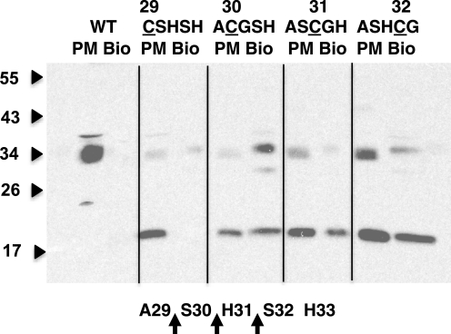
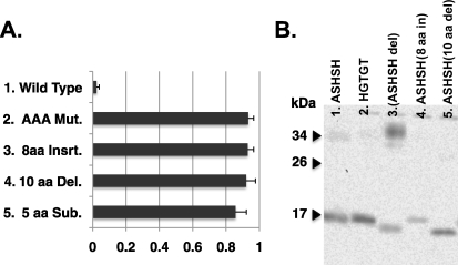
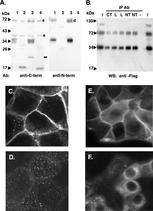
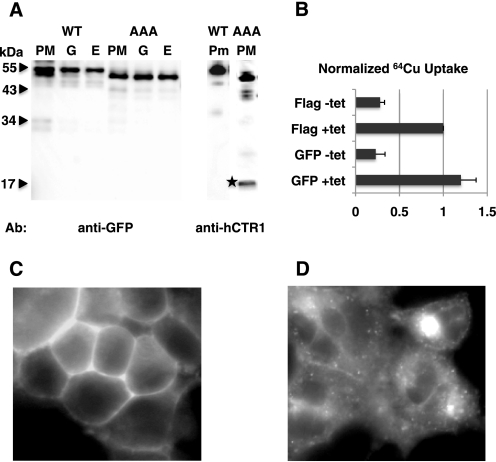
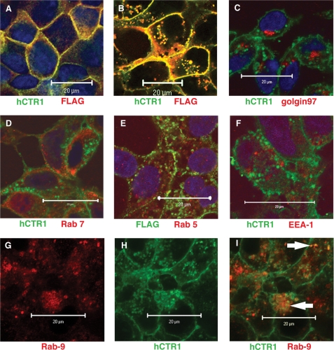
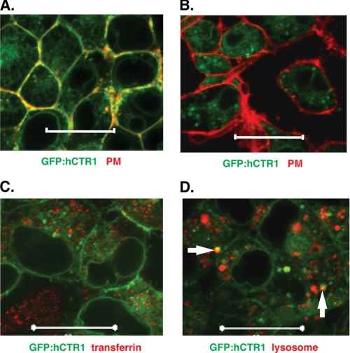
The human copper transporter hCTR1 is a homotrimer composed of a plasma membrane protein of 190 amino acids that contains three transmembrane segments. The extracellular 65-amino acid amino terminus of hCTR1 contains both N-linked (at Asn(15)) and O-linked (at Thr(27)) sites of glycosylation. If O-glycosylation at Thr(27) is prevented, hCTR1 is efficiently cleaved, removing approximately 30 amino acids from the amino terminus. We have now investigated (i) the site of this cleavage, determining which peptide bonds are cleaved, (ii) the mechanism by which glycosylation prevents cleavage, and (iii) where in the cell the proteolytic cleavage takes place. Cleavage occurs in the sequence Ala-Ser-His-Ser-His (residues 29-33), which does not contain previously recognized protease cleavage sites. Using a series of hCTR1 mutants, we show that cleavage occurs preferentially between residues Ala(29)-Ser(30)-His(31). We also show that the O-linked polysaccharide at Thr(27) blocks proteolysis due to its proximity to the cleavage site. Moving the cleavage site away from the Thr(27) polysaccharide by insertion of as few as 5 amino acids allows cleavage to occur in the presence of glycosylation. Imaging studies using immunofluorescence in fixed cells and a functional green fluorescent protein-tagged hCTR1 transporter in live cells showed that the cleaved peptide accumulates in punctate structures in the cytoplasm. These puncta overlap compartments were stained by Rab9, indicating that hCTR1 cleavage occurs in a late endosomal compartment prior to delivery of the transporter to the plasma membrane.


Figures

FIGURE 1.
FIGURE 1.
Extracellular amino terminus of hCTR1. Location of N- and O-linked glycosylation at Asn15 and Thr27, and the end points of 3 truncation mutants in gray: H22, A29, and G34. In the absence of O-glycosylation at Thr27, hCTR1 is efficiently cleaved between A29 and G34 (black triangles). Location of the FLAG epitope tag is shown. Inset shows the complete 190-amino acid hCTR1 protein, with extracellular NH2 terminus, three membrane spanning domains, intracellular loop, and COOH-terminal tail. The 5 amino acids in which cleavage occurs are shown in black. Three hCTR1 polypeptides form a symmetrical trimer in the copper transporter (8, 9).
FIGURE 2.
FIGURE 2.
O-Glycosidase treatment of hCTR1 proteins. Wild-type, H22, A29, and G34 proteins were overexpressed in HEK-293 cells and recovered in plasma membrane fractions. Arrows show the shift of wild-type and H22 after treatment with a mixture of glycosylases specific for O-linked polysaccharides (GC, see Ref. 17). The A29 mutant, which is not O-glycosylated, is mostly cleaved to yield a 17-kDa fragment migrating close to the G34 truncation. The uncleaved A29 mutant contains a FLAG tag. G34 has an initiator methionine at the amino terminus. All lanes were taken from the same gel.
FIGURE 3.
FIGURE 3.
Effect on cleavage of hCTR1 of inserting amino acids between Thr27 and the ASHSH cleavage site. A, example Western blot used to determine the extent of cleavage. The blot was probed with anti-COOH-terminal hCTR1 antibody (6). Plasma membranes (10 μg of protein) from cells expressing: lane 1, wild-type hCTR1; lane 2, AAA mutant hCTR1; lane 3, hCTR1 mutant having a 3-amino acid insertion (CGT) between the O-glycosylation site and ASHSH cleavage site; and lane 4, hCTR1 mutant with a 5-amino acid insertion (CGTGT). B, extent of cleavage of the hCTR1 proteins. The AAA mutant lacking O-glycosylation is shown at top. Wild-type hCTR1 is shown at the bottom. Between are mutants with insertions between Thr27 and the ASHSH cleavage site. Inserted amino acids are boxed in 4 mutants. Error bars represent the S.D. of three to five determinations for each mutant.
FIGURE 4.
FIGURE 4.
Extent of cleavage of hCTR1 mutants within the ASHSH cleavage site. Each mutation in ASHSH has in addition an AAA substitution for the TTS tripeptide that contains the site of O-linked glycosylation in wild-type. Wild-type is shown at the top. Below the wild-type is the AAA mutant with ASHSH (mutant 2). Mutant 3 is a deletion of ASHSH. Mutants 4–11 are various substitutions within the ASHSH cleavage site, as shown, in addition to the AAA mutation. Error bars represent the S.D. of three to six determinations for each mutant.
FIGURE 5.
FIGURE 5.
Determination of cleaved peptide bonds. Cells expressing each hCTR1 Cys substitution mutant were biotinylated with a sulfhydryl reagent, lysed, and supernatants were mixed with avidin beads to recover labeled proteins (lanes marked Bio). 10 μg of plasma membrane protein from the same cells was run to the left of each biotinylated sample (PM). The presence of the 17-kDa band in lanes marked Bio indicates cleavage of hCTR1 to the left of the single cysteine in each mutant, as shown below in the diagram. Lines were drawn on the gel image for clarity.
FIGURE 6.
FIGURE 6.
A, cleavage of mutants on the carboxyl side of the ASHSH cleavage site. Rows 1 and 2, wild-type and AAA mutant hCTR1 proteins, respectively; row 3, 8 Ins., the peptide APARPNAG was inserted between G35 and G36 (see Fig. 1); row 4, 10-amino acid del, the peptide SSMMMMPMTF, amino acids 38–47, were deleted in this mutant; row 5, 5-amino acids sub, the methionine cluster MMMMPM (amino acids 40–45) was changed to AAAAPA. Error bars represent the S.D. of three determinations for each mutant. B, the 17-kDa cleavage products of deletion and insertion mutants. Migration of these fragments is altered based on the insertion or loss of amino acids compared with fragments from substitution mutants shown in lanes 1 or 2.
FIGURE 7.
FIGURE 7.
Protease inhibitors and hCTR1 cleavage. Cleavage of hCTR1 AAA mutant protein expressed in HEK-293 cells that were exposed to the indicated protease inhibitors during a 48-h tet induction. Concentration of inhibitors used are shown: aprotinin, 100 units/ml; leupeptin, 20 μm; pepstatin, 10 μm; GM6001, 20 μm; TAPI-1, 10 μm; MG132, 2 μm. Higher concentrations of MG132 caused cell death. Error bars represent the S.D. of three to six determinations for each mutant (*, p < 0.05; **, p < 0.01, t test).
FIGURE 8.
FIGURE 8.
Anti-hCTR1 amino-terminal antibody. A, Western blots using anti-hCTR1 antibodies raised against either the amino or carboxyl terminus of hCTR1, as indicated below each blot. 10 μg of plasma membrane protein from cells overexpressing hCTR1 variants were run in each lane. Lane 1, FLAG-tagged WT hCTR1; lane 2, AAA mutant hCTR1 (mostly cleaved); lane 3, WT hCTR1; lane 4, G34, a truncated hCTR1 lacking the first 33 amino acids of hCTR1. *, nonspecific cross-reactive bands. Arrow, proteolytic fragment sometimes observed in wild-type hCTR1 preparations. d, position of dimer form of wild-type. Faint bands in lane 2 are monomer and dimer forms of unglycosylated AAA mutant. B, IP of monomer and dimer forms of FLAG-tagged, overexpressed hCTR1. hCTR1 membrane proteins were precipitated with hCTR1 antibodies raised against the carboxyl terminus (CT), the intracellular loop (L), or the amino terminus (NT). −, no antibody control. Lanes marked I are 50% of input protein amount for each IP. C–F, immunostaining with anti-hCTR1 amino-terminal antibody. MDCK cells expressing wild-type hCTR1 (C) or AAA mutant hCTR1 (D) and HEK-293 cells expressing wild-type (E) or AAA mutant hCTR1 (F) are shown.
FIGURE 9.
FIGURE 9.
GFP-hCTR1 fusion proteins. A, Western blot analysis of overexpressed GFP-hCTR1 fusion proteins. Cells expressing either GFP-WT hCTR1 fusion protein or AAA mutant fusion were fractionated, and membranes enriched for endoplasmic reticulum (E), golgi (G), or plasma membranes (PM) were probed with anti-GFP antibodies (left panel). Plasma membrane fractions were also probed with anti-COOH-terminal hCTR1 antibody on the right. The two lanes shown are from the same gel. The star shows the 17-kDa cleavage product. B, 64Cu uptake in cells expressing tet-regulated FLAG-tagged wild-type hCTR1 or GFP-tagged hCTR1. Values are normalized to tet-induced FLAG-tagged hCTR1 to correct for differences in expression. Shown are the averages of three uptake experiments, with error bars showing S.D. C, live cell image of HEK-293 cells expressing a GFP-hCTR1 wild-type fusion protein; D, live cell image of HEK-293 cells expressing a GFP-hCTR1 AAA mutant fusion protein.
FIGURE 10.
FIGURE 10.
Double labeling immunofluorescence with anti-hCTR1 (or anti-FLAG) and organelle marker antibodies. A, HEK-293 and MDCK cells expressing wild-type hCTR1 and B, HEK-293 cells expressing AAA mutant hCTR1 co-stained with anti-amino-terminal hCTR1 antibody and anti-FLAG antibody. C–F, HEK-293 cells expressing AAA mutant hCTR1. In each panel, anti-hCTR1 (or anti-FLAG in E) is in green, whereas organelle-specific antibodies are in red. Organelle-specific antibodies are: C, golgin97 (cis, medial golgi); D, Rab7, (early, late endosome); E, Rab5 (early endosome, recycling endosome, plasma membrane); and F, EEA-1 (early endosome). G–I, HEK-293 cells expressing AAA mutant hCTR1 stained with: Rab9 (G) (late endosome, in red), anti-hCTR1 (H) in green, and merged images (I). White arrows in I show the overlap in yellow. Scale bars, 20 μm.
FIGURE 11.
FIGURE 11.
Live cell imaging of GFP-hCTR1 fusion proteins with cell markers. A, GFP-hCTR1 wild-type fusion expressed in HEK-293 cells stained with plasma membrane dye, in red; B, GFP-hCTR1 AAA mutant fusion protein with plasma membrane dye; C, GFP-hCTR1 AAA mutant fusion protein in cells labeled with (endocytosed) fluorescent transferrin; D, GFP-hCTR1 AAA mutant fusion protein in cells labeled with a lysosome-specific stain. White arrows indicate overlap between the GFP and lysosome, stained in yellow. Scale bars, 20 μm.

Similar articles

Cited by

References

    1. Petris M. J. (2004) Pflugers Arch. 447, 752–755 - PubMed
    1. Linder M. C., Wooten L., Cerveza P., Cotton S., Shulze R., Lomeli N. (1998) Am. J. Clin. Nutr. 67, Suppl. 5, 965S–971S - PubMed
    1. Maryon E. B., Molloy S. A., Zimnicka A. M., Kaplan J. H. (2007) Biometals 20, 355–364 - PubMed
    1. Kim B. E., Nevitt T., Thiele D. J. (2008) Nat. Chem. Biol. 4, 176–185 - PubMed
    1. Huffman D. L., O'Halloran T. V. (2001) Annu. Rev. Biochem. 70, 677–701 - PubMed

Publication types

MeSH terms