. 2009 Oct 23;284(43):29437-45.
doi: 10.1074/jbc.M109.048181. Epub 2009 Aug 14.

Radial spoke protein 3 is a mammalian protein kinase A-anchoring protein that binds ERK1/2

Affiliations

Radial spoke protein 3 is a mammalian protein kinase A-anchoring protein that binds ERK1/2

Arif Jivan et al. J Biol Chem. .

Abstract

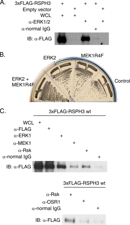
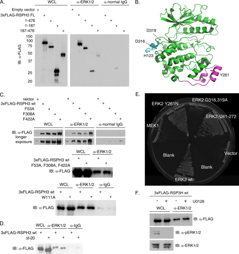
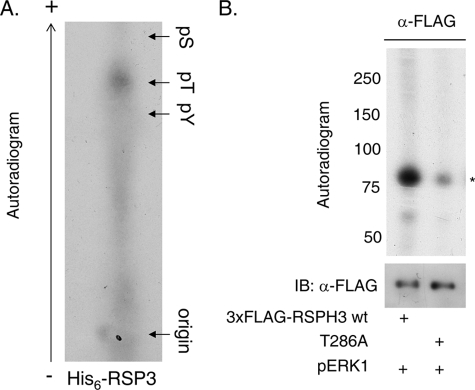
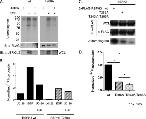
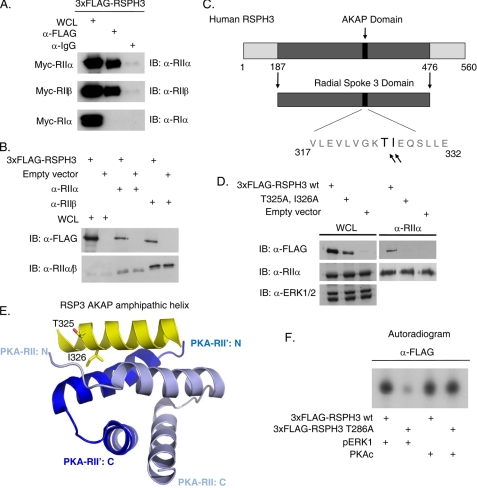
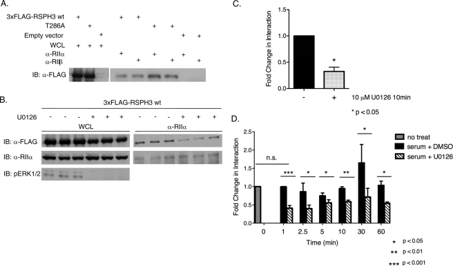
Initially identified in Chlamydomonas, RSP3 (radial spoke protein 3) is 1 of more than 20 identified radial spoke structural components of motile cilia and is required for axonemal sliding and flagellar motility. The mammalian orthologs for this and other radial spoke proteins, however, remain to be characterized. We found mammalian RSP3 to bind to the MAPK ERK2 through a yeast two-hybrid screen designed to identify interacting proteins that have a higher affinity for the phosphorylated, active form of the protein kinase. Consistent with the screening result, the human homolog, RSPH3, interacts with and is a substrate for ERK1/2. Moreover, RSPH3 is a protein kinase A-anchoring protein (AKAP) that scaffolds the cAMP-dependent protein kinase holoenzyme. The binding of RSPH3 to the regulatory subunits of cAMP-dependent protein kinase, RIIalpha and RIIbeta, is regulated by ERK1/2 activity and phosphorylation. Here we describe an ERK1/2-interacting AKAP and suggest a mechanism by which cAMP-dependent protein kinase-AKAP binding can be modulated by the activity of other enzymes.


Figures

FIGURE 1.
FIGURE 1.
RSPH3 binds to ERK1/2 and other cascade components. A, endogenous ERK1/2 were immunoprecipitated from HEK293 cell lysates expressing 3xFLAG-RSPH3 or empty vector (negative control) and immunoblotted (IB) with the anti-FLAG antibody. Lanes labeled whole cell lysate (WCL) contain 10% of the input for immunoprecipitation. Immunoprecipitation with normal rabbit IgG was used as a negative control. B, yeast were co-transformed with plasmids containing mouse RSP3 and ERK2, MEK1R4F, a co-expression plasmid containing ERK2 plus MEK1R4F, or empty vector as a control. C, endogenous ERK1/2, MEK1, and Rsk (top) or Rsk and OSR1 (bottom) were immunoprecipitated from cells transfected with 3xFLAG-RSPH3. Precipitates were immunoblotted with anti-FLAG antibodies. Normal IgG was immunoprecipitated as control. The whole cell lysate lane contains 5% of the input for immunoprecipitation. Experiments in A–C were repeated 10, 2, and 3 times.
FIGURE 2.
FIGURE 2.
ERK CD region is involved in the RSPH3 interaction. A, endogenous ERK1/2 was immunoprecipitated from HEK293 cells expressing 3xFLAG-tagged RSPH3 truncation mutants, residues 1–476, 1–187, and 187–476. Immunoprecipitation with normal rabbit IgG was used as a negative control. Precipitates were immunoblotted (IB) with anti-FLAG antibody. The whole cell lysate (WCL) lane contains 5% of the input for the immunoprecipitations. B, ribbon representation of ERK2 (green) bound via its CD domain to a peptide derived from the hematopoietic tyrosine phosphatase HePTP (cyan) (Protein Data Bank code 2gph). The MAPK insert is highlighted in magenta, with Tyr-261 indicated. C, three putative FXF motifs within 3xFLAG-RSPH3 were mutated singly (top panel) or in combination (middle panel), and a WXY motif within 3xFLAG-RSPH3 was mutated (bottom), and all were tested for ERK1/2 binding. Endogenous ERK1/2 immunoprecipitates were immunoblotted with anti-FLAG. Immunoprecipitation from cells transfected with empty vector or with normal rabbit IgG were used as negative controls. The whole cell lysate lane contains 5% of the input for immunoprecipitation. D, experiment as in C with a truncation of the first 20 residues of 3xFLAG-RSPH3. E, pairwise yeast two-hybrid analysis of mouse RSP3 interaction with wild-type (wt) ERK2 or ERK2 mutants. Yeast containing RSP3 were co-transformed with wild-type MEK1 or empty vector as controls. F, HEK293 cells expressing 3xFLAG-RSPH3 were treated with 10 μm U0126 or diluent for 10 min prior to lysis. Endogenous ERK1/2 were immunoprecipitated, and the precipitates were immunoblotted (IB) with anti-FLAG. The whole cell lysate lane contains 5% of the input. Phosphorylated and total ERK1/2 were immunoblotted in the lysates. n = 3. Experiments in each panel were performed three times, except the two-hybrid test which was performed twice.
FIGURE 3.
FIGURE 3.
ERK1/2 phosphorylate RSPH3. A, phosphoamino acid analysis of His6-RSP3 phosphorylated in vitro with pERK1. An autoradiogram is shown. B, 3xFLAG-RSP3H wild-type or T286A was immunoprecipitated from HEK293 cells and used as substrate for an in vitro kinase reaction with purified, recombinant pERK1. Mutating T286A decreased 32P incorporation by ∼70% (n = 6). pS, phosphoserine; pT, phosphothreonine; pY, phosphotyrosine.
FIGURE 4.
FIGURE 4.
RSPH3 phosphorylation dependent on the ERK1/2 pathway. A, HEK293 expressing 3xFLAG-RSP3H wild-type (wt) or T286A were placed in serum-free medium for 20 h and then labeled with 1 mCi/ml [32P]orthophosphate with 10 μm U0126 or diluent for 60 min, and then the indicated groups were treated with 10 ng/ml EGF for 10 min. Autoradiograms of the 3xFLAG-RSP3H immunoprecipitates are shown. n = 3. IB, immunoblot; WCL, whole cell lysate. B, quantification of 32P incorporation into the RSP3H bands from one experiment. Incorporation was normalized to the U0126-treated RSPH3 wild-type sample and adjusted for differences in protein expression as assessed by immunoblotting and densitometry. C and D, 3xFLAG-RSP3H wild-type, T286A, or T243V/T286A was immunoprecipitated from HEK293 cells and used as substrate for in vitro kinase assays with pERK1. Graph represents fold 32P incorporation into RSP3H wild-type compared with the mutants (mean + S.E.). Protein amount in the immunoprecipitates was measured via densitometric analysis. Incorporation was normalized to that into the wild-type protein and corrected for differences in protein amount. n = 3. Mean values for T286A and T243V/T286A RSPH3 were 0.32 and 0.21, respectively. Statistical significance was evaluated with a nonparametric Kruskal-Wallis analysis.
FIGURE 5.
FIGURE 5.
RSPH3 is AKAP. A, 3xFLAG-RSPH3 was co-expressed with Myc-tagged RIIα, RIIβ, or RIα PKA regulatory subunits in HEK293 cells. RSPH3 was immunoprecipitated with the anti-FLAG antibody and immunoblotted (IB) with antibodies against the regulatory subunits. n = 3. WCL, whole cell lysate. B, endogenous RIIα and RIIβ were immunoprecipitated from HEK293 cells expressing 3xFLAG-RSPH3. Co-immunoprecipitation was detected by immunoblotting with anti-FLAG antibodies. Empty vector transfected lysates were used as a negative control. The whole cell lysate lane contains 10% of the input. n = 4. C, schematic of human RSPH3 showing the RII-binding sequence of the AKAP domain, based on sequence similarity to the Chlamydomonas ortholog. D, endogenous RIIα was immunoprecipitated from the HEK293 cells expressing 3xFLAG-RSPH3 wild type (wt) or the AKAP-defective mutant, T325A/I326A. Samples were immunoblotted with the indicated antibodies. n = 4. E, ribbon diagram depicting the amphipathic AKAP helix in RSPH3 that mediates the interaction with the PKA regulatory subunit dimerization interface. F, overexpressed 3xFLAG-RSP3H wild type or T286A was immunoprecipitated from HEK293 cells and used as substrate for an in vitro kinase reaction with purified recombinant pERK1 or purified catalytic subunit of PKA (PKAc). n = 3.
FIGURE 6.
FIGURE 6.
ERK1/2 activity modulates the AKAP function of RSPH3. A, endogenous RIIα or RIIβ was immunoprecipitated from HEK293 cells expressing 3xFLAG-RSPH3 wild type (wt), T286A, or empty vector. Samples were immunoblotted (IB) with anti-FLAG antibody. Fold change in interaction (as described under “Results”) was determined using a ratiometric analysis of the intensity of the RSPH3 band relative to the whole cell lysate (WCL) for each protein. The experiment was performed three times each for RIIa and for RIIb. Statistical significance of the fold change in interaction was determined using a two-tailed t test, p value = 0.011. B, HEK293 cells expressing wild-type 3xFLAG-RSP3H were placed in serum-free medium for 12 h prior to stimulating with serum in the presence of 10 μm U0126 or diluent for 10 min. Endogenous RIIα was immunoprecipitated from the lysates, and the material was immunoblotted with the indicated antibodies. Data of B are shown in C as mean + S.E., n = 3. The fold change in interaction was based on the ratiometric analysis, and values were normalized to the DMSO-treated control. Statistical significance was analyzed using a two-tailed t test, p value = 0.0139. D, HEK293 cells expressing 3xFLAG-RSP3H were stimulated with serum with 10 μm U0126 or DMSO for the indicated times. RIIα was immunoprecipitated, and the precipitates were blotted with anti-FLAG. Fold change was determined as above. Samples were normalized to the untreated control. Data represent mean + S.E., n = 3. Statistical significance was analyzed using a two-tailed t test with p values as indicated.

Similar articles

Cited by

  • General and specific promotion of flagellar assembly by a flagellar nucleoside diphosphate kinase.
    Zhu X, Poghosyan E, Gopal R, Liu Y, Ciruelas KS, Maizy Y, Diener DR, King SM, Ishikawa T, Yang P. Zhu X, et al. Mol Biol Cell. 2017 Nov 1;28(22):3029-3042. doi: 10.1091/mbc.E17-03-0156. Epub 2017 Sep 6. Mol Biol Cell. 2017. PMID: 28877983 Free PMC article.
  • Complex modular architecture around a simple toolkit of wing pattern genes.
    Van Belleghem SM, Rastas P, Papanicolaou A, Martin SH, Arias CF, Supple MA, Hanly JJ, Mallet J, Lewis JJ, Hines HM, Ruiz M, Salazar C, Linares M, Moreira GRP, Jiggins CD, Counterman BA, McMillan WO, Papa R. Van Belleghem SM, et al. Nat Ecol Evol. 2017;1(3):52. doi: 10.1038/s41559-016-0052. Epub 2017 Jan 30. Nat Ecol Evol. 2017. PMID: 28523290 Free PMC article.
  • The mouse radial spoke protein 3 is a nucleocytoplasmic shuttling protein that promotes neurogenesis.
    Yan R, Hu X, Zhang W, Song L, Wang J, Yin Y, Chen S, Zhao S. Yan R, et al. Histochem Cell Biol. 2015 Oct;144(4):309-19. doi: 10.1007/s00418-015-1338-y. Epub 2015 Jun 17. Histochem Cell Biol. 2015. PMID: 26082196
  • Genome-wide interaction study of smoking and bladder cancer risk.
    Figueroa JD, Han SS, Garcia-Closas M, Baris D, Jacobs EJ, Kogevinas M, Schwenn M, Malats N, Johnson A, Purdue MP, Caporaso N, Landi MT, Prokunina-Olsson L, Wang Z, Hutchinson A, Burdette L, Wheeler W, Vineis P, Siddiq A, Cortessis VK, Kooperberg C, Cussenot O, Benhamou S, Prescott J, Porru S, Bueno-de-Mesquita HB, Trichopoulos D, Ljungberg B, Clavel-Chapelon F, Weiderpass E, Krogh V, Dorronsoro M, Travis R, Tjønneland A, Brenan P, Chang-Claude J, Riboli E, Conti D, Gago-Dominguez M, Stern MC, Pike MC, Van Den Berg D, Yuan JM, Hohensee C, Rodabough R, Cancel-Tassin G, Roupret M, Comperat E, Chen C, De Vivo I, Giovannucci E, Hunter DJ, Kraft P, Lindstrom S, Carta A, Pavanello S, Arici C, Mastrangelo G, Karagas MR, Schned A, Armenti KR, Hosain GM, Haiman CA, Fraumeni JF Jr, Chanock SJ, Chatterjee N, Rothman N, Silverman DT. Figueroa JD, et al. Carcinogenesis. 2014 Aug;35(8):1737-44. doi: 10.1093/carcin/bgu064. Epub 2014 Mar 24. Carcinogenesis. 2014. PMID: 24662972 Free PMC article.
  • Heterogeneity of radial spoke components in Tetrahymena cilia.
    Bicka M, Ghanaeian A, Black C, Joachimiak E, Osinka A, Majhi S, Konopka A, Bulska E, Bui KH, Wloga D. Bicka M, et al. Cell Mol Life Sci. 2025 Aug 31;82(1):329. doi: 10.1007/s00018-025-05871-x. Cell Mol Life Sci. 2025. PMID: 40886204 Free PMC article.

References

    1. Raman M., Chen W., Cobb M. H. (2007) Oncogene 26, 3100–3112 - PubMed
    1. Elion E. A. (2001) J. Cell Sci. 114, 3967–3978 - PubMed
    1. Kolch W. (2005) Nat. Rev. Mol. Cell Biol. 6, 827–837 - PubMed
    1. Morrison D. K., Davis R. J. (2003) Annu. Rev. Cell Dev. Biol. 19, 91–118 - PubMed
    1. Rubin C. S. (1994) Biochim. Biophys. Acta 1224, 467–479 - PubMed

Publication types

MeSH terms

Substances