. 2009 Oct;191(20):6425-35.
doi: 10.1128/JB.00644-09. Epub 2009 Aug 14.

Structural and mutational analysis of band 7 proteins in the cyanobacterium Synechocystis sp. strain PCC 6803

Affiliations

Structural and mutational analysis of band 7 proteins in the cyanobacterium Synechocystis sp. strain PCC 6803

Marko Boehm et al. J Bacteriol. 2009 Oct.

Abstract

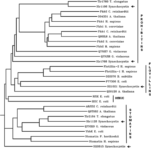
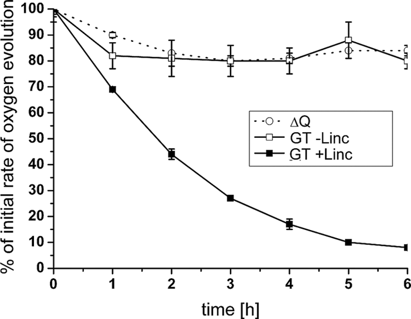
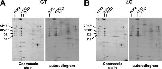
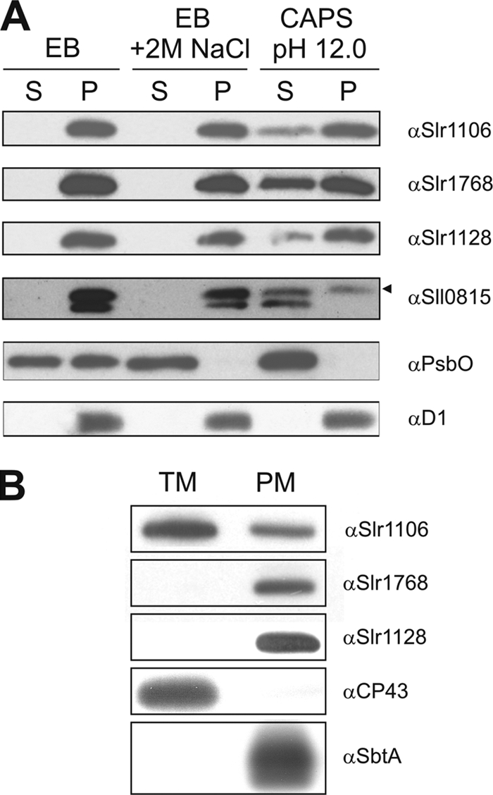
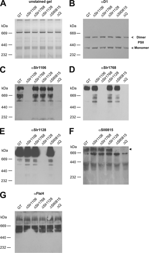
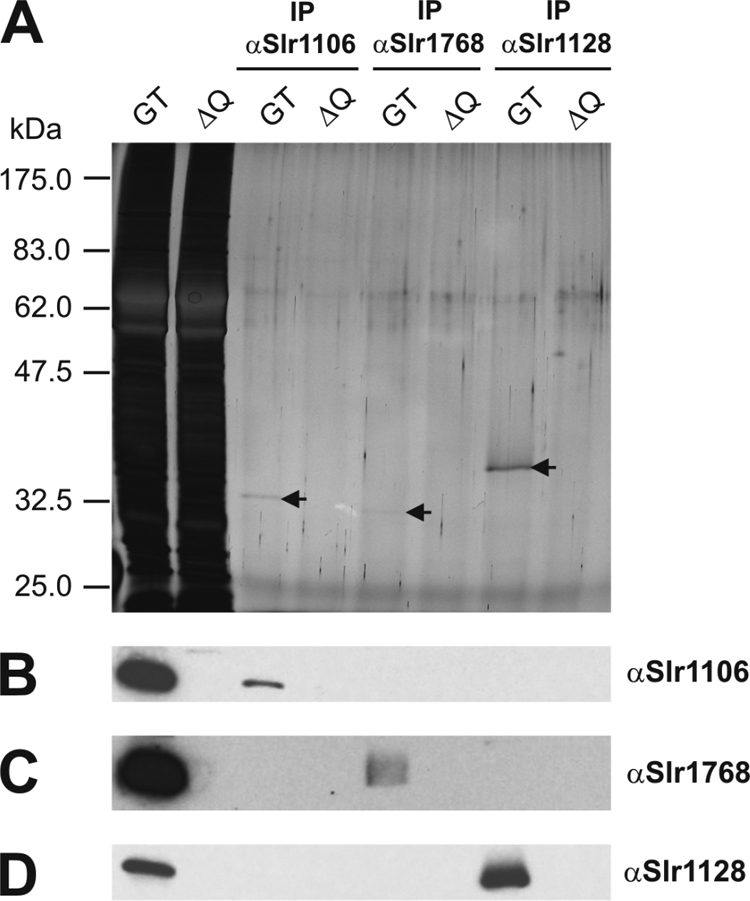
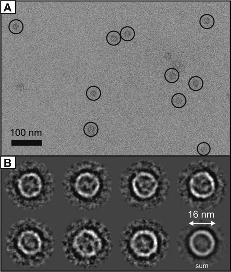
Band 7 proteins, which encompass members of the stomatin, prohibitin, flotillin, and HflK/C protein families, are integral membrane proteins that play important physiological roles in eukaryotes but are poorly characterized in bacteria. We have studied the band 7 proteins encoded by the cyanobacterium Synechocystis sp. strain PCC 6803, with emphasis on their structure and proposed role in the assembly and maintenance of the photosynthetic apparatus. Mutagenesis revealed that none of the five band 7 proteins (Slr1106, Slr1128, Slr1768, Sll0815, and Sll1021) was essential for growth under a range of conditions (including high light, salt, oxidative, and temperature stresses), although motility was compromised in an Slr1768 inactivation mutant. Accumulation of the major photosynthetic complexes in the thylakoid membrane and repair of the photosystem II complex following light damage were similar in the wild type and a quadruple mutant. Cellular fractionation experiments indicated that three of the band 7 proteins (Slr1106, Slr1768, and Slr1128) were associated with the cytoplasmic membrane, whereas Slr1106, a prohibitin homologue, was also found in the thylakoid membrane fraction. Blue native gel electrophoresis indicated that these three proteins, plus Sll0815, formed large (>669-kDa) independent complexes. Slr1128, a stomatin homologue, has a ring-like structure with an approximate diameter of 16 nm when visualized by negative stain electron microscopy. No evidence for band 7/FtsH supercomplexes was found. Overall, our results indicate that the band 7 proteins form large homo-oligomeric complexes but do not play a crucial role in the biogenesis of the photosynthetic apparatus in Synechocystis sp. strain PCC 6803.


Figures

FIG. 1.
FIG. 1.
Phylogenetic analysis of selected band 7 proteins. Selected protein sequences were aligned using CLUSTALW, and a rooted phylogenetic tree was generated using PHYLIP/DRAMGRAM. Band 7 proteins of the following organisms were included in this analysis (in alphabetical order): Arabidopsis thaliana, Bacillus subtilis (subsp. subtilis strain 168), Chlamydomonas reinhardtii, Escherichia coli (strain K-12), Gloeobacter violaceus, Homo sapiens, Pyrococcus horikoshii, Saccharomyces cerevisiae (baker's yeast), Synechocystis sp. strain PCC 6803, Thermosynechococcus elongatus BP-1. Arrows indicate the band 7 proteins of Synechocystis sp. strain PCC 6803.
FIG. 2.
FIG. 2.
Photoinhibition analyses of the Synechocystis sp. strain PCC 6803 GT and the ΔQ quadruple mutant. Cells at a Chl a concentration of 20 μg ml−1 were treated with high light (1,200 μE m−2 s−1) at 29°C over a period of 6 hours. Oxygen evolution of whole cells was assessed for the Synechocystis sp. strain PCC 6803 GT and the ΔQ quadruple mutant strains in the presence or absence of 100 μg ml−1 lincomycin at the indicated time points in the presence of 2 mM DCBQ and 1 mM K3Fe(CN) using a Hansatech DW2 oxygen electrode (Hansatech Instruments Ltd., United Kingdom). Oxygen evolution rates (in μmol oxygen per mg of Chl a per h) were normalized (value at t = 0 was 100%) and plotted as a function of time. Error bars represent standard deviation from the means of three measurements. The initial, absolute rates of oxygen evolution for the respective strains were as follows: 324 (ΔQ without Linc), 316 (GT without Linc), and 350 (GT +Linc) (all in μmol oxygen per mg of Chl a per h).
FIG. 3.
FIG. 3.
Comparative 2-D BN/SDS-PAGE analysis of pulse-labeled crude membrane extracts isolated from the Synechocystis sp. strain PCC 6803 GT and the quadruple mutant strain, ΔQ. Coomassie blue-stained gels and autoradiograms of the 2-D BN/SDS-PAGE analysis of pulse-labeled samples (an amount of 6 μg of Chl a was loaded per sample) were prepared as described by Sobotka et al. (43). The positions of PSII dimers (RCC2), PSII monomers (RCC1), and RC47 protein complexes (RC47, PSII core complexes lacking CP43) as well as those of the D1, D2, CP43, and CP47 PSII subunits are indicated.
FIG. 4.
FIG. 4.
Membrane association and localization of band 7 proteins of Synechocystis sp. strain PCC 6803. (A) Differential membrane protein extraction from Synechocystis sp. strain PCC 6803 GT crude membrane isolations. After two consecutive freeze-thaw cycles (30 min at −80°C and 20 min at RT) in the indicated buffers (EB, 20 mM Tricine, pH 8.0; EB with 2 M NaCl; or 20 mM CAPS, pH 12.0), soluble (S) and pellet (P) fractions were generated by ultracentrifugation. An amount corresponding to 1 μg of Chl a was loaded per lane. The arrowhead indicates the antibody signal that corresponds to the Sll0815 protein. (B) Purified thylakoid membrane (TM) and plasma membrane (PM) fractions were generated by aqueous polymer two-phase partitioning, and 5 μg of protein was loaded in each lane. All samples in panels A and B were analyzed by 1-D SDS PAGE followed by immunoblotting with the indicated antibodies. (A) PsbO and D1 were used as markers for peripheral and integral membrane proteins, respectively. (B) CP43 and SbtA were used as markers for the purity of the thylakoid and plasma membrane fractions, respectively.
FIG. 5.
FIG. 5.
Band 7 protein complexes of Synechocystis sp. strain PCC 6803. Crude membrane isolations (samples contained 1 μg of Chl a) of the Synechocystis sp. strain PCC 6803 GT and various band 7 single and multiple gene disruption mutant strains were analyzed by 1-D BN-PAGE on a 5 to 12.5% (wt/vol) linear gradient polyacrylamide gel and immunoblotted with the indicated antibodies. (B) The positions of monomeric and dimeric PSII protein complexes are indicated. (F) The arrowhead indicates the antibody signal that corresponds to the Sll0815 protein.
FIG. 6.
FIG. 6.
Immunoprecipitation of band 7 proteins of Synechocystis sp. strain PCC 6803. Crude membrane isolations of the Synechocystis sp. strain PCC 6803 GT and the quadruple mutant strain ΔQ (initial samples contained 40 μg of Chl a) were used for immunoprecipitation with anti-Slr1106, anti-Slr1768, or anti-Slr1128 antibodies coupled to protein A-Sepharose beads. Crude membranes of both strains (0.25 μg Chl a) and eluted fractions (7.5 μl of a total volume of 80 μl) were analyzed by 1-D SDS-PAGE (A) and immunoblotted with indicated antibodies (B to D).
FIG. 7.
FIG. 7.
Single-particle analysis of Slr1128 protein complexes of Synechocystis sp. strain PCC 6803. (A) Selected region of an electron micrograph that shows Slr1128 protein complexes (circled). Bar, 100 nm. (B) A subpopulation of 499 particles was classified as being the largest projections observed within a total data set of 3,708, and these were assigned as a top views. These were further classified into seven classes, as shown, with a total average (last image) of ∼16 nm in diameter. Protein is appears as white, and stain is black.

References

    1. Bairoch, A., R. Apweiler, C. H. Wu, W. C. Barker, B. Boeckmann, S. Ferro, E. Gasteiger, H. Huang, R. Lopez, M. Magrane, M. J. Martin, D. A. Natale, C. O'Donovan, N. Redaschi, and L. S. Yeh. 2005. The Universal Protein Resource (UniProt). Nucleic Acids Res. 33:D154-D159. - PMC - PubMed
    1. Berger, K. H., and M. P. Yaffe. 1998. Prohibitin family members interact genetically with mitochondrial inheritance components in Saccharomyces cerevisiae. Mol. Cell. Biol. 18:4043-4052. - PMC - PubMed
    1. Blum, H., H. Beier, and H. J. Gross. 1987. Improved silver staining of plant proteins, RNA and DNA in polyacrylamide gels. Electrophoresis 8:93-99.
    1. Borner, G. H., D. J. Sherrier, T. Weimar, L. V. Michaelson, N. D. Hawkins, A. Macaskill, J. A. Napier, M. H. Beale, K. S. Lilley, and P. Dupree. 2005. Analysis of detergent-resistant membranes in Arabidopsis. Evidence for plasma membrane lipid rafts. Plant Physiol. 137:104-116. - PMC - PubMed
    1. Bourges, I., C. Ramus, B. Mousson de Camaret, R. Beugnot, C. Remacle, P. Cardol, G. Hofhaus, and J. P. Issartel. 2004. Structural organization of mitochondrial human complex I: role of the ND4 and ND5 mitochondria-encoded subunits and interaction with prohibitin. Biochem. J. 383:491-499. - PMC - PubMed

Publication types

Substances

LinkOut - more resources