. 2009 Nov;297(5):L846-55.
doi: 10.1152/ajplung.90590.2008. Epub 2009 Aug 14.

Modulation of reactive oxygen species by Rac1 or catalase prevents asbestos-induced pulmonary fibrosis

Affiliations

Modulation of reactive oxygen species by Rac1 or catalase prevents asbestos-induced pulmonary fibrosis

Shubha Murthy et al. Am J Physiol Lung Cell Mol Physiol. 2009 Nov.

Abstract

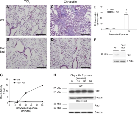
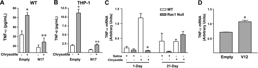
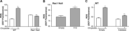
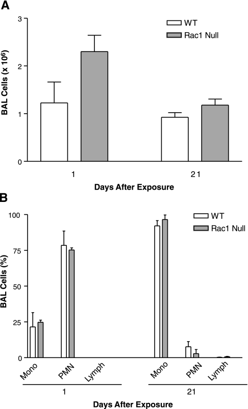
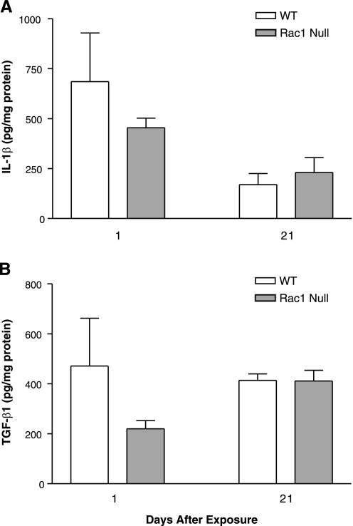
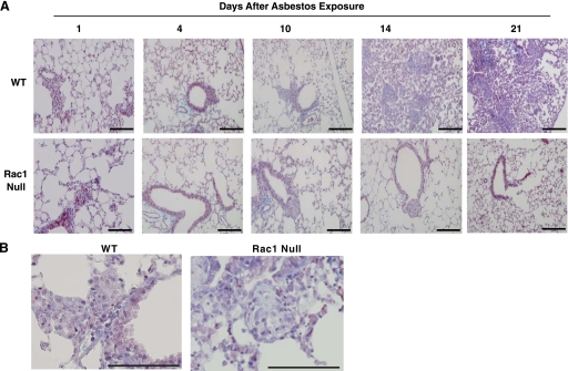
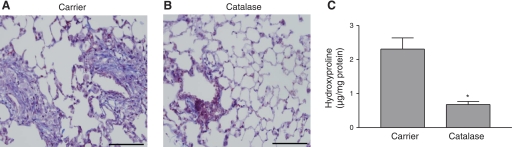
The release of reactive oxygen species (ROS) and cytokines by alveolar macrophages has been demonstrated in asbestos-induced pulmonary fibrosis, but the mechanism linking alveolar macrophages to the pathogenesis is not known. The GTPase Rac1 is a second messenger that plays an important role in host defense. In this study, we demonstrate that Rac1 null mice are protected from asbestos-induced pulmonary fibrosis, as determined by histological and biochemical analysis. We hypothesized that Rac1 induced pulmonary fibrosis via generation of ROS. Asbestos increased TNF-alpha and ROS in a Rac1-dependent manner. TNF-alpha was elevated only 1 day after exposure, whereas ROS generation progressively increased in bronchoalveolar lavage cells obtained from wild-type (WT) mice. To determine whether ROS generation contributed to pulmonary fibrosis, we overexpressed catalase in WT monocytes and observed a decrease in ROS generation in vitro. More importantly, administration of catalase to WT mice attenuated the development of fibrosis in vivo. For the first time, these results demonstrate that Rac1 plays a crucial role in asbestos-induced pulmonary fibrosis. Moreover, it suggests that a simple intervention may be useful to prevent progression of the disease.


Figures

Fig. 1.
Fig. 1.
Rac1 null mice are protected from developing asbestos-induced pulmonary fibrosis. TiO2 (A and B) or chrysotile asbestos (C and D) was administered intratracheally at 100 μg in 50 μl of normal saline to wild-type (WT) and Rac1 null C57BL/6 mice, and animals were euthanized 21 days later. Lungs were removed and processed for collagen deposition using Masson's trichrome stain. Representative micrographs of 1 of 5 animals are shown. Scale bars, 200 μm. E: 1 and 21 days after chrysotile asbestos exposure, WT and Rac1 null mice were subjected to bronchoalveolar lavage (BAL), and hydroxyproline concentration was determined in BAL fluid. *Significantly different from Rac1 null at 21 days. F: equivalent amounts of protein from WT and Rac1 null mouse monocytes were separated by SDS-PAGE, and Western blot analysis was performed with Rac1 and β-actin monoclonal antibodies to determine the presence and equal loading of the proteins, respectively. G: WT and Rac1 null monocytes were exposed to chrysotile asbestos (10 μg/cm2) for 0–60 min. Activated Rac1 was determined by binding of Rac1 to PAK-PBD beads immobilized in a 96-well plate using GLISA. Rac1 activation is expressed as relative light units (RLU) at each time point normalized to control values. H: Rac1 protein levels and equal loading of proteins in lysates from each time point in F were determined by Western blot analysis using Rac1 and β-actin monoclonal antibodies, respectively.
Fig. 2.
Fig. 2.
Rac1 activation is necessary for asbestos-induced TNF-α production. A and B: WT mouse monocytes and THP-1 cells were infected for 48 h with Ad5.CMV containing either empty vector or dominant-negative N17-Rac1 vector at 500 multiplicity of infection (moi). For the last 24 h, cells were exposed to chrysotile asbestos (10 μg/cm2). Conditioned medium was analyzed for TNF-α by ELISA. Values are means ± SE; n = 2. *Significantly different from chrysotile(−) Empty. **Significantly different from chrysotile(+) Empty. C: BAL cells were collected from WT and Rac1 null mice 1 and 21 days after saline or chrysotile asbestos exposure, and TNF-α mRNA was determined by real-time PCR. Values are means ± SE; n = 2. *Significantly different from WT 1 day. D: Rac1 null monocytes were infected for 48 h with Ad5.CMV containing either empty vector or Ad5.V12-Rac1 vector at 500 moi, and TNF-α mRNA was determined by real-time PCR. Values are means ± SE; n = 2. *Significantly different from Empty.
Fig. 3.
Fig. 3.
Rac1 activation is necessary for asbestos-induced generation of reactive oxygen species (ROS). Dichlorofluorescin diacetate (DCFH-DA, 20 μM) was added to cells during the last 20 min, and intracellular ROS levels were determined by counting the number of cells in which DCFH was oxidized to its fluorescent analog DCF by flow cytometry. A: WT and Rac1 null monocytes were exposed to chrysotile asbestos (10 μg/cm2) for 24 h, and ROS generation was determined by measurement of cells labeled with DCF by flow cytometry. Values are means ± SE of relative fluorescence of DCF; n = 3 wells per treatment. *Significantly different from chrysotile(−) WT. **Significantly different from chrysotile(+) WT. B: Rac1 null monocytes were infected with Ad5.CMV containing either empty vector or constitutive active V12-Rac1 vector at 500 moi. After 24 h, cells were exposed to chrysotile asbestos (10 μg/cm2) for 24 h, and ROS generation was determined as described in A. Values are means ± SE; n = 3. *Significantly different from Empty. C: WT monocytes were infected for 48 h with Ad5.CMV containing either empty vector or a catalase vector at 500 moi. Cells were exposed to chrysotile asbestos (10 μg/cm2) for the last 24 h, and ROS generation was determined as described in A. Values are means ± SE; n = 3. *Significantly different from chrysotile(−) Empty. **Significantly different from chrysotile(+) Catalase.
Fig. 4.
Fig. 4.
BAL cell counts (A) and cell differential (B) are similar in WT and Rac1 null mice after asbestos exposure. WT or Rac1 null mice were exposed to 100 μg of chrysotile asbestos in 50 μl of saline. After 1 and 21 days, animals were euthanized, and BAL was performed. BAL cells were counted to determine total cell count and cell differential was determined with Wright-Giemsa stain. Values are means ± SE; n = 3 animals per group. Mono, mononuclear; PMN, polymorphonuclear; Lymph, lymphocytes.
Fig. 5.
Fig. 5.
IL-1β and transforming growth factor (TGF)-β1 in BAL are not regulated by Rac1. Mice were exposed to 100 μg of chrysotile asbestos, and BAL was performed 1 and 21 days later. BAL fluid was analyzed for IL-1β (A) and TGF-β1 (B) by ELISA. Values, normalized to BAL total protein, are means ± SE; n = 3 animals per group.
Fig. 6.
Fig. 6.
Rate of development of pulmonary fibrosis after asbestos exposure. A: WT or Rac1 null mice were exposed to 100 μg of asbestos in 50 μl of saline and euthanized 1, 4, 10, 14, and 21 days later. Lungs were removed and processed for collagen deposition using Masson's trichrome stain. Representative serial sections from 1 of 3 animals per time point are shown. Scale bars, 200 μm. B: high-power (×40) field of inflammatory cell aggregates in WT and Rac1 null mice. Scale bars, 100 μm.
Fig. 7.
Fig. 7.
Exogenous catalase prevents pulmonary fibrosis. WT mice were exposed to asbestos, and, on each day for the next 20 days, carrier (A) or catalase (2,000 U; B) was administered intratracheally. Animals were euthanized on day 21, and lungs were removed and processed for collagen deposition using Masson's trichrome stain. Representative sections of 1 of 6 mice are shown. Scale bars, 200 μm. C: hydroxyproline concentrations determined in BAL fluid collected from mice treated with carrier (water) or catalase for 20 days after chrysotile asbestos exposure. Values are means ± SE; n = 4. *Significantly different from carrier.

References

    1. Adam O, Frost G, Custodis F, Sussman MA, Schafers HJ, Bohm M, Laufs U. Role of Rac1 GTPase activation in atrial fibrillation. J Am Coll Cardiol 50: 359–367, 2007 - PubMed
    1. . Diagnosis and initial management of nonmalignant diseases related to asbestos. Am J Respir Crit Care Med 170: 691–715, 2004 - PubMed
    1. Begin R, Martel M, Desmarais Y, Drapeau G, Boileau R, Rola-Pleszczynski M, Masse S. Fibronectin and procollagen 3 levels in bronchoalveolar lavage of asbestos-exposed human subjects and sheep. Chest 89: 237–243, 1986 - PubMed
    1. Bokoch GM. Regulation of innate immunity by Rho GTPases. Trends Cell Biol 15: 163–171, 2005 - PubMed
    1. Brenner DA, O'Hara M, Angel P, Chojkier M, Karin M. Prolonged activation of jun and collagenase genes by tumour necrosis factor-α. Nature 337: 661–663, 1989 - PubMed

Publication types

MeSH terms