. 2009 Aug;21(8):2323-40.
doi: 10.1105/tpc.109.067819. Epub 2009 Aug 14.

MATE transporters facilitate vacuolar uptake of epicatechin 3'-O-glucoside for proanthocyanidin biosynthesis in Medicago truncatula and Arabidopsis

Affiliations

MATE transporters facilitate vacuolar uptake of epicatechin 3'-O-glucoside for proanthocyanidin biosynthesis in Medicago truncatula and Arabidopsis

Jian Zhao et al. Plant Cell. 2009 Aug.

Erratum in

  • Plant Cell. 2010 Mar;22(3):991

Abstract

Expression of the Arabidopsis thaliana MYB transcription factor TRANSPARENT TESTA 2 (TT2) in Medicago trunculata hairy roots induces both proanthocyanidin accumulation and the ATP-dependent vacuolar/vesicular uptake of epicatechin 3'-O-glucoside; neither process is active in control roots that do, however, possess anthocyanidin 3-O-glucoside vacuolar uptake activity. A vacuolar membrane-localized multidrug and toxic compound extrusion (MATE) transporter, Medicago MATE1, was identified at the molecular level and shown to preferentially transport epicatechin 3'-O-glucoside. Genetic evidence has implicated TT12, a tonoplastic MATE transporter from Arabidopsis, in the transport of precursors for proanthocyanidin biosynthesis in the seed coat. However, although Arabidopsis TT12 facilitates the transport of cyanidin 3-O-glucoside into membrane vesicles when expressed in yeast, there is no evidence that cyanidin 3-O-glucoside is converted to proanthocyanidins after transport into the vacuole. Here, we show that Arabidopsis TT12, like Medicago MATE1, functions to transport epicatechin 3'-O-glucoside as a precursor for proanthocyanidin biosynthesis, and Medicago MATE1 complements the seed proanthocyanidin phenotype of the Arabidopsis tt12 mutant both quantitatively and qualitatively. On the basis of biochemical properties, tissue-specific expression pattern, and genetic loss-of-function analysis, we conclude that MATE1 is an essential membrane transporter for proanthocyanidin biosynthesis in the Medicago seed coat. Implications of these findings for the assembly of oligomeric proanthocyanidins are discussed.


Figures
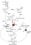
Figure 1.
Figure 1.
Diagrammatic Representation of Key Reactions for PA Precursor Synthesis and Transport in Arabidopsis. Enzymes are as follows: DFR, dihydroflavonol reductase; LDOX, leucoanthocyanidin dioxygenase; UGT, uridine diphosphate glycosyltransferase; MRP, multidrug resistance-associated protein. [See online article for color version of this figure.]
Figure 2.
Figure 2.
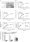
Uptake of Cy3G, E3′G, and Daidzin (D7) by Vacuole-Enriched Membrane Vesicles from M. truncatula Hairy Roots. (A) Protein gel blot analysis of fractions from sucrose density gradients (numbers show percentage of sucrose, w/v) probed with antibodies against V-H+-ATPase (vacuole marker), Arabidopsis SEC12 (endoplasmic reticulum marker), and PM-H+-ATPase (plasma membrane marker). (B) to (D) Time-dependent uptake of (iso)flavonoid glucosides into vacuole-enriched vesicles from hairy roots transformed with Arabidopsis TT2 (solid circles), empty vector (open squares), or TT2 but with no ATP in the uptake assay (open triangles). Substrate concentration was 50 μM. Results are mean and sd of three replicate uptake assays. D7G, daidzein 7-O-glucoside (daidzin). (E) and (F) Concentration dependence of uptake of Cy3G and E3′G into membrane vesicles from hairy roots transformed with TT2 (solid circles) or empty vector (empty circles). (G) Inhibition of Cy3G, E3′G, and D7G uptake (50 μM, 20-min assays) by inhibitors of membrane transport. Inhibitor concentrations were 1 mM (vanadate), 0.1 μM (bafilomycin A1), 5 mM (NH4Cl), and 5 μM (gramicidin D). Results are mean and sd of five replicate uptake assays. No-ATP values are essentially zero.
Figure 3.
Figure 3.
Phylogeny and Expression Pattern of Medicago MATE1. (A) Phylogenetic tree of MATE transporters from different plant species. Protein sequences of the known anthocyanin MATE transporters Arabidopsis TT12, V. vinifera AM1 and AM3, Zea mays (maize) MTP77, a nicotine transporter from tobacco, and predicted MATE transporters XP_002282932 and CAO69962 from V. vinifera, XP_002307572 from P. trichocarpa, ACJ36213 from field mustard (Brassica rapa), and MATE1 from M. truncatula were aligned with ClustalW, and the nonrooted neighbor-joining tree was generated by the PAUP 4.0 program. Numbers at branch points indicate bootstrap support. (B) Validation of Medicago MATE1 expression in TT2-expressing M. truncatula hairy roots by RT-PCR. Expression of TT2 also turns on the expression of ANR, but not of another Medicago MATE transporter gene, AC121237_16.5. Photos show representatives of three similar replicates. (C) qRT-PCR analysis of the expression level (relative to Medicago ACTIN) of MATE1 in different tissues of M. truncatula. Pods were analyzed at different times from 3 to 36 d after fertilization. Data are means and sd from three biological replicates.
Figure 4.
Figure 4.
Uptake of Cy3G and E3′G by Yeast Microsomal Vesicles Expressing Medicago MATE1. (A) and (B) Time-dependent uptake into vesicles from yeast cells transformed with MATE1 (closed squares) or empty vector (open squares). Substrate concentration was 100 μM. Results are mean and sd of three replicate uptake assays from three independent membrane preparations. (C) and (D) Concentration dependence of uptake of Cy3G and E3′G into vesicles from yeast expressing MATE1. (E) and (F) Double reciprocal plots of initial rate data at different concentrations of Cy3G and E3′G.
Figure 5.
Figure 5.
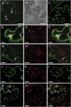
Subcellular Localization of MATE1-GFP. MATE1-GFP driven by the cauliflower mosaic virus 35S promoter was transiently expressed in tobacco leaf epidermal cells and viewed by confocal microscopy. (A) GFP fluorescence image of cells expressing MATE1-GFP. The large arrow shows the nucleus, and the arrowheads show prevacuolar or nuclear membranes. Bar =50 μm. (B) Differential interference contrast image of the same epidermal cell expressing MATE1-GFP as in (A). Bar = 50 μm. (C) FM1-43–labeled plasma membrane. Bar = 25 μm. (D) to (F) Fluorescence images of a tobacco cell expressing free GFP. Bars = 20 μm. (D) GFP fluorescence. (E) Chloroplast autofluorescence image. (F) Merged GFP image and chloroplast autofluorescence image. Arrow indicates the nucleus. (G) to (I) Fluorescence images of a tobacco cell expressing MATE1-GFP. Bars = 20 μm. (G) GFP fluorescence. (H) Chloroplast autofluorescence image. (I) Merged GFP image and chloroplast autofluorescence image. Arrow shows the position of the nucleus. (J) to (L) Fluorescence images of a tobacco epidermal cell stained with the plasma membrane–specific dye FM1-43. Bars = 10 μm. (J) FM1-43 fluorescence. (K) Chloroplast autofluorescence image. (L) Merged image of FM1-43–labeled plasma membrane (green fluorescence) and chloroplasts (red autofluorescence). (M) to (O) Fluorescence images of a tobacco epidermal cell expressing MATE1-GFP and stained with the plasma membrane–specific dye FM4-64. Bars = 25 μm. (M) GFP fluorescence. (N) FM4-64–labeled plasma membrane. (O) Merged image of (M) and (N). The arrows show the nucleus outside the vacuole, and the arrowheads show prevacuole-like vesicles.
Figure 6.
Figure 6.
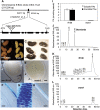
Loss-of-Function Analysis of MATE1 in M. truncatula. (A) The MATE1 gene and the position of the Tnt1 retrotransposon insertion of line mate1 (NF2629). Positions of introns (lines) and exons (black boxes) are shown. (B) to (E) Seed phenotypes of wild-type M. truncatula R108 (left) and retrotransposon insertion line mate1-1 (right). (B) and (C) Whole seeds stained with DMACA in (C). Bars = 2 mm. (D) and (E) Cross sections of seeds stained with DMACA. Arrows show seed coats stained blue in R108 seed (D), with almost no staining in the mate1 seed (E). Bars = 0.2 mm. (F) to (H) Enlarged view of seed coat staining and cross section of stained seed coat cells showing location of PAs in the vacuoles. Blue color shows PA accumulation inside vacuoles. Bars = 40 μm. (I) Levels of extractable PAs (soluble and insoluble) from seed of R108 and mate1. Values are mean and sd from three biological replicates. (J) to (L) Analysis of size distribution of PAs in Medicago lines. Soluble PAs were resolved by normal phase HPLC with postcolumn derivatization with DMACA reagent and monitoring at 640 nm. (J) Standards of monomer (catechin) and dimer (procyanidin B1). (K) Soluble PAs from wild-type M. truncatula R108 seeds. (L) PAs from the M. truncatula Tnt1 mutant mate1.
Figure 7.
Figure 7.
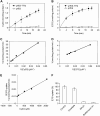
Uptake of Cy3G and E3′G by Yeast Microsomal Vesicles Expressing Arabidopsis TT12. (A) and (B) Time-dependent uptake into vesicles from yeast cells transformed with TT12 (closed squares) or empty pYES vector (open squares). Substrate concentration was 100 μM. Results are mean and sd of at least three replicate uptake assays from three independent membrane preparations. (C) and (D) Double reciprocal plots of initial rate data at different concentrations of Cy3G and E3′G. (E) Plot showing inhibition of E3′G uptake by Cy3G. (F) Inhibition of E3′G uptake (100 μM, 8-min assays) by inhibitors of membrane transport. Inhibitor concentrations were 1 mM (vanadate), 5 mM (NH4Cl), and 0.1 μM (bafilomycin A1). Results are mean and sd from three replicate uptake assays.
Figure 8.
Figure 8.
Complementation of the Arabidopsis tt12 Mutation by Medicago MATE1. (A) RT-PCR screening to indicate the presence of Medicago MATE1 transcripts in three independent Arabidopsis transformants. Arabidopsis ACTIN expression was used as an internal control. (B) Seed phenotypes of the tt12 mutant, wild-type Arabidopsis ecotype Ws, and tt12 complemented with MATE1. The three bottom panels show seed after staining with DMACA. Bars = 0.3 mm. (C) Levels of extractable PAs (soluble and insoluble) from seeds of the tt12 mutant, wild-type Arabidopsis ecotype Ws, and tt12 complemented with MATE1. Values are mean and sd from three biological replicates. (D) to (F) Soluble PA size distribution in seeds of Arabidopsis. Soluble PAs were resolved by normal-phase HPLC with postcolumn derivatization with DMACA reagent and monitoring at 640 nm. (E) PAs from wild-type Ws seeds. (F) PAs from tt12 seeds. (G) PAs from tt12/MATE1 seeds.

References

    1. Abrahams, S., Lee, E., Walker, A.R., Tanner, G.J., Larkin, P., and Ashton, A.R. (2003). The Arabidopsis TDS4 gene encodes leucoanthocyanidin dioxygenase (LDOX) and is essential for proanthocyanidin synthesis and vacuole development. Plant J. 35: 624–636. - PubMed
    1. Abrahams, S., Tanner, G.J., Larkin, P.J., and Ashton, A.R. (2002). Identification and biochemical characterization of mutants in the proanthocyanidin pathway in Arabidopsis. Plant Physiol. 130: 561–576. - PMC - PubMed
    1. Ariga, T., Asao, Y., Sugimoto, H., and Yokotuska, T. (1981). Occurrence of astringent oligomeric proanthocyanidins in legume seeds. Agric. Biol. Chem. 45: 2705–2708.
    1. Bartholomew, D.M., Van Dyk, D.E., Lau, S.M., O'Keefe, D.P., Rea, P.A., and Viitanen, P.V. (2002). Alternate energy-dependent pathways for the vacuolar uptake of glucose and glutathione conjugates. Plant Physiol. 130: 1562–1572. - PMC - PubMed
    1. Baxter, I.R., Young, J.C., Armstrong, G., Foster, N., Bogenschutz, N., Cordova, T., Peer, W.A., Hazen, S.P., Murphy, A.S., and Harper, J.F. (2005). A plasma membrane H+-ATPase is required for the formation of proanthocyanidins in the seed coat endothelium of Arabidopsis thaliana. Proc. Natl. Acad. Sci. USA 102: 2649–2654. - PMC - PubMed

Publication types

MeSH terms

Associated data