. 2009 Sep;11(9):1150-6.
doi: 10.1038/ncb1930. Epub 2009 Aug 16.

Silencing by small RNAs is linked to endosomal trafficking

Affiliations

Silencing by small RNAs is linked to endosomal trafficking

Young Sik Lee et al. Nat Cell Biol. 2009 Sep.

Erratum in

  • Nat Cell Biol. 2009 Dec;11(12):1495

Abstract

Small RNAs direct RNA-induced silencing complexes (RISCs) to regulate stability and translation of mRNAs. RISCs associated with target mRNAs often accumulate in discrete cytoplasmic foci known as GW-bodies. However, RISC proteins can associate with membrane compartments such as the Golgi and endoplasmic reticulum. Here, we show that GW-bodies are associated with late endosomes (multivesicular bodies, MVBs). Blocking the maturation of MVBs into lysosomes by loss of the tethering factor HPS4 (ref. 5) enhances short interfering RNA (siRNA)- and micro RNA (miRNA)-mediated silencing in Drosophila melanogaster and humans. It also triggers over-accumulation of GW-bodies. Blocking MVB formation by ESCRT (endosomal sorting complex required for transport) depletion results in impaired miRNA silencing and loss of GW-bodies. These results indicate that active RISCs are physically and functionally coupled to MVBs. We further show that MVBs promote the competence of RISCs in loading small RNAs. We suggest that the recycling of RISCs is promoted by MVBs, resulting in RISCs more effectively engaging with small RNA effectors and possibly target RNAs. It may provide a means to enhance the dynamics of RNA silencing in the cytoplasm.


Figures
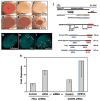
Figure 1
Figure 1. Loss of HPS4 enhances miRNA- and siRNA-mediated silencing
a–c, Silencing of the white gene by GMR-wIR hairpin RNA is enhanced in a complementation group of mutations that maps to the dHPS4 locus. Eyes from a wild-type adult (a), a white(RNAi) adult (b), a white(RNAi) adult in a mutant dHPS4 background (c). Stronger silencing generates a white eye. d–f, Silencing of the Csk gene by Csk hairpin RNA is enhanced in dHPS4 mutants. Eyes from a Csk(RNAi) adult (d), a Csk(RNAi) adult in a mutant dicer-2 (dcr-2) background (e), and a Csk(RNAi) adult in a mutant dHPS4 background (f). The mispatterning of the eye caused by Csk(RNAi) was restored in a dcr-2 mutant lacking RNAi, and was strongly enhanced in the dHPS4 mutant. g–i, Eyes from adults expressing GFP under control of the Brd 3′ UTR. (g) A non-mosaic wildtype eye. (h) Mosaic eye with presumed dicer-1 (dcr-1) mutant cells as outlined in yellow, and other cells are wildtype. Impaired miRNA processing in dcr-1 cells leads to de-repressed GFP expression. (i) Mosaic eye with presumed dHPS4 mutant cells as outlined in yellow, and other cells are wildtype. GFP expression is more strongly repressed. j, The dHPS4 gene and protein. Top, the dHPS4 transcription unit. Positions of start and stop codons are indicated by vertical blue and red lines, respectively. Bottom, the protein products showing conserved domains. The SNARE-like longin domain is found within the conserved amino-terminal domain. The positions of the five mutations are indicated, all generating premature stop codons (X). Vertebrate orthologues are shown below, with conserved domains and percent amino acid identity to the Drosophila protein indicated. k, Depletion of HPS4 enhances siRNA-mediated silencing in HeLa cells. Silencing of a Pp-luciferase reporter was triggered by an shRNA vector (left), and silencing of a Renilla-luciferase reporter gene was triggered by a CXCR4 siRNA (right). They were co-transfected with a control siRNA. Alternatively, they were co-transfected with either siRNA HPS4-882 (left) or siRNAs HPS1-242 and HPS4-1348 (right). Error bars represent one standard deviation.
Figure 2
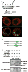
Figure 2. Characterization of dHPS4 and dHPS1
a, Top, Western blot of dHPS4 protein from wildtype adult head extract after fractionation by OptiPrep density gradient centrifugation. Selected fractions are shown. Fraction 1 at the top contains membrane-associated proteins, as indicated by the selective presence of dGM130 Golgi protein in Fraction 1 of parallel blots. Bottom, Western blot of dHPS4 protein from wildtype (+) and dHPS4 mutant (−) head extract. b, Two representative S2 cells visualized by confocal microscopy for HA-dHPS4 (red) and GFP-Me31b (green) proteins. GFP-Mei31b highlights GW-bodies. Confocal sections are high resolution (2048×2048 pixels from 100X objective) and ultra-thin (0.4μm). Parallel analysis of the same stable cell line without induction and naive S2 cells with induction confirmed that the anti-HA staining was dependent upon the presence of the MT>HA-dHPS4 gene and the metal inducer. These two cells represent a n>50 sample set. c, Two representative S2 cells visualized by confocal microscopy for HA-dHPS4 (red) and Lamp1-GFP (green) proteins. The dHPS4 protein surrounds Lamp1-positive endosomes and lysosomes. d, Domain structure of the HPS1 protein family. A conserved domain unique to the family is shown in green, and percent amino acid sequence identity between each orthologue and Drosophila HPS1 (CG12855) is shown in parentheses. Lengths of vertebrate orthologues are given in amino acids (aa). dHPS1 is 596 amino acids in length. e, Co-immunoprecipitation (Co-IP) of epitope-tagged dHPS4 and dHPS1 from transfected S2 cells. All IPs were performed on HA-tagged proteins, and FLAG-tagged proteins were probed for co-IP. HA-GFP was used as a control for IP specificity.
Figure 3
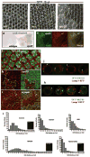
Figure 3. MVBs are sites of miRNA-mediated silencing
a–c, Expression of the GFP::Brd reporter of miRNA silencing in pupal eyes that contain clones of hrsD28 (a,b), and vps25A3 (c) mutant cells. Presumed clone boundaries are marked in yellow. Two hrs clones are shown at different z-planes to highlight GFP up-regulation in cone cells (a) and pigment cells (b). d–e, Expression of a lacZ::E(spl)m8 reporter of miRNA silencing in larval eye discs that contain vps25A3 clones. (d) Whole eye discs that were stained with X-Gal, with the left disc lacking any clones and the right disc containing vps25A3 clones. (e) A region of an eye disc with vps25A3 clones stained for the lacZ::E(spl)m8 reporter protein (green) and Ago1 protein (red). Note the correlation between enhanced lacZ::E(spl)m8 expression and Ago1 concentration in the presumed clones. f–i, Larval eye discs were stained for Ago1 protein (red) to detect miRISC distribution. Magnified views of wildtype (f) and dHPS4W515X mutant (g) eye discs counterstained for nuclei (green). Perinuclear Ago1 localization in wildtype cells is highlighted with arrowheads. This localization is lacking in the mutant cells. Eye discs containing clones of vps25A3 (h), and myopicT612 (i) mutant cells. In these genetically mosaic eyes, mutant cells are visualized by lack of expression of a marker gene (green). Yellow lines mark boundaries of clones. Mutant cells exhibit concentrated Ago1 around large vesicles (arrows). j–k, S2 cells showing Lamp1-RFP (red) that marks MVBs and lysosomes. Cells have been transfected with YFP-GW182 (j) or GFP-Me31b (k) to highlight GW-bodies (green). Note selected examples (arrows) of bodies juxtaposed with Lamp1-positive membranes. l, Distributions of Ago2-GFP cytoplasmic bodies per cell in HeLa cell populations that underwent different siRNA treatments. Indicated in each plot are the siRNAs that were incubated with cells prior to counting GW-body numbers. Inset images are single HeLa cells from selected treatments showing Ago2-GFP bodies.
Figure 4
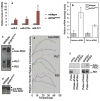
Figure 4. RISC loading of small RNAs is dependent upon MVBs
a, Quantitated levels of miR-8, miR-276a, and miR-277 miRNAs that co-immunoprecipitated with Ago1 protein from extracts of wildtype and dHPS4W515X mutants. RNAs were detected by splinted ligation and were normalized to the amount of Ago1 immunoprecipitated in each sample. Data represent the mean ± standard error of the normalized values from four independent experiments, relative to wildtype. b, Activity of RISC complexes in head extracts that were loaded with exogenous bantam miRNAs or Pp-luc siRNAs. Cleavage of target mRNA with a sequence perfectly complementary to either the miRNA or siRNA was quantified. End-point cleavage activity of dHPS4 mutant extracts is expressed relative to wildtype extracts. Error bars show standard deviations. c, Loading of holo-RISC in wildtype and dHPS4W515X mutant head extracts after addition of labelled Pp-luc siRNA. RDI, RLC and holo-RISC complexes are indicated in this native gel. An asterisk marks material too large to enter the gel. d, Loading of pre-RISC in dHPS4W515X mutant head extract after addition of a labelled siRNA that contains a single base mismatch. This siRNA enables detection of pre-RISC, which is otherwise not observed in the assay. e, Time course following RDI, RLC, and holo-RISC loading with wildtype (blue diamonds) and dHPS4W515X mutant (green squares) extracts. f, Pulse (purified RDI added with labelled siRNA) and Chase (head extract added with excess unlabelled siRNA) mixtures were added to reactions in order as indicated. Native gel analysis visualized RDI and holo-RISC complexes associated with labelled siRNA. Genotypes of extracts are indicated above.
Figure 5
Figure 5. Protein ubiquitination is required for RNA silencing and RISC loading
a–b, Eyes of white+ adult flies with one copy of GMR-wIR. (a) The white gene is partially silenced, resulting in an orange eye colour in a dFBX011+ background. (b) A fly homozygous for dFBX011Q803R showing a redder eye colour that indicates impaired siRNA-mediated silencing. c–d, Eyes of adults with one copy of GMR-wIR and mutant for dcr-1. (c) Loss of dcr-1 results in a smaller eye and partially impaired silencing of white. (d) An eye homozygous for dcr-1 dFBX011Q803R shows greater impairment of white silencing and enhanced eye reduction due to genetic interaction with dcr-1. e–f, Eyes of adults carrying the GFP::Brd reporter for miRNA-mediated silencing. (e) An adult showing normal GFP expression when silenced. (f) An adult with clones of dFBX011Q803R mutant cells in its eye. GFP silencing is inhibited in dFBX011Q803R mutant clones, resulting in strongly variegated GFP expression. g, Northern blot for white siRNAs from adults with indicated genotypes. The dFBX011 mutant allele used was dFBX011Q803R. h, Northern blot for miR-2 RNAs from adult heads with indicated dFBX011 genotypes. There are three isoforms of miR-2, which can be seen on the blot. i, siRISC activity in wildtype and dFBX011 mutant extracts that were loaded with exogenously added Pp-luc siRNA. The unreacted Pp-luc mRNA substrate and the 5′ cleavage product are indicated by an arrow and arrowhead, respectively. j, A ubiquitin mutant (I44A) protein with defects in protein-protein interactions exerts a dominant-negative effect on loading of siRISC with labelled Pp-luc siRNA. The “tailless” negative control carries a four-amino-acid C-terminal truncation and is unable to enter the ubiquitin conjugation pathway.

Comment in

References

    1. Zamore PD, Haley B. Ribo-gnome: the big world of small RNAs. Science. 2005;309:1519–24. - PubMed
    1. Carthew RW, Sontheimer EJ. Origins and mechanisms of miRNAs and siRNAs. Cell. 2009;136:642–55. - PMC - PubMed
    1. Filipowicz W, Bhattacharyya SN, Sonenberg N. Mechanisms of post-transcriptional regulation by microRNAs: are the answers in sight? Nat Rev Genet. 2008;9:102–14. - PubMed
    1. Cikaluk DE, et al. GERp95, a membrane-associated protein that belongs to a family of proteins involved in stem cell differentiation. Mol Biol Cell. 1999;10:3357–72. - PMC - PubMed
    1. Li W, et al. Murine Hermansky-Pudlak syndrome genes: regulators of lysosome-related organelles. Bioessays. 2004;26:616–28. - PubMed

Publication types

LinkOut - more resources