. 2009 Sep;8(9):736-41.
doi: 10.1038/nmat2515. Epub 2009 Aug 16.

Nanolitre liquid patterning in aqueous environments for spatially defined reagent delivery to mammalian cells

Affiliations

Nanolitre liquid patterning in aqueous environments for spatially defined reagent delivery to mammalian cells

H Tavana et al. Nat Mater. 2009 Sep.

Abstract

Microscale biopatterning enables regulation of cell-material interactions and cell shape, and enables multiplexed high-throughput studies in a cell- and reagent-efficient manner. The majority of available techniques rely on physical contact of a stamp, pin, or mask with mainly a dry surface. Inkjet and piezoelectric printing is carried out in a non-contact manner but still requires a substantially dry substrate to ensure fidelity of printed patterns. These existing methods, therefore, are limited for patterning onto delicate surfaces of living cells because physical contact or substantially dry conditions are damaging to them. Microfluidic patterning with laminar streams does enable non-contact patterning in fully aqueous environments but with limited throughput and reagent diffusion across interfacial flows. Here, we describe a polymeric aqueous two-phase system that enables patterning nanolitres of a reagent-containing aqueous phase, in arbitrary shapes, within a second aqueous phase covering a cell monolayer. With the appropriate medium formulation, reagents of interest remain confined to the patterned phase without significant diffusion. The fully aqueous environment ensures high reagent activity and cell viability. The utility of this strategy is demonstrated with patterned delivery of genetic materials to mammalian cells for phenotypic screening of gene expression and gene silencing.


Conflict of interest statement

Competing interests statement

The authors declare no competing financial interests.

Figures
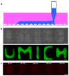
Figure 1
Figure 1. Polymeric aqueous two-phase systems generate user-defined patterns of a reagent on a cell monolayer
(a) Schematic representation of patterning aqueous DEX phase (blue) on a cell monolayer covered with the PEG phase (pink). (b) Bright-field and fluorescent images of patterned DEX phase on HEK293H cells spelling “UMICH”. The shapes were generated by continuous horizontal movement of a pipette tip filled with FITC-labeled DEX solution over cells covered with the PEG phase. (c) Complexes of genetic materials and the transfection reagent, Lipofectamine 2000, partition well to the DEX phase and remain within the dispensed drop over a 4hrs imaging period. Scale bar, 1 mm in a,b, 500 μm in c.
Figure 2
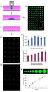
Figure 2. Addressable delivery of nucleic acids to cells using two-phase patterned microarrays
(a) Cells cultured to desired confluence are covered with the PEG phase (pink). Slot pins filled with transfection complexes suspended in the DEX phase (blue) are slowly lowered into this solution using a micromanipulator that controls the vertical motion. Pins dispense their contents onto the cell monolayer and small droplets of transfection complexes form on discrete groups of cells. (b) Fluorescent micrograph of a 12×8 microarray of FITC-DEX solution droplets patterned on a cell monolayer covered with PEG solution. All droplets were formed in one printing step using 500 nl dispensing slot pins that rest on a 1536-well plate format fixture. (c) Only cells confined to the DEX droplets become transfected and exhibit the corresponding phenotype. (d) Fluorescent micrograph of a 6×4 microarray of HEK293H cell clusters expressing eGFP. (e) The highest eGFP transfection efficiency as measured by fluorescence intensity in the cell clusters is obtained using a complex preparation with a 1/1 (μg/μl) ratio of plasmid DNA/transfection reagent. Each bar represents the mean value of five sets of data and the error bars represent ±SD. (f) The level of protein expression in the microarray of transfected cells initially increases with the amount of plasmid DNA and then levels off. Each bar is the mean value of five different data sets and the error bars represent ±SD. (g) Fluorescent micrograph of arrays of cells transfected with plasmid DNAs for eGFP, dsRed, or both. Green and red cell clusters correspond to the eGFP and dsRed transfected cells, respectively. Co-transfected cells are shown in yellow and are obtained by superimposition of green and red fluorescent signals. (h) Assuming that each droplet is a spherical cap, the drop volume follows a quadratic relation with the contact diameter of the drop (VD2). Therefore, the contact diameter of DEX drops and hence the size of transfected cell clusters approximately change linearly with the square root of the drop volume according to the following relation that represents a trend line obtained from experimental diameter and volume data: D = 43.2V 0.5 + 211.2. The goodness of fit, R2, is 0.992. This is useful to pre-determine the number of treated cells from the dispensing pin volume within the drop volume range 20–1000 nl. Scale bar, 700 μm in b,d,g,h.
Figure 3
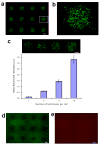
Figure 3. Patterned microarrays of lentiviral-mediated gene expression and gene knockdown
(a) Fluorescent image of a 3×4 array of MDA-MB-231 human breast cancer cells transduced with a lentiviral vector containing eGFP gene. The infected cells are well-contained to patterns of DEX droplets during transduction process. (b) A magnified view of the boxed spot in (a). (c) Dose-dependent infection of cells with lentiviral vectors. Increasing the number of lentiviruses from 1 to 10 per each cell results in increase in the intensity of the eGFP signal. (d) A 3×4 microarray of localized eGFP gene knockdown obtained with patterned infection of cells with lentiviruses encoding eGFP shRNA. (e) Cells in the spots express similar levels of the mPlum (red) gene compared to cells outside the spots. This confirms that silencing of the eGFP gene is due to the specificity of the eGFP shRNA and not due to interference of shRNA with gene expression. Scale bar, 500 μm in a,c,d,e.
Figure 4
Figure 4. Patterned microarrays facilitate phenotypic screening of function of different genes in cell cultures on soft substrates
(a) Schematic representation of localized degradation of collagen I only by MT1-MMP-expressing cells. Cells expressing MMP2 or control eGFP are incapable of collagenolysis. (b) Fluorescent micrographs of HEK293H cells cultured on Alexa fluor 594-labeled collagen I and transfected with expression constructs for MT1-MMP (bottom row), MMP2 (middle row), and eGFP (top row). MT1-MMP expressing cells showed collagenolytic activity and degraded their subjacent ECM whereas cells expressing MMP2 pro-enzyme or eGFP lacked this invasive phenotype. (c) Fluorescent micrograph of cells stained with antibody for MT1-MMP (green) grown on fluorescently labeled collagen (red). MT1-MMP protein expression correlates with the degradation of collagen. Scale bar, 700 μm in b,c.

Comment in

References

    1. Chen CS, Mrksich M, Huang S, Whitesides GM, Ingber DE. Geometric control of cell life and death. Science. 1997;276:1425–1428. - PubMed
    1. Khetani SR, Bhatia SN. Microscale culture of human liver cells for drug development. Nature Biotech. 2008;26:120–126. - PubMed
    1. Singhvi R, et al. Engineering cell shape and function. Science. 1994;264:696–698. - PubMed
    1. Anderson DG, Levenberg S, Langer R. Nanoliter-scale synthesis of arrayed biomaterials and application to human embryonic stem cells. Nature Biotech. 2004;22:863–866. - PubMed
    1. Anderson DG, Putnam D, Lavik E, Mahmood T, Langer R. Biomaterial microarrays: Rapid, microscale screening of polymer-cell interaction. Biomater. 2005;26:4892–4897. - PubMed

Publication types