. 2009 Sep;12(9):1129-35.
doi: 10.1038/nn.2372. Epub 2009 Aug 16.

Rapamycin activation of 4E-BP prevents parkinsonian dopaminergic neuron loss

Affiliations

Rapamycin activation of 4E-BP prevents parkinsonian dopaminergic neuron loss

Luke S Tain et al. Nat Neurosci. 2009 Sep.

Abstract

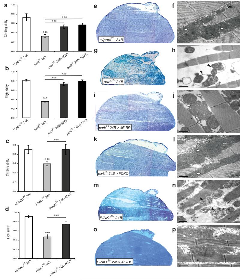
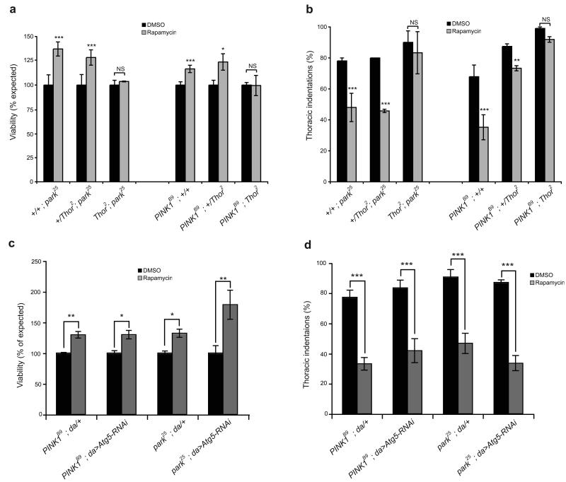
Mutations in PINK1 and PARK2 cause autosomal recessive parkinsonism, a neurodegenerative disorder that is characterized by the loss of dopaminergic neurons. To discover potential therapeutic pathways, we identified factors that genetically interact with Drosophila park and Pink1. We found that overexpression of the translation inhibitor Thor (4E-BP) can suppress all of the pathologic phenotypes, including degeneration of dopaminergic neurons in Drosophila. 4E-BP is activated in vivo by the TOR inhibitor rapamycin, which could potently suppress pathology in Pink1 and park mutants. Rapamycin also ameliorated mitochondrial defects in cells from individuals with PARK2 mutations. Recently, 4E-BP was shown to be inhibited by the most common cause of parkinsonism, dominant mutations in LRRK2. We also found that loss of the Drosophila LRRK2 homolog activated 4E-BP and was also able to suppress Pink1 and park pathology. Thus, in conjunction with recent findings, our results suggest that pharmacologic stimulation of 4E-BP activity may represent a viable therapeutic approach for multiple forms of parkinsonism.


Figures

Figure 1
Figure 1. 4E-BP overexpression suppresses parkin/PINK1 locomotor deficits and muscle degeneration
Overexpression of 4E-BP suppresses climbing and flight defects of (a, b) parkin and (c, d) PINK1 mutants. Overexpression of FOXO also rescued locomotor deficits in parkin mutants (a, b). (e-p) Toluidine blue stained sections of adult thorax and TEM images of muscle show 4E-BP or FOXO overexpression suppresses muscle degeneration and mitochondrial defects. Abnormal mitochondrial morphology in mutants (black arrowheads) is restored (white arrowheads). Scale bars show 1μm. Charts show mean and SEM. Significance was determined by one-way ANOVA with Bonferroni correction (*** P<0.001).
Figure 2
Figure 2. Overexpression of 4E-BP can suppress degeneration of dopaminergic neurons in parkin/PINK1 mutants
Quantification of anti-tyrosine hydroxylase positive stained dopaminergic neurons in the PPL1 cluster. (a) Effect of 4E-BP or FOXO overexpression in parkin or PINK1 mutants. Control genotype: park25/+; THG4/+. (b) Thor2 mutants show loss of dopaminergic neurons after 30 days compared to a revertant control. Charts show mean and SEM, n ≥ 10. Significance was determined by one-way ANOVA with Bonferroni correction (*** P<0.001, * P<0.05).
Figure 3
Figure 3. Post-translational state of 4E-BP activity in parkin/PINK1 mutants
(a) Western blot analysis of phosphorylated and non-phosphorylated 4E-BP levels in adult tissue from control, parkin, and PINK1 mutant flies. Thor2 mutant flies were used as a negative control. (b) Relative levels of phosphorylated and non-phosphorylated 4E-BP after normalization for total levels of 4E-BP. (c) Western blots for phosphorylated and non-phosphorylated Akt1 in control, parkin and PINK1 mutant adult tissue. (d) Quantified proportion of phospho-Akt1 relative to total Akt1. Charts show mean and SEM of at least three independent experiments. Significance was determined by Student’s t-test with Bonferroni correction (***P<0.001, *P<0.05).
Figure 4
Figure 4. Pharmacological suppression of parkin/PINK1 mutant phenotypes by rapamycin
(a) Thoracic indentations, (b) climbing ability, and (c) number of dopaminergic neurons in parkin and PINK1 mutants fed rapamycin or vehicle (DMSO). (d-f) TEM of muscle sections from control (DMSO) treated wild type, parkin and PINK1 mutants. (g-i) TEM of muscle sections from rapamycin fed wild type, parkin and PINK1 mutants Scale bars show 2μm. Wild types are out-crossed heterozygous mutations. Charts show mean and SEM. Significance determined by Student’s t-test (*** P<0.001, ** P<0.01).
Figure 5
Figure 5. Rapamycin rescues mitochondrial defects in parkin-deficient Drosophila and human cells
Analysis of mitochondrial aspect ratio (length) in (a) parkin RNAi treated Drosophila cells, and (b) fibroblasts from individuals with parkin mutations after exposure to rapamycin or vehicle. (c) Mitochondrial membrane potential in human parkin-deficient fibroblasts after rapamycin or control treatment. Charts show mean and SEM. Statistical analyses were performed using Kruskal-Wallis test with Dunn’s comparison for Drosophila cells or two-way ANOVA for fibroblast lines (*** P<0.001, ** P<0.01, * P<0.05).
Figure 6
Figure 6. Rapamycin suppression of parkin/PINK1 phenotypes is dependent on 4E-BP but does not require autophagy
Effects of rapamycin on (a,c) viability or (b,d) thoracic indentations in parkin and PINK1 mutants in combination with Thor2 mutations or RNAi-mediated knockdown of Atg5. Charts show mean and SEM of triplicate experiments. Significance determined by Mann-Whitney tests (*** P<0.001, ** P<0.01, * P<0.05).
Figure 7
Figure 7. GstS1 levels are increased by 4E-BP activation
(a) Western blot analysis of GstS1 levels in WT (w; 24B-GAL4/+), transgenic 4E-BP overexpression (w; 24B-GAL4/UAS-4E-BP), and wild type flies treated with rapamycin or vehicle. (b) Quantified GstS1 protein levels are normalized to a Tubulin loading control. Charts show mean and SEM of at least three replicates. Statistical analysis was Student’s t-test (**P<0.01, *P<0.05).
Figure 8
Figure 8. Loss of Drosophila LRRK increases the hypo-phosphorylated 4E-BP and partially suppresses parkin and PINK1 mutant phenotypes
(a) Western blot analysis of levels of phosphorylated and non-phosphorylated 4E-BP. (b) Quantification of relative amounts of phosphorylated and non-phosphorylated 4E-BP after normalization for total levels of 4E-BP. The mean and SEM of three independent experiments are represented. Analysis of (c) dopaminergic neurons, (d) flight and (e) climbing in mutant combinations. Charts show mean and SEM. Wild type is park25/+; LRRK e03680/+.

References

    1. Abou-Sleiman PM, Muqit MM, Wood NW. Expanding insights of mitochondrial dysfunction in Parkinson’s disease. Nat. Rev. Neurosci. 2006;7:207–219. - PubMed
    1. Farrer MJ. Genetics of Parkinson disease: paradigm shifts and future prospects. Nat. Rev. Genet. 2006;7:306–318. - PubMed
    1. Clark IE, et al. Drosophila pink1 is required for mitochondrial function and interacts genetically with parkin. Nature. 2006;441:1162–1166. - PubMed
    1. Park J, et al. Mitochondrial dysfunction in Drosophila PINK1 mutants is complemented by parkin. Nature. 2006;441:1157–1161. - PubMed
    1. Yang Y, et al. Mitochondrial pathology and muscle and dopaminergic neuron degeneration caused by inactivation of Drosophila Pink1 is rescued by Parkin. Proc. Natl. Acad. Sci. USA. 2006;103:10793–10798. - PMC - PubMed

Publication types

MeSH terms