. 2009 Sep;41(9):1022-6.
doi: 10.1038/ng.425. Epub 2009 Aug 16.

Mutation of SHOC2 promotes aberrant protein N-myristoylation and causes Noonan-like syndrome with loose anagen hair

Affiliations

Mutation of SHOC2 promotes aberrant protein N-myristoylation and causes Noonan-like syndrome with loose anagen hair

Viviana Cordeddu et al. Nat Genet. 2009 Sep.

Abstract

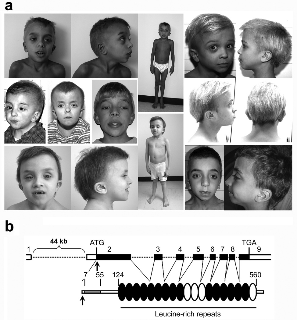
N-myristoylation is a common form of co-translational protein fatty acylation resulting from the attachment of myristate to a required N-terminal glycine residue. We show that aberrantly acquired N-myristoylation of SHOC2, a leucine-rich repeat-containing protein that positively modulates RAS-MAPK signal flow, underlies a clinically distinctive condition of the neuro-cardio-facial-cutaneous disorders family. Twenty-five subjects with a relatively consistent phenotype previously termed Noonan-like syndrome with loose anagen hair (MIM607721) shared the 4A>G missense change in SHOC2 (producing an S2G amino acid substitution) that introduces an N-myristoylation site, resulting in aberrant targeting of SHOC2 to the plasma membrane and impaired translocation to the nucleus upon growth factor stimulation. Expression of SHOC2(S2G) in vitro enhanced MAPK activation in a cell type-specific fashion. Induction of SHOC2(S2G) in Caenorhabditis elegans engendered protruding vulva, a neomorphic phenotype previously associated with aberrant signaling. These results document the first example of an acquired N-terminal lipid modification of a protein causing human disease.


Figures

Figure 1
Figure 1. The germline 4A>G mutation in the SHOC2 gene underlies a distinctive phenotype of the neuro-cardio-facial-cutaneous syndrome family
(a) Representative phenotypic features of affected subjects carrying the SHOC2 mutation. Common features include macrocephaly, high forehead, hypertelorism, palpebral ptosis, low-set/posteriorly rotated ears, short and webbed neck, and pectus anomalies. Affected subjects also exhibited easily pluckable, sparse, thin, slow-growing hair. (b) SHOC2 genomic organization and protein structure. The coding exons are shown at the top as numbered filled boxes. Intronic regions are represented by dotted lines. SHOC2’s motifs comprise an N-terminal lysine-rich region (grey coloured; Prosite motif score = 8.8) followed by 19 leucine-rich repeats (Pfam hits with an E-value <0.5 are black coloured, while those with an E-value >1 are represented in white). Numbers above the domain structure indicate the amino acid boundaries of those domains.
Figure 2
Figure 2. The disease-causing 4A>G change in SHOC2 promotes protein myristoylation and cell membrane targeting
(a) [3H]myristic acid incorporation (middle) occurs in SHOC2S2G but not in SHOC2wt or SHOC2S2A. Equivalent levels of SHOC2 proteins in immunoprecipitates (left) and [3H]myristic acid incorporation in cells (right) are shown. (b) SHOC2wt is uniformly present in the cytoplasm and nucleus in starved Cos-1 cells (upper left) and is restricted to the nucleus following EGF stimulation (upper right), while SHOC2S2G is targeted to the cell membrane basally (lower left) and after stimulation (lower right). Confocal microscopy visualized SHOC2 (anti-V5 monoclonal antibody, then Alexa Fluor-594 goat anti-mouse antibody; red), actin cytoskeleton (Alexa Fluor 488-phalloidin; green) and nuclei (DAPI; blue). (c) Cell fractioning assay documenting preferential membrane targeting of SHOC2S2G. Transiently transfected cells were serum-starved or stimulated with EGF, and lysates were fractionated to separate membrane-associated proteins. ERBB2 is shown to demonstrate equivalent fractionation efficiency, while anti-V5 blot from cell lysates show equivalent transfection efficiency. (d) Co-localization of V5-tagged SHOC2S2G and ganglioside M1 to the plasma membrane in Cos-1 cells. Subcellular localization of V5-tagged wild type SHOC2 (left) and V5-tagged SHOC2S2G (right) is shown. Ganglioside M1 was detected by using the Vybrant Lipid Raft Labeling kit (green). SHOC2 proteins and nuclei are visualized as reported above. Cells were cultured in DMEM supplemented with 10% heat-inactivated FBS. (e) Subcellular localization of the endogenous SHOC2wt protein in primary skin fibroblasts, basally (left) and following stimulation (right). Confocal microscopy was performed using an anti-SHOC2 polyclonal antibody, followed by Alexa Fluor 594 goat anti-rabbit antibody (red), while actin cytoskeleton was detected by Alexa Fluor 488-phalloidin (green). All images represent single optical sections representative of > 50 transfected cells observed in each experiment. Bars indicate 20 µm (b and d) or 40 µm (e).
Figure 3
Figure 3. Functional characterization of the disease-causing 4A>G change in SHOC2
(a) Subcellular localization of co-expressed SHOC2wt (green) and SHOC2S2G (red) documenting that SHOC2S2G does not impair EGF-stimulated SHOC2wt translocation to the nucleus. Imaging of V5-tagged (anti-V5 monoclonal antibody, then Alexa Fluor-594 goat anti-mouse antibody) and Myc-tagged (anti-Myc antibody, then Alex Fluor 488 goat anti-rabbit antibody) SHOC2 proteins and nuclei (DAPI, blue). Panels above show Myc-tagged SHOC2wt and V5-tagged SHOC2S2G and below show V5-tagged SHOC2wt and Myc-tagged SHOC2S2G. Cells were imaged basally (left) and following EGF stimulation (right). Bars indicate 20 µm. (b) Lysates of Cos-1 cells co-expressing Myc-tagged SHOC2wt and V5-tagged SHOC2S2G were immunoprecipitated using anti-Myc (above panel) or anti-V5 (below panel) antibody, and immunoprecipitated proteins were visualized by western blotting. These results indicate that SHOC2 proteins do not form heterodimers. (c, d) ERK phosphorylation in V5-tagged SHOC2wt or SHOC2S2G transiently expressed Neuro2A cells basally or following EGF stimulation. Phosphorylation levels are reported as a multiple of basal ERK phosphorylation in cells not transfected with a SHOC2 construct, averaged from four replicates ± s.d. (c). Results for cells expressing the S2G mutant were compared with those overexpressing the wild type protein (below) or untransfected cells (above) at the same time points using two-tailed t-test. * indicates P < 0.05, ** indicates P < 0.01, *** indicates P < 0.001. Representative blots are also shown (d).
Figure 4
Figure 4. Consequences of SHOC2S2G expression in C. elegans vulva development
(a–d) Nomarski images of vulvas of adult animals. A normal vulva is observed in animals expressing SHOC2wt (a), while in worms expressing SHOC2S2G (b and c) or myr::SHOC2wt (d) a protrusion of the vulva is visible. (e–j) Subcellular localization of V5-tagged SHOC2 proteins in C. elegans cells. In excretory canal cells (e–g) and intestinal cells (h–j), SHOC2wt protein is present throughout the cytoplasm (e and h), while both SHOC2S2G (f and i) and myr::SHOC2wt (g and j) are enriched in or restricted to the plasma membrane. Anti-V5 antibody (red) was used to visualize SHOC2 proteins. In intestinal cells, nuclei express GFP due to pelt-2::GFP plasmid used as a marker for transformation. (k–n) Nomarski images of vulval precursor cells at L3 stage. In animals expressing SHOC2wt only P6.p descendant invaginate (k), while in SHOC2S2G (l and m) and myr::SHOC2wt (n) expressing animals also P5.p (l to n) and P7.p descendants (m and n) detach from the cuticle. Black arrowheads point to P6.p descendant invagination, while white arrowheads point to P5.p and P7.p descendant invagination. Anterior is to the left and dorsal is up in all images.

References

    1. Resh MD. Trafficking and signaling by fatty-acylated and prenylated proteins. Nat Chem Biol. 2006;2:584–590. - PubMed
    1. Farazi TA, Waksman G, Gordon JI. The biology and enzymology of protein N-myristoylation. J Biol Chem. 2001;276:39501–39504. - PubMed
    1. Selfors LM, Schutzman JL, Borland CZ, Stern MJ. soc-2 encodes a leucine-rich repeat protein implicated in fibroblast growth factor receptor signaling. Proc Natl Acad Sci USA. 1998;95:6903–6908. - PMC - PubMed
    1. Sieburth DS, Sun Q, Han M. SUR-8, a conserved Ras-binding protein with leucine-rich repeats, positively regulates Ras-mediated signaling in C. elegans. Cell. 1998;94:119–130. - PubMed
    1. Li W, Han M, Guan KL. The leucine-rich repeat protein SUR-8 enhances MAP kinase activation and forms a complex with Ras and Raf. Genes Dev. 2000;14:895–900. - PMC - PubMed

Publication types