

Comparative pathogenesis of an avian H5N2 and a swine H1N1 influenza virus in pigs
- PMID: 19684857
- PMCID: PMC2722722
- DOI: 10.1371/journal.pone.0006662
Comparative pathogenesis of an avian H5N2 and a swine H1N1 influenza virus in pigs
Abstract
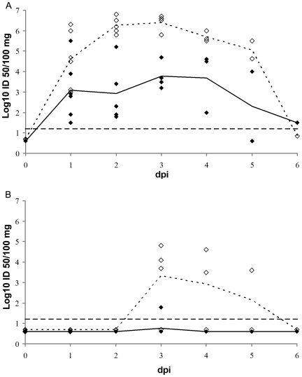
Pigs are considered intermediate hosts for the transmission of avian influenza viruses (AIVs) to humans but the basic organ pathogenesis of AIVs in pigs has been barely studied. We have used 42 four-week-old influenza naive pigs and two different inoculation routes (intranasal and intratracheal) to compare the pathogenesis of a low pathogenic (LP) H5N2 AIV with that of an H1N1 swine influenza virus. The respiratory tract and selected extra-respiratory tissues were examined for virus replication by titration, immunofluorescence and RT-PCR throughout the course of infection. Both viruses caused a productive infection of the entire respiratory tract and epithelial cells in the lungs were the major target. Compared to the swine virus, the AIV produced lower virus titers and fewer antigen positive cells at all levels of the respiratory tract. The respiratory part of the nasal mucosa in particular showed only rare AIV positive cells and this was associated with reduced nasal shedding of the avian compared to the swine virus. The titers and distribution of the AIV varied extremely between individual pigs and were strongly affected by the route of inoculation. Gross lung lesions and clinical signs were milder with the avian than with the swine virus, corresponding with lower viral loads in the lungs. The brainstem was the single extra-respiratory tissue found positive for virus and viral RNA with both viruses. Our data do not reject the theory of the pig as an intermediate host for AIVs, but they suggest that AIVs need to undergo genetic changes to establish full replication potential in pigs. From a biomedical perspective, experimental LP H5 AIV infection of pigs may be useful to examine heterologous protection provided by H5 vaccines or other immunization strategies, as well as for further studies on the molecular pathogenesis and neurotropism of AIVs in mammals.
Conflict of interest statement
Figures
References
-
- Van Reeth K. Avian and swine influenza viruses: our current understanding of the zoonotic risk. Vet Res. 2007;38:243–260. - PubMed
Publication types
MeSH terms
Substances
Grants and funding
LinkOut - more resources
Full Text Sources
