

Expression, localisation and functional activation of NFAT-2 in normal human skin, psoriasis, and cultured keratocytes
- PMID: 19684889
- PMCID: PMC2719706
Expression, localisation and functional activation of NFAT-2 in normal human skin, psoriasis, and cultured keratocytes
Abstract
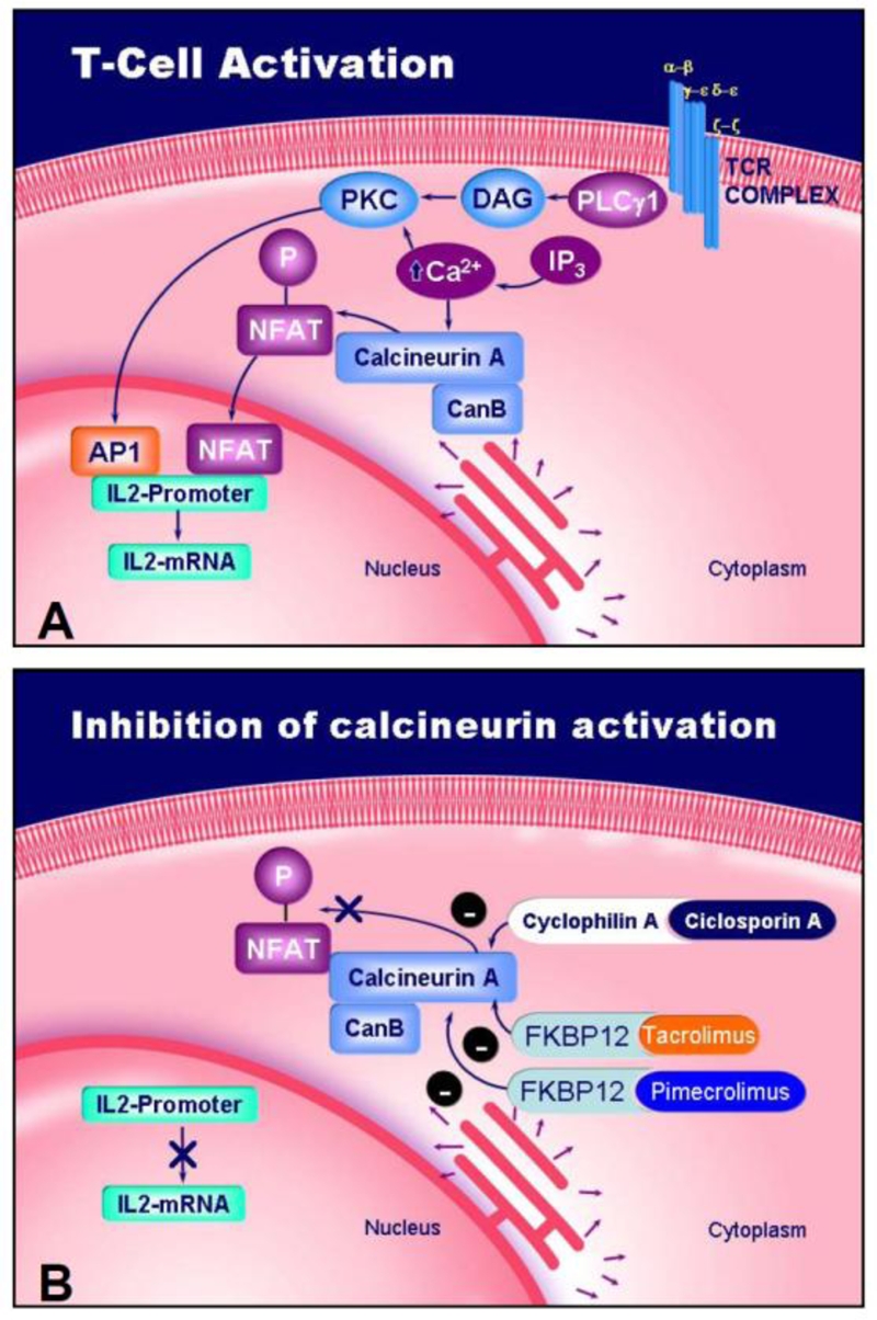
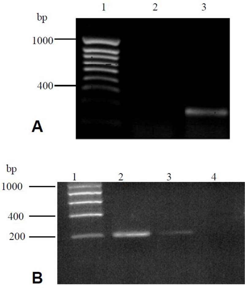
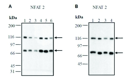
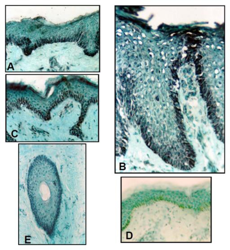
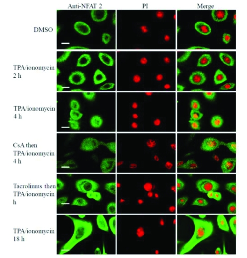
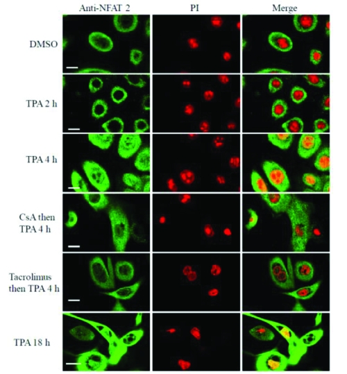
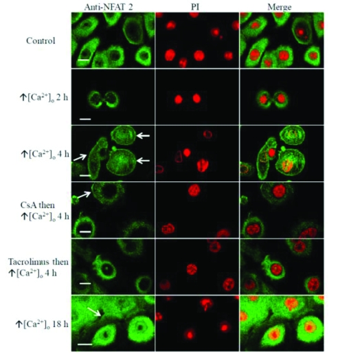
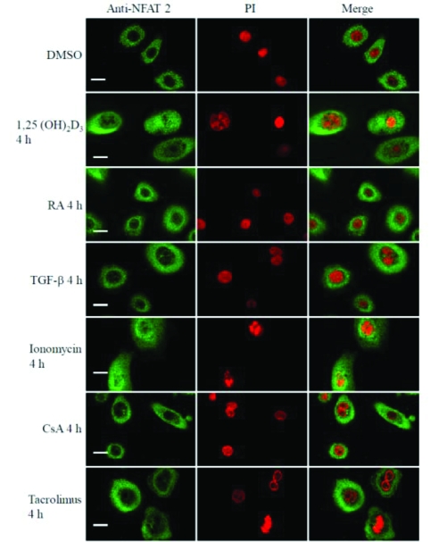
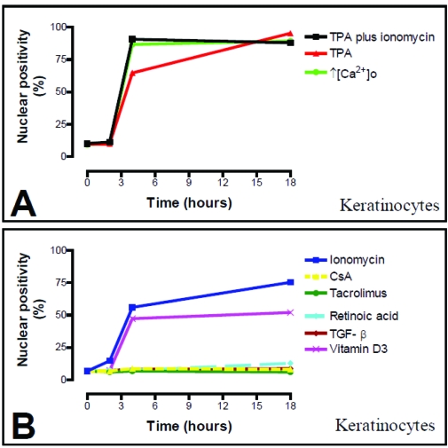
Ciclosporin A (CsA) is widely utilized for the treatment of inflammatory skin diseases such as psoriasis. The therapeutic effects of CsA are thought to be mediated via its immunosuppressive action on infiltrating lymphocytes in skin lesions. CsA and tacrolimus block T cell activation by inhibiting the phosphatase calcineurin and preventing translocation from the cytoplasm to the nucleus of the transcription factor Nuclear Factor of Activated T cells (NFAT). As calcineurin and NFAT 1 have been shown to be functionally active in cultured human keratocytes, expression of other NFAT family members such as NFAT-2 and possible functional activation was investigated in human keratocytes. RT-PCR and Western Analysis were used to investigate the presence of NFAT-2 mRNA and protein in human keratocytes. Tissue culture of human keratocytes and immunostaining of cells on coverslips and confocal microscopy were used to assess the degree of nuclear localisation of NFAT-2 in cultured cells. Keratome biopsies were taken from patients with psoriasis (lesional and non-lesional skin) and normal skin and immunohistochemistry was used to assess the NFAT-2 localisation in these biopsies using a well characterized anti-NFAT-2 antibody. The NFAT-2 mRNA and protein expression was demonstrated using RT-PCR and Western blotting. Moreover, the expression of NFAT-2 in normal skin, non-lesional and lesional psoriasis showed a striking basal staining suggesting a role for NFAT-2 in keratocytes proliferation. A range of cell types in the skin express NFAT-2. The expression of NFAT-2 in human keratocytes and response to different agonists provides perhaps a unique opportunity to examine the regulation, subcellular localization and kinetics of translocation of different NFATs in primary cultured human cells. In these experiments the author assessed the expression, localization of NFAT-2 in cultured human keratocytes and measured the degree of nuclear localisaion of NFAT-2 using immunofluorescence and confocal microscopy and whether CsA and tacrolimus inhibit NFAT-2 nuclear translocation. As with NFAT 1, differentiation-promoting agents that increase intracellular calcium concentration induced nuclear translocation of NFAT-2 in cultured keratocytes but with different kinetics. These data provide the first evidence of that NFAT-2 is expressed in normal skin, psoriasis and that NFAT-2 functionally active in human keratocytes and that nuclear translocation of NFAT-2 in human skin cells has different kinetics than NFAT 1 suggesting that NFAT-2 may play an important role in regulation of keratocytes proliferation and differentiation at a different stage. Inhibition of this pathway in human epidermal keratocytes many account, in part for the therapeutic effects of CsA and tacrolimus in skin disorders such as psoriasis. Thus, supporting our previous work data that calcineurin/NFAT is functionally active not only in T cells, but in skin cells.
Keywords: Ciclosporin A (CsA); NFAT-2; Tacrolimus; calcineurin; intracellular calcium; keratocytes differentiation; psoriasis.
Figures
References
-
- Hodge MR, Chun HJ, Rengarajan J, Alt A, Lieberson R, Glimcher LH. NF-AT-Driven interleukin-4 transcription potentiated by NIP45. Science. 1996;274:1903–5. - PubMed
-
- Kiani A, Viola JP, Lichtman AH, Rao A. Down-regulation of IL-4 gene transcription and control of Th2 cell differentiation by a mechanism involving NFAT1. Immunity. 1997;7:849–60. - PubMed
-
- Oukka M, Ho IC, de la Brousse FC, Hoey T, Grusby MJ, Glimcher LH. The transcription factor NFAT4 is involved in the generation and survival of T cells. Immunity. 1998;9:295–304. - PubMed
-
- Ranger AM, Oukka M, Rengarajan J, Glimcher LH. Inhibitory function of two NFAT family members in lymphoid homeostasis and Th2 development. Immunity. 1998;9:627–35. - PubMed
-
- Ranger AM, Hodge MR, Gravallese EM, et al. Delayed lymphoid repopulation with defects in IL-4-driven responses produced by inactivation of NF-ATc. Immunity. 1998;8:125–34. - PubMed
LinkOut - more resources
Full Text Sources
Other Literature Sources
Research Materials
Miscellaneous