. 2009 Sep 29;48(38):8985-93.
doi: 10.1021/bi900833f.

The role of arginine-127 at the proximal NO-binding site in determining the electronic structure and function of 5-coordinate NO-heme in cytochrome c' of Rhodobacter sphaeroides

Affiliations

The role of arginine-127 at the proximal NO-binding site in determining the electronic structure and function of 5-coordinate NO-heme in cytochrome c' of Rhodobacter sphaeroides

Byunghoon Lee et al. Biochemistry. .

Abstract

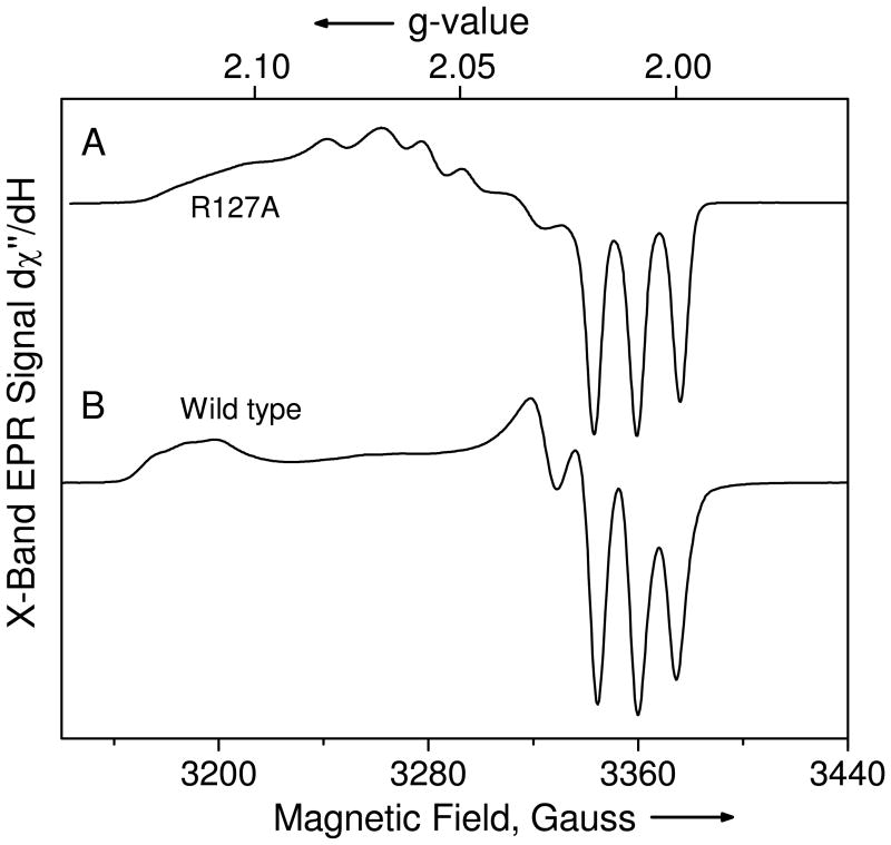
Cytochrome c' is a heme protein from a denitrifying variant of Rhodobacter sphaeroides which may serve to store and transport metabolic NO while protecting against NO toxicity. Its heme site bears resemblance through its 5-coordinate NO-binding capability to the regulatory site in soluble guanylate cyclase. A conserved arginine (Arg-127) abuts the 5-coordinate NO-heme binding site, and the alanine mutant R127A provided insight into the role of the Arg-127 in establishing the electronic structure of the heme-NO complex and in modifying the heme-centered redox potential and NO-binding affinity. By comparison to R127A, the wild-type Arg-127 was determined to increase the heme redox potential, diminish the NO-binding affinity, perturb and diminish the 14NO hyperfine coupling determined by ENDOR (electron nuclear double resonance), and increase the maximal electronic g-value. The larger isotropic NO hyperfine and the smaller maximal g-value of the R127A mutant together predicted that the Fe-N-O bond angle in the mutant is larger than that of the Arg-127-containing wild-type protein. Deuterium ENDOR provided evidence for exchangeable H/D consistent with hydrogen bonding of Arg-127, but not Ala-127, to the O of the NO. Proton ENDOR features previously assigned to Phe-14 on the distal side of the heme were unperturbed by the proximal side R127A mutation, implying the localized nature of that mutational perturbation at the proximal, NO-binding side of the heme. From this work two functions of positively charged Arg-127 emerged: the first was to maintain the KD of the cytochrome c' in the 1 microM range, and the second was to provide a redox potential that enhances the stability of the ferrous heme.


Figures

Figure 1
Figure 1
This figure provides a scheme of the locale of the heme group in Cyt c′, showing the NO, the arginine near the NO (Arg-127 in R. sphaeroides) and the occluding distal Phe-14. Since no crystal structure of NO-Cyt c′ is available from R. sphaeroides, this composite graphic was created from the heme, NO, and arginine coordinates of the NO-Cyt c′ of A. xylosoxidans (1E85) and the Phe-14 and heme coordinates of ferric Cyt c′ of R. sphaeroides (1GQA) by application of Swiss PdB Viewer™ software. The numbering system is that of R. sphaeroides. There were two crystallographically resolved orientations of the NO, and the closer one to Arg-127 is shown. The putative positions of protons were provided by Swiss PdB Viewer™ software. Plausible hydrogen bonds from the nearest guanidinium protons of Arg-127 to the O of NO are schematically indicated.
Figure 2
Figure 2
UV-Vis absorption spectra of (A) mutant (R127A) and (B) wild type R. sphaeroides Cyts c′. Heme concentration is 10μM; in 100 mM phosphate buffer, pH 7. Black spectrum is ferric, red spectrum is ferrous, green spectrum is the NO-bound ferrous, and blue spectrum is the CO-bound ferrous.
Figure 3
Figure 3
X-band EPR spectra of the ferrous 14NO derivatives of (A) the R127A mutant and (B) wild type Cyt c′. The spectra were recorded at T = 77 K, 3 G field modulation, 100 s of signal averaging, 2 mW microwave power, EPR frequency νEPR = 9.446 GHz.
Figure 4
Figure 4
Nitrogen ENDOR spectra of the 14NO-ligated R127A mutant (black) and wild type Cyt c′ (red) at the following magnetic fields: 1.158 T (g = 2.104), 1.168 T (g = 2.086), 1.178 T (g = 2.069), 1.208 T (g =2.018), and 1.212 T (g = 2.010). Arrows indicate 14N nuclear Zeeman splitting of 2 14ν. Conditions were adiabatic rapid passage, T = 2 K, microwave power = 0.22 nW, 100 kHz field modulation = 5 G ptp, a system time constant = 160 ms, RF power ≈ 20 W, radio frequency sweep rate = 2 MHz/s, overall signal averaging time = 400 s, vEPR = 34.10 GHz, RF power was pulsed with a 100/900 μs duty cycle.
Figure 5
Figure 5
This figure provides a comparison of the strongly coupled proton ENDOR spectra of the 14NO-bound ferrous forms of the R127A mutant (black) and wild type (red) at the g-values indicated. Protons α, α′ were previously assigned as the nearest protons of Phe-14. Data were obtained under the conditions: adiabatic rapid passage, T = 2 K, microwave power = 0.22 nW, 100 kHz field modulation = 2 G ptp, a system time constant = 160 ms, RF power ≈ 20 W, radio frequency sweep rate = 2 MHz/s, overall signal averaging time = 400 s, vEPR = 34.10 GHz, RF power was pulsed with a 100/900 μs duty cycle.
Figure 6
Figure 6
ENDOR spectra of D2O-exchanged NO-Cyt c′ from wild type and R127A mutant (black) and wild type (red). These spectra were collected near the free deuterium ENDOR frequency of 7.9 MHz which occurs at the magnetic field of 1.208 T (g = 2.017). Experimental conditions were: adiabatic rapid passage, T = 2 K, microwave power = 0.22 nW, 100 kHz field modulation = 0.15 G ptp, a system time constant = 320 ms, RF power ≈ 20 W, RF sweep rate = 0.06 MHz/s, overall signal averaging time = 1200 s, vEPR = 34.10 GHz, RF power was pulsed with a 100/900 μs duty cycle.
Figure 7
Figure 7
These Nernst plot traces show redox titrations in the form of Nernst Plots for the heme group of Cyt c′ belonging to wild type (triangles) and R127A (squares). The midpoint potential for wild type protein was +10 ± 3 mV. For R127A the midpoint potential was −24 ± 3 mV.
Figure 8
Figure 8
UV-Vis titrations of (A) mutant Cyt c′ (R127A) and (B) ferrous wild type Cyt c′, 10 μM protein in 100 mM phosphate buffer at pH 7.05. For (A), [NO] (X 10−7 M): 0.41 (blue), 0.80 (cyan), 1.13 (magenta), 1.44 (yellow), 1.77 (dark yellow), 2.24 (navy), 2.71 (purple), 3.22 (wine), 3.82 (olive), 4.45 (dark cyan), 5.12 (royal), 5.82 (orange), and for (B), [NO] (X 10−7 M): 1.33 (green), 2.74 (blue), 4.37 (cyan), 5.99 (magenta), 7.645 (yellow), 9.29 (dark yellow), 10.9 (navy), 12.5 (purple), 14.0 (wine), 19.3 (royal), 23.0 (violet).
Figure 9
Figure 9
Fractional heme NO saturation levels (Y) are presented for ferrous myoglobin (squares), R127A (triangles), and wild type Cyt c′ (diamonds) as a function of free NO concentration. The values of Y were determined from the NO-sensitive UV-Vis spectra at 450.0 nm for myoglobin and at 430.6 nm for both Cyts c′. Free NO concentration was determined from the difference between added NO and protein bound NO, as described in the Methods Section.
Scheme A
Scheme A
This scheme is taken from (8). Left: Energy levels, d-centered wavefunctions, and spin population of the low spin d6 ferroheme. Center: Perturbed d-centered energy levels and wavefunctions after bonding to NO and bending of the Fe-N-O. The lowest energy orbital has primarily n character, the next six orbitals, including the SOMO, have primarily d character, and the two highest unfilled orbitals have primarily π* character. The energy levels are schematic. Right: Important orbitals of the nitrosyl ligand.

Similar articles

Cited by

References

    1. Lawson DM, Stevenson CE, Andrew CR, Eady RR. Unprecedented proximal binding of nitric oxide to heme: implications for guanylate cyclase. Embo J. 2000;19:5661–5671. - PMC - PubMed
    1. Lawson DM, Stevenson CE, Andrew CR, George SJ, Eady RR. A two-faced molecule offers NO explanation: the proximal binding of nitric oxide to haem. Biochem Soc Trans. 2003;31:553–557. - PubMed
    1. Ramirez LM, Axelrod HL, Herron SR, Rupp B, Allen JP, Kantardjieff KA. High resolution crystal structure of ferricytochrome c′ from Rhodobacter sphaeroides. Journal of Chemical Crystallography. 2003;33:413–424.
    1. Choi PS, Grigoryants VM, Abruna HD, Scholes CP, Shapleigh JP. Regulation and function of cytochrome c′ in Rhodobacter sphaeroides 2.4.3. J Bacteriol. 2005;187:4077–4085. - PMC - PubMed
    1. Choi PST. Ph D Thesis, Dept of Microbiology. Cornell; Ithaca, NY: 2004.

Publication types

MeSH terms